Abelson tyrosine-protein kinase 2 regulates myoblast proliferation and controls muscle fiber length
- NYU Medical School, United States
Figures
Figure 1 with 1 supplement

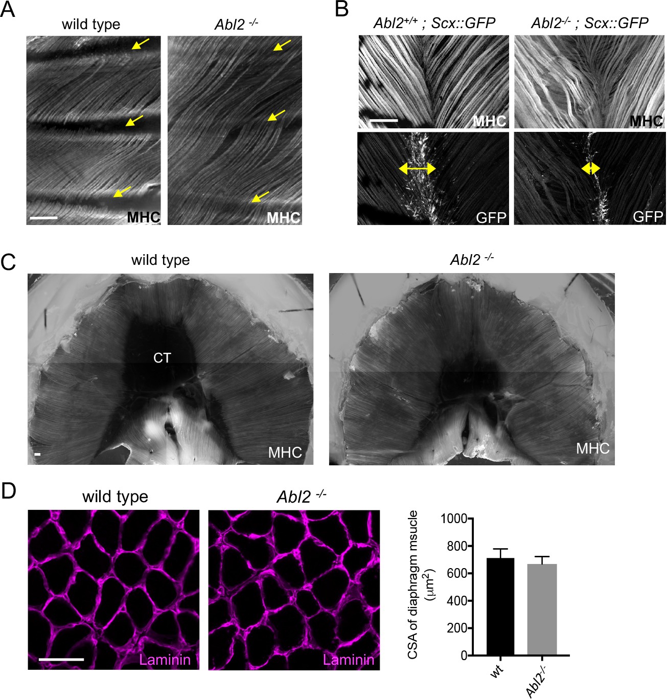
Diaphragm muscle fibers in E18.5 Abl2 mutant mice are extraordinarily long, and the central tendon is diminished in size.
Whole mounts of muscle were stained with antibodies to myosin heavy chain (MHC). (A) Costal muscle fibers in E18.5 embryonic diaphragm muscle normally extend from the ribcage and attach medially to the central tendon (CT). (B) The mean myofiber length, measured in the ventral quadrant of the costal diaphragm muscle, is ~1.7 fold longer in E18.5 Abl2 mutant than in wild type (wt) mice. (C,D) The area of the central tendon, circumscribed by Scx RNA expression, was reduced in Abl2−/− mice. **p<0.01, ****p<0.001.
Figure 1—figure supplement 1
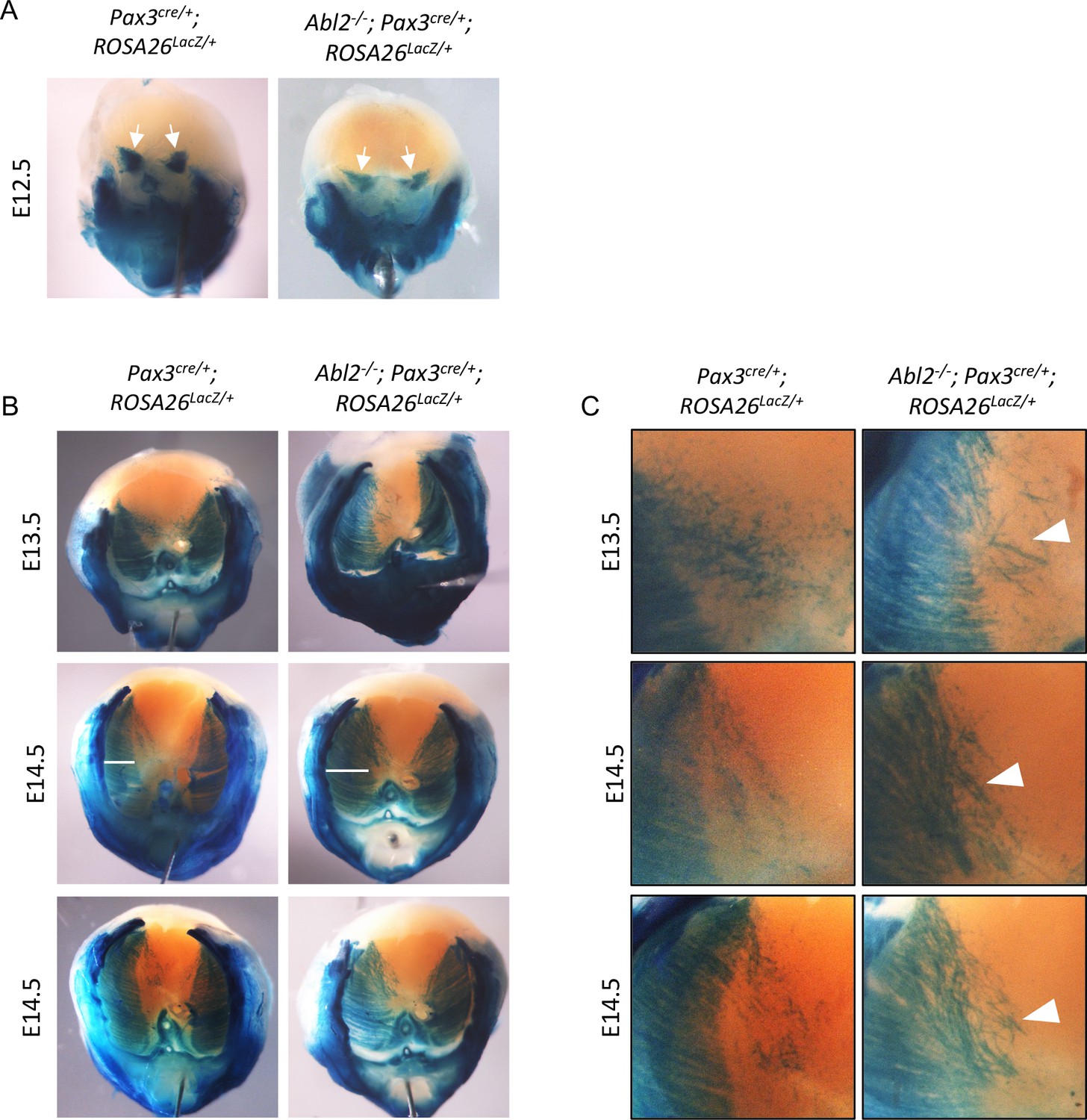
Embryonic development of Abl2−/− diaphragm.
Embryos were collected between E12.5-E14.5 to detect differences in diaphragm muscle development between Abl2 mutant and control mice. β-galactosidase activity was detected in myoblasts and muscle fibers from Abl2−/−; Pax3cre/+; ROSA26LacZ/+ mutant and Pax3cre/+; ROSA26LacZ/+ control littermates. (A) At E12.5, Pax3+ cells accumulated at the dorsal edge of the developing diaphragm (white arrows) in both Abl2 mutant and control mice. (B) At 14.5, the muscle domain appears larger in Abl2 mutant mice (white line) compared to control mice. (C) Higher magnification images of the ventral left quadrant of the diaphragm from E13.5–14.5 mice. Muscle fibers begin to extend into the central tendon domain in Abl2 mutant mice (white arrowheads), whereas muscle fibers are largely excluded from the central tendon domain in control mice.
Figure 2 with 1 supplement
Muscle fibers are excessively long in intercostal, levator auris, and diaphragm muscles in adult Abl2 mutant mice.
Whole mounts of adult muscle were stained with antibodies to myosin heavy chain (MHC) and GFP. Cross-sections of muscle were stained with antibodies to Laminin. (A) Intercostal muscle fibers normally extend from one rib to the adjacent rib (arrows) but extend and cross over one or more ribs in Abl2 mutant mice. (B) Muscle fibers in the levator auris muscle appear wavy in Abl2 mutant mice. Moreover, the midline tendon, marked by Scx::GFP, is reduced in size (double headed arrows). (C) Muscle fibers in the diaphragm muscle remain longer and the central tendon is reduced in size in adult Abl2 mutant mice. (D) The cross-sectional area of myofibers in adult, 8 week old, Abl2−/− mice is normal. Scale bar is 250 µm in A and B, and 50 µm in D.
Figure 2—figure supplement 1
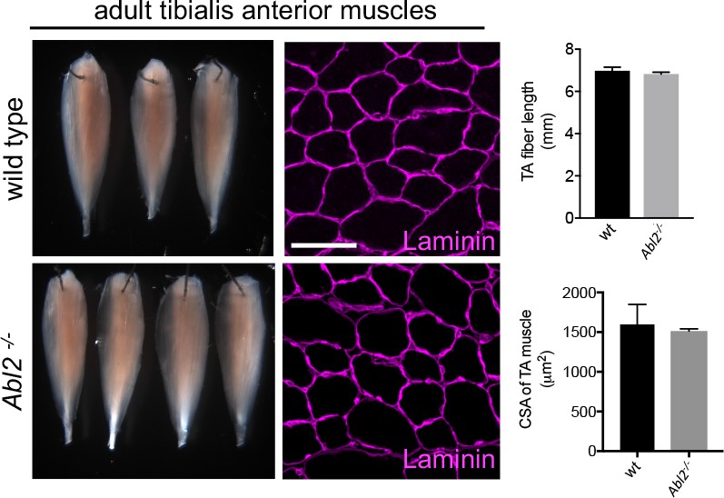
Limb muscles appear normal in muscle length and cross-sectional area.
Hind limbs were fixed in 4% PFA, and tibialis anterior (TA) muscles were dissected at full length from three wild type and 3 Abl2−/− mice. Sections were stained with antibodies to Laminin to outline the muscle fibers. The length and cross-sectional area of Abl2−/− TA muscles are normal. The means ± SEMs from the left TA muscles of 3–4 mice in each group are shown. Scale bar is 50 µm.
Figure 3
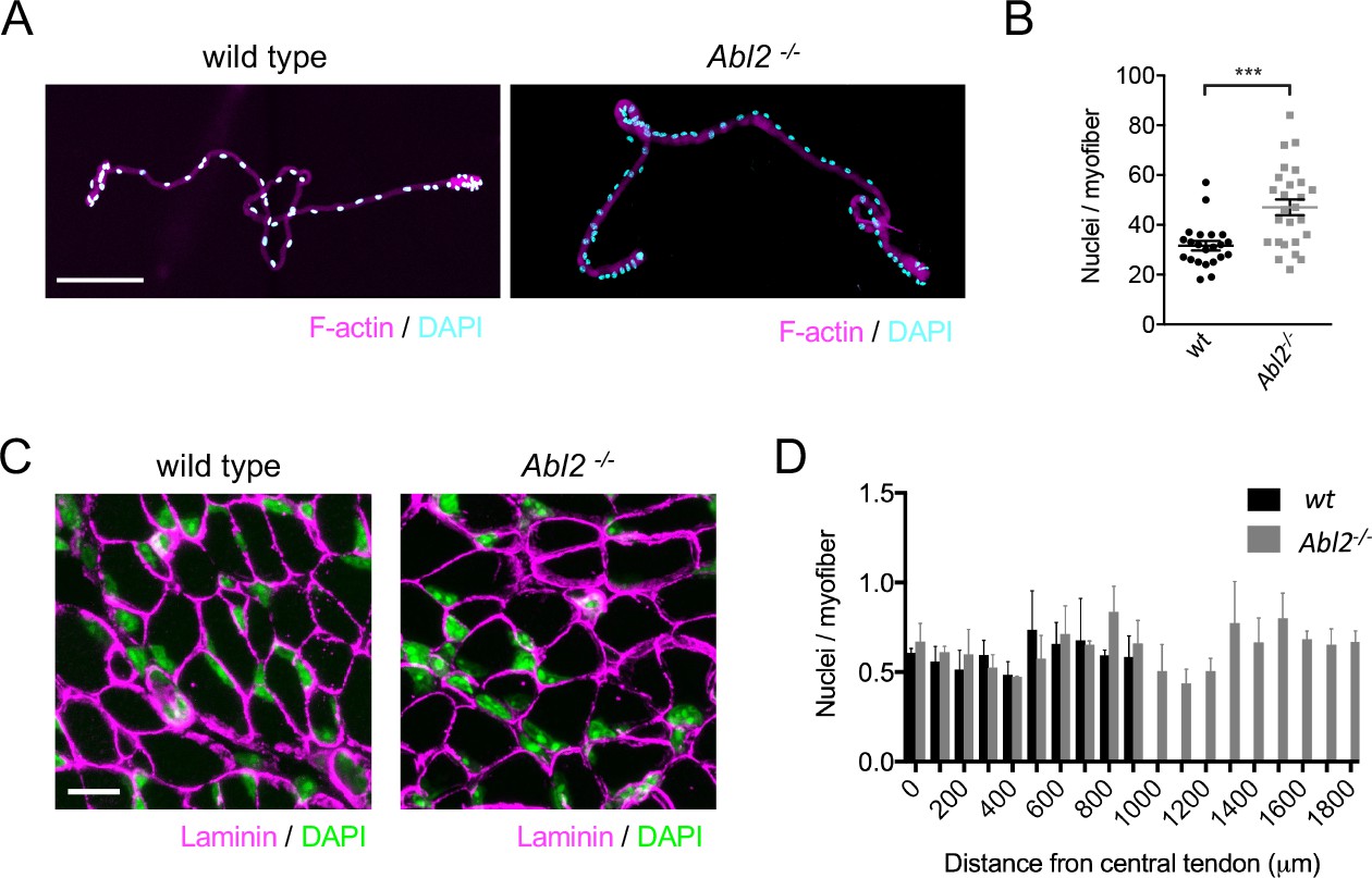
The increase in muscle fiber length is accompanied by an increase in myonuclei number.
(A) Single fibers dissociated from the ventral quadrant of the costal diaphragm muscle were stained with Phalloidin to label F-actin and DAPI to label nuclei. (B) The scatter plot shows the number of nuclei per myofiber for individual dissociated myofibers, dissected from three individual Abl2 mutant and three wild type (wt) mice, as well as the mean ±SEM. (C) Representative images from serial cross sections of the diaphragm muscle, stained for DAPI to label nuclei and with antibodies to Laminin to outline muscle fibers. (D) The number of myonuclei in each section was divided by the number of muscle fibers. Myonuclei are similarly spaced along the entire length of muscle from wild type and Abl2 mutant mice. The graph shows the mean ±SEM per field of view in serial cross sections taken every 100 µm from the central tendon to the ribcage of 3 wild type and 3 Abl2 mutant mice. Scale bar is 150 µm in A and 10 µm in C. ***p<0.005.
Figure 4 with 1 supplement
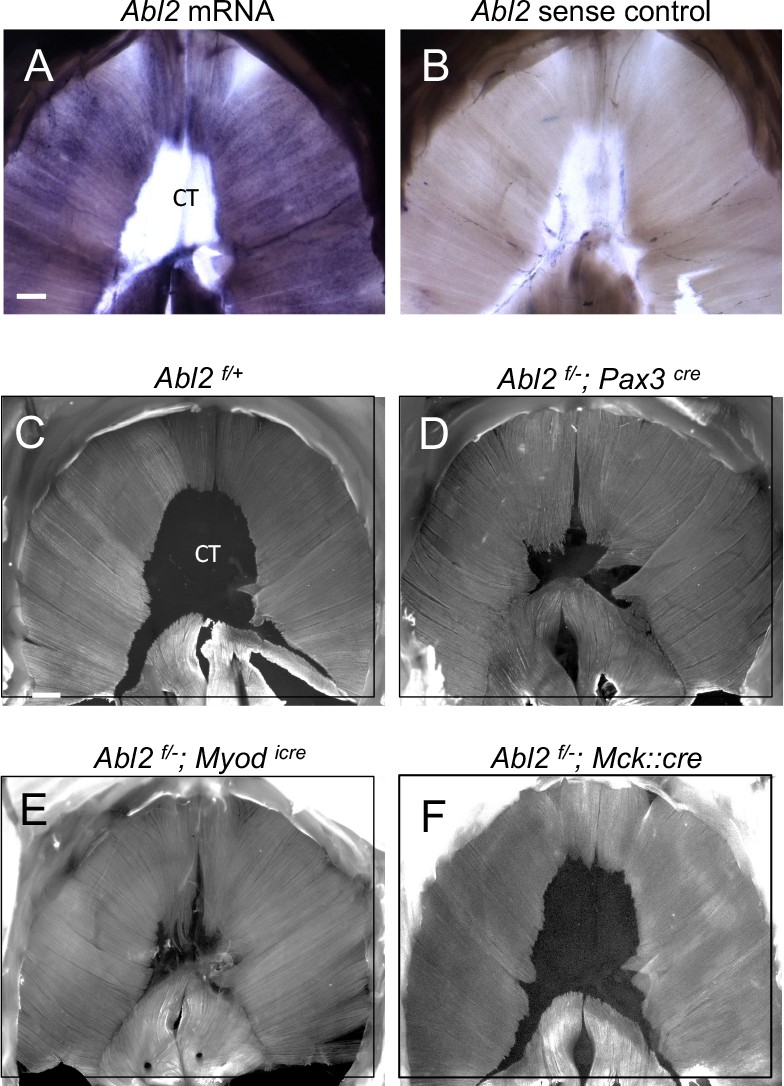
Abl2 acts in myoblasts to regulate muscle development.
(A,B) Abl2 mRNA is highly expressed in the muscle but not within the central tendon (CT) of the diaphragm. (C) Muscle and tendon development appear normal in Abl2flox/+ control mice (Abl2f/+). Muscle length is increased and tendon size is reduced by conditionally inactivating Abl2 in (D) muscle precursors (Abl2f/−; Pax3cre), or (E) committed myoblasts (Abl2f/−; Myodicre). (F) Muscle and tendon development appear normal by inactivating Abl2 in mature myotubes (Abl2f/−; Mck::cre). Scale bars are 500 µm in A and C.
Figure 4—figure supplement 1
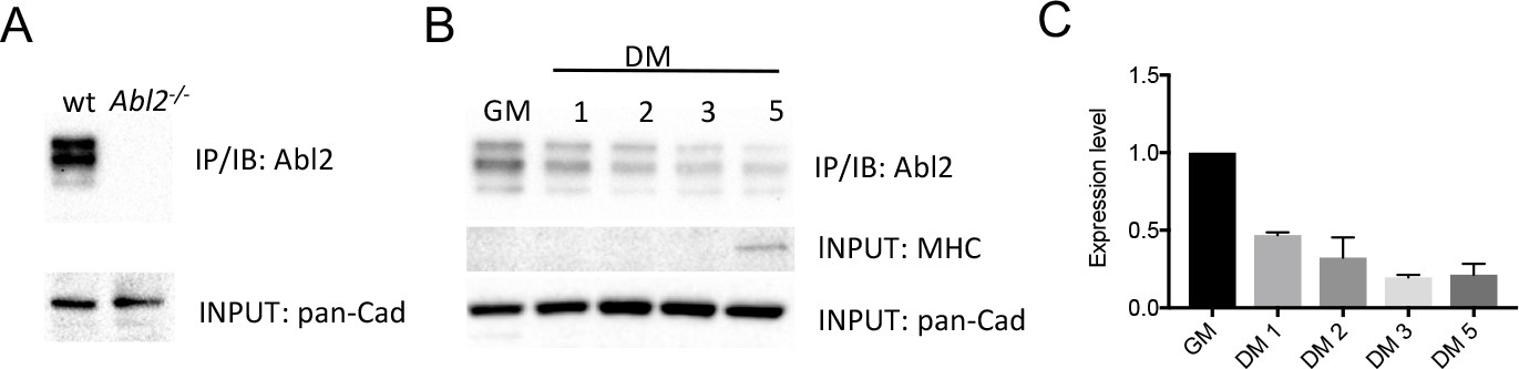
Abl2 expression is enriched in proliferating myoblasts.
(A) Western blot analysis of wild type (wt) and Abl2−/− primary cultures. Cadherin levels (pan-Cad) were measured in cell lysates, and Abl2 was immunoprecipitated (IP) from lysates containing equal levels of pan-Cad in wt and Abl2−/− primary cultures to validate the IP and immunoblotting (IB) antibodies. (B) C2C12 cells collected at sequential time points during differentiation. Abl2 was immunoprecipitated from lysates of C2C12 cultures grown in growth medium (GM) or in differentiation medium (DM) for the indicated number of days. Abl2 is highly expressed in C2C12 cells in growth media and migrates as three distinct bands between 100–150 kDa. (C) Abl2 expression declines during differentiation. Abl2 expression in GM was assigned a value of 1.0, and all other values are expressed relative to GM.
Figure 5 with 1 supplement

The exercise endurance of Abl2 null and Abl2 muscle-conditional mutant mice is impaired.
(A) During a twenty-hour period, Abl2−/− and Abl2f/−; MyodiCre conditionally mutant mice run less than wild type (wt) mice. (B) The limb strength of Abl2−/−, Abl2f/−; MyodiCre and wild type mice are similar. The graph and scatter plot show the values for individual mice and the mean values together with the SEMs. All Abl2 null and conditional Abl2 mutant mice were tested at 18 weeks with littermate controls. (C) Cross-sections of the diaphragm muscle were stained with Laminin and myosin heavy chain Type I (MHC-slow) to identify slow-twitch muscle fibers. The number of slow-twitch fibers is normal in Abl2 mutant mice. The scatter plot shows the percentage of myofibers in cross-sections of muscle that were stained by antibodies to MHC-I. The mean values and SEMs from three control (Abl2f/+) and four muscle-conditional Abl2 mutant mice (Abl2f/−; MyodiCre) are shown. Scale bar is 50 µm.
Figure 5—figure supplement 1

Ultrastructural appearance of muscle-tendon junctions and muscle fibers is normal in Abl2 mutant mice.
(A) Deep muscle membrane specializations form at the myotendinous junction in wild type and Abl2−/− mice. (B) The scatter plot shows the sarcomere lengths for 12 fibers from two wild type and two Abl2−/− mice. The mean ±SEMs are also shown.
Figure 6 with 1 supplement
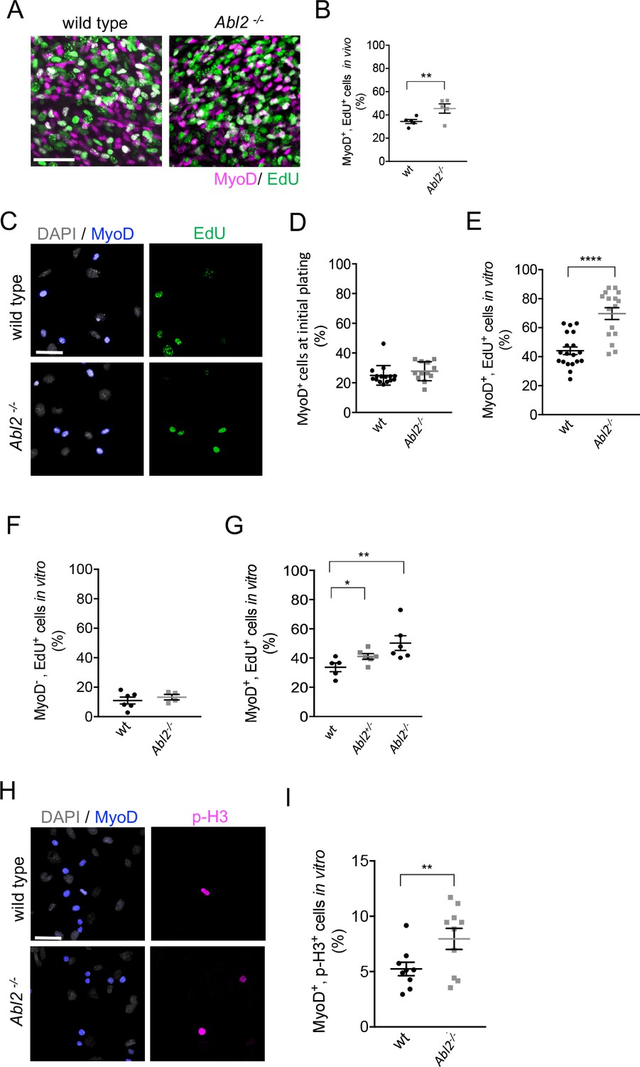
Myoblast proliferation is enhanced by a loss of Abl2.
(A,B) In vivo labeling of E13.5-E14.5 diaphragm muscles shows that EdU incorporation is greater in Abl2−/− myoblasts than wild type (wt) myoblasts. (C) Representative images of cultured diaphragm cells stained with MyoD to label myoblasts, DAPI to label all cells and EdU to label proliferating cells. (D) At initial plating, a similar number of MyoD+ cells are isolated from Abl2−/− and wild type mice. (E) Cultured MyoD+ myoblasts from Abl2−/− diaphragm muscles showed increased EdU incorporation. (F) In contrast, non-muscle cells (MyoD−) from Abl2 mutant and wild type mice proliferate at similar rates. (G) Abl2 heterozygous myoblasts proliferated at a rate that was intermediate between wild type and Abl2 homozygous mutant myoblasts. (H) Representative images of cultured diaphragm cells stained with MyoD to label myoblasts, DAPI to label all cells, and phospho-Histone H3 (pHH3), a marker for mitotic cells. (I) MyoD+ myoblasts, cultured from the diaphragm muscle of Abl2−/− mice, showed a greater percentage of mitotic figures than myoblasts isolated from wild type mice. *p=0.1, **p<0.05, ****p<0.001.
Figure 6—figure supplement 1
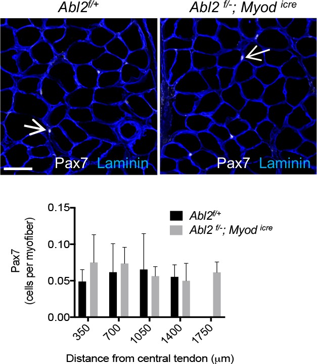
The number of Pax7+cells is increased in proportion to the increased length of myofibers.
Serial cross-sections of the diaphragm were stained with antibodies to Laminin to outline muscle fibers and with antibodies to Pax7 to label satellite cells. The number of Pax7+ cells was divided by the total number of muscle fibers in each visual field. Longer muscle fibers in Abl2−/− mice have proportionally more Pax7+ cells. The graph shows the mean (±s.d.) number of Pax7+ cells in serial cross sections from three mice in each group. Scale bar is 50 µm.
Figure 7
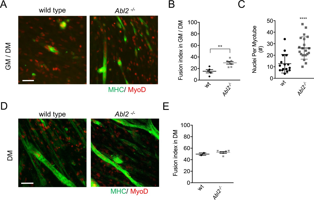
Enhanced myoblast proliferation in Abl2−/− mice leads to enhanced myoblast fusion.
Primary cultures from the diaphragm muscle of E18.5 mice were stained with antibodies to myosin heavy chain (MHC) to label differentiated muscle fibers and MyoD to label myoblasts. (A) Representative images of diaphragm muscle cells, which were proliferated for 2 days before a switch to differentiation medium. (B) Under these conditions, Abl2 mutant myoblasts displayed enhanced myotube formation. (C) The number of nuclei per myotube was quantified. Differentiated Abl2 mutant myotubes incorporated more nuclei per myotube. (D) Representative images of Abl2 mutant myoblasts, which were plated at confluent density and directly into differentiation media. (E) Confluent cultures, which did not have an opportunity to proliferate, formed myotubes like wild type myoblasts. Scale bars are 50 µm in A,I, and L. **p<0.01, ****p<0.001.
Figure 8 with 1 supplement
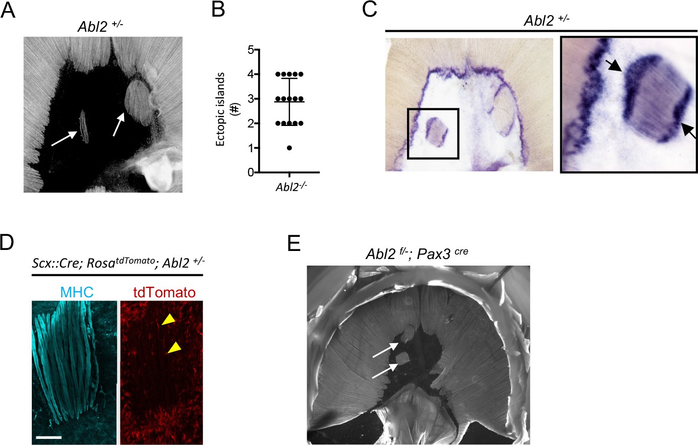
Ectopic muscle islands, which are found in the central tendon of mice that are heterozygous for Abl2, induce tendon cell differentiation.
(A,B) Whole mount diaphragms, stained with antibodies to myosin heavy chain (MHC), revealed 1 to 4 ectopic islands in the central tendon of Abl2+/− mice. (C) Scx RNA expression is enhanced at the ends of muscle fibers in the ectopic islands (black arrows) as well as at the normal MTJ in the diaphragm muscle. (D) Lineage-tracing experiments in Abl2+/− mice reveal that tendon cells, marked by tdTomato (arrowheads), intercalate between MHC-stained muscle fibers within the ectopic islands but do not contribute to myofibers. (E) Whole mount images reveal ectopic muscles in the central tendon of muscle conditional Abl2 heterozygous mice (Abl2f/+; Pax3cre). Scale bar is 250 µm in D.
Figure 8—figure supplement 1
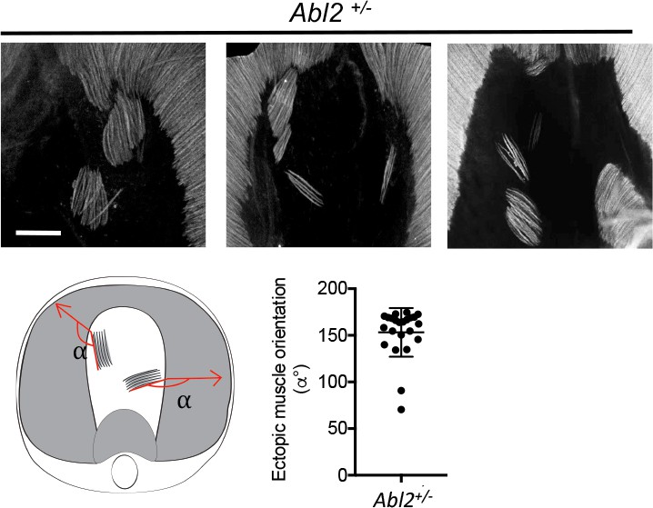
The orientation of ectopic muscle fibers correlates with the orientation of myofibers in the nearby main costal diaphragm muscle.
Representative images of the central tendon area in whole mounts of the diaphragm from Abl2+/− mice. The cartoon shows the method for measuring and quantifying the orientation of myofibes in ectopic muscle islands, compared to myofibers in the nearest costal diaphragm. The scatter plot shows the angle of each ectopic island in relation to the orientation of the myofibers in the nearest costal diaphragm muscle. The mean value and ±s.d. are shown. Scale bar is 500 µm.
Tables
Key resources table
| Reagent type (species) or resource | Designation | Source or reference | Identifiers | Additional information |
|---|---|---|---|---|
| gene (Mus musculus) | Abl2 | The Jackson Laboratory | RRID:MGI:2653897 | |
| gene (Mus musculus) | Myod iCre | The Jackson Laboratory | RRID:IMSR_JAX:014140 | |
| gene (Mus musculus) | Mck-Cre | The Jackson Laboratory | RRID:IMSR_JAX:006475 | |
| gene (Mus musculus) | Pax3 Cre | The Jackson Laboratory | RRID:IMSR_JAX:005549 | |
| gene (Mus musculus) | Rosa 26 LacZ | The Jackson Laboratory | RRID:IMSR_JAX:003474 | |
| gene (Mus musculus) | Scx::Cre | MGI:5317938 | n/a | Ronen Schweitzer lab |
| gene (Mus musculus) | Scx::GFP | PMID: 17497702 | n/a | Ronen Schweitzer lab |
| strain, strain background (Mus musculus) | C57BL/6J | The Jackson Laboratory | RRID:IMSR_JAX000664 | |
| genetic reagent (Mus musculus) | Targeting Vector: Abl2tm1a(EUCOMM)Hmgu | EUCOMM | RRID:SCR_003104 | European Conditional Mouse Mutagenesis Program |
| cell line (Mus musculus) | C2C12 skeletal muscle cells | Burden lab/ATCC | Cat# CRL-1772, RRID:CVCL_0188 | |
| antibody | Rabbit anti-Abl2 | Proteintech Group | Cat# 17693–1-AP RRID:AB_2289025 | 1:1000 |
| antibody | Goat anti-Arg (C-20) | Santa Cruz Biotechnology | Cat# sc-6356 RRID:AB_2221106 | 1:200 |
| antibody | Rabbit anti-β-Actin (13E5) | Cell Signaling Technology | Cat # 4970 | 1:1000 |
| antibody | Mouse anti-Myosin (MY-32) | Sigma-Aldrich | Cat# M7523 RRID:AB_260649 | 1:1000 |
| antibody | Chicken anti-GFP | Abcam | Cat# ab92456 RRID:AB_10561923 | 1:3000 |
| antibody | Mouse anti-Myosin (NOQ7.5.4D) | Sigma-Aldrich | Cat# M8421 RRID:AB_477248 | 1:1000 |
| antibody | Rabbit anti-Laminin | Sigma-Aldrich | Cat# L9393 RRID:AB_477163 | 1:1000 |
| antibody | Mouse anti-Pax7 | Santa Cruz Biotechnology | Cat# sc-81648 RRID:AB_2159836 | 1:500 (bioreactor) |
| antibody | Rabbit anti-MyoD C-20 | Santa Cruz Biotechnology | Cat# sc-304 RRID:AB_631992 | 1:1000 |
| antibody | Mouse anti-MyoD (5.8A) | ThermoFisher Scientific | Cat# MA1-21816 RRID:AB_560242 | 1:1000 |
| antibody | Donkey anti-mouse (H + L) Highly Cross-Adsorbed Secondary Antibody Alexa Fluor 594 | ThermoFisher Scientific | Cat# A-21203 RRID:AB_2535789 | 1:2000 |
| antibody | Donkey anti-rabbit (H + L) Highly Cross-Adsorbed Secondary Antibody Alexa Fluor 594 | ThermoFisher Scientific | Cat# A-21207 also A21207 RRID:AB_141637 | 1:2000 |
| antibody | Donkey anti-mouse (H + L) Highly Cross-Adsorbed Secondary Antibody Alexa Fluor 488 | ThermoFisher Scientific | Cat# A-21202 RRID:AB_141607 | 1:2000 |
| antibody | Donkey anti-rabbit (H + L) Highly Cross-Adsorbed Secondary Antibody Alexa Fluor 488 | ThermoFisher Scientific | Cat# A-21206 also A21206 RRID:AB_2535792 | 1:2000 |
| antibody | Goat anti-Rat IgG (H + L) Cross-adsorbed Secondary Antibody Alexa Fluor 488 | ThermoFisher Scientific | Cat# A-11006 also A11006 RRID:AB_2534074 | 1:2000 |
| commercial assay or kit | Click-iT Plus EdU Alexa Fluor 488 Imaging Kit | ThermoFisher Scientific | Cat# C10637 | |
| software, algorithm | Prism 7.0 | http://www.graphpad.com/scientific-software/prism/ | RRID:SCR_002798 |
Additional files
-
Transparent reporting form
- https://doi.org/10.7554/eLife.29905.016
Download links
A two-part list of links to download the article, or parts of the article, in various formats.
Downloads (link to download the article as PDF)
Open citations (links to open the citations from this article in various online reference manager services)
Cite this article (links to download the citations from this article in formats compatible with various reference manager tools)
Abelson tyrosine-protein kinase 2 regulates myoblast proliferation and controls muscle fiber length
eLife 6:e29905.
https://doi.org/10.7554/eLife.29905


