Molecular coordination of Staphylococcus aureus cell division
- University of Sheffield, United Kingdom
- University of Bonn, Germany
- University of York, United Kingdom
Figures
Figure 1 with 4 supplements

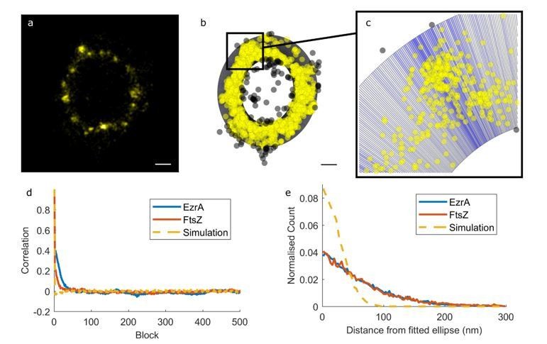
Distribution of cell division components during septation.
(a) Examples of EzrA distributions obtained using localisation microscopy of SH4388 (ezrA-eyfp ΔezrA). Scale bars 200 nm. (b) Examples of FtsZ distributions obtained using localisation microscopy of SH4665 (pCQ11-FtsZ-eYFP) grown with 50 µM IPTG. Scale bars 200 nm. (c) Simulated distributions of localisations randomly distributed by angle with different radii (r), number of localisations (n) and random error from a normal distribution with standard deviation (σ) [i] r = 440 nm, n = 1118, σ = 20 nm, [ii] r = 440 nm, n = 1118, σ = 40 nm, [iii] r = 440 nm, n = 1118, σ = 80 nm, [iv] r = 440 nm, n = 145, σ = 20 nm, [v] r = 440 nm, n = 2010, σ = 20 nm. Scale bars 200 nm. (d) An enlarged example of EzrA-eYFP distribution. Scale bar 200 nm. (e) The distribution from ‘d’ plotted as a scatter graph, and as histograms of number of localisations with respect to angle and distance from centre. (f) Mean angular autocorrelations of 14 EzrA, 19 FtsZ and 15 simulated distributions. Autocorrelation drops less quickly for EzrA and FtsZ than for simulations where angle is randomised. This shows that neither EzrA or FtsZ are randomly distributed by angle. (g) Histograms of localisations with respect to distance from the centre of a fitted circle with varying localisation precision. Data for EzrA and FtsZ are spread more widely than simulated data with poor localisation precision.
Figure 1—figure supplement 1
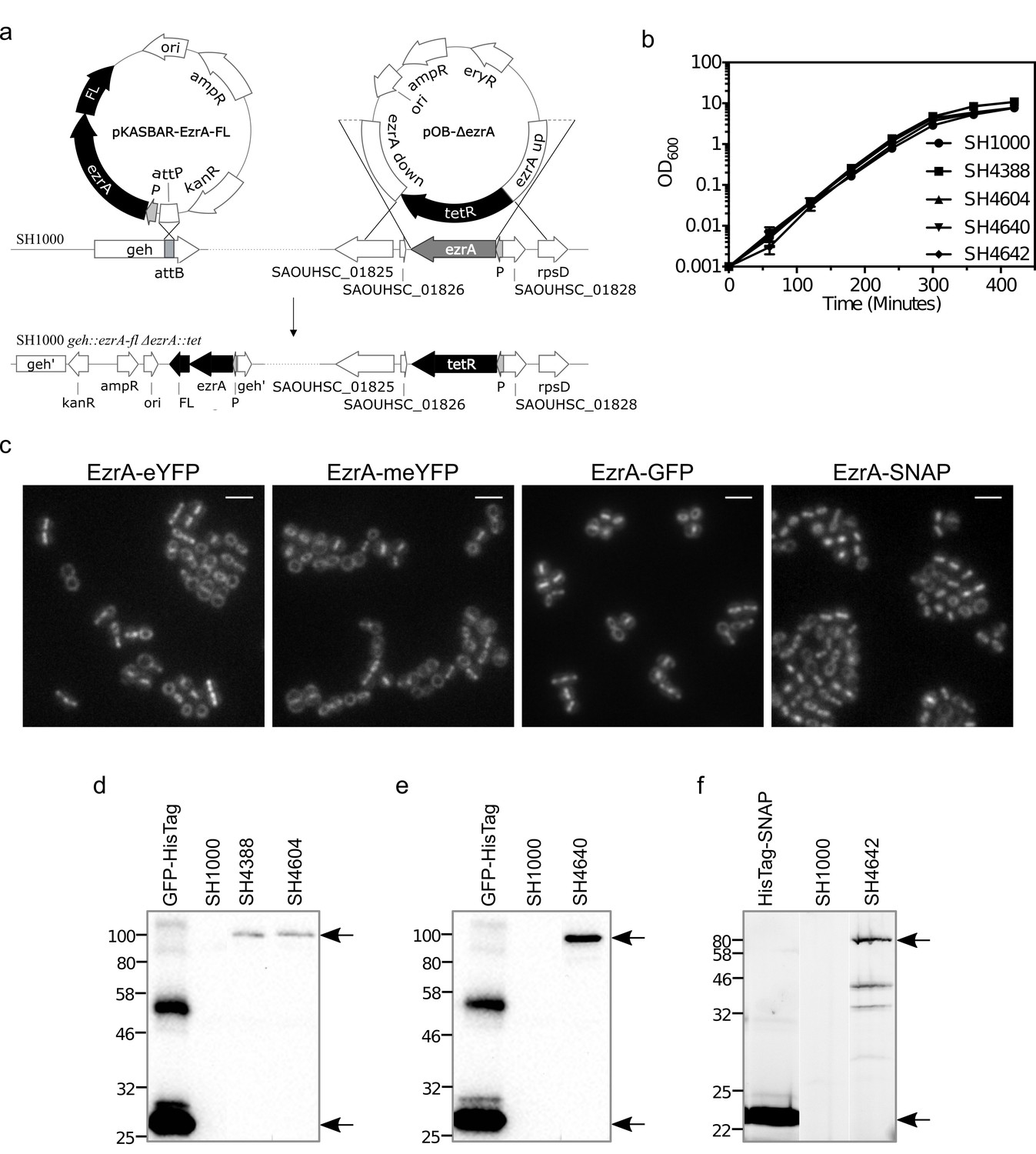
EzrA fusions are functional.
(a) Construction of S. aureus strains in which the only copy of ezrA is tagged (FL). Integration of pKASBAR-EzrA-FL at S. aureus lipase (geh) resulted in an ectopic copy of ezrA-fl under the control of the native ezrA promoter (P). A double-crossover event of pOB-ΔezrA allowed for marked with a tetracycline resistance (tetR) gene deletion of ezrA from its native chromosomal location. FL represents either eYFP, meYFP, GFP or SNAP. Not to scale. (b) Growth rates of ezrA fusions. EzrA-eYFP, EzrA-meYFP, EzrA-GFP and EzrA-SNAP complement native ezrA knock-out in SH4388 (ezrA-eyfp ΔezrA), SH4604 (ezrA-meyfp ΔezrA), SH4640 (ezrA-gfp ΔezrA) and SH4642 (ezrA-snap ΔezrA), respectively. The mutant strains (doubling time 24 min) showed similar growth to the wild type strain, SH1000 (doubling time 25 min). Growth rates were obtained by fitting an exponential growth equation to the most linear region of growth curves (R2 >0.98). Bacterial cultures were prepared in triplicate and the error bars represent standard deviation from the mean. (c) Epifluorescence microscopy images of EzrA-eYFP in SH4388 (ezrA-eyfp ΔezrA), EzrA-meYFP in SH4604 (ezrA-meyfp ΔezrA), EzrA-GFP in SH4640 (ezrA-gfp ΔezrA) and SNAP-Cell TMR-Star labelled EzrA-SNAP in SH4642 (ezrA-snap ΔezrA). Images are maximum intensity fluorescence projections of z stacks. Scale bars 3 μm. (d) EzrA-eYFP in SH4388 (ezrA-eyfp ΔezrA) and EzrA-meYFP in SH4604 (ezrA-meyfp ΔezrA) were detected by western blot analysis of total protein extracts using anti-GFP antibodies. Whole cell lysate of SH1000 and a recombinant GFP-HisTag protein were used as controls. Bands detected at ~95 kDa (EzrA-eYFP and EzrA-meYFP) and ~28 kDa (GFP-HisTag) are indicated with black arrows. Sizes of a protein ladder are shown in kDa. (e) EzrA-GFP in SH4640 (ezrA-gfp ΔezrA) was detected by immunoblot analysis of total protein extract using anti-GFP antibodies. Whole cell lysate of SH1000 and a recombinant GFP-HisTag protein were used as controls. Bands detected at ~95 kDa (EzrA-GFP) and ~28 kDa (GFP-HisTag) are indicated with black arrows. Sizes of a protein ladder are shown in kDa. (f) Whole cell lysate of SNAP-Cell TMR-Star labelled SH4642 (ezrA-snap ΔezrA) was resolved by 10% (w/v) SDS-PAGE and visualised by fluorescence detection. Whole cell lysate of SNAP-Cell TMR-Star labelled SH1000 and a purified SNAP-Cell TMR-Star labelled HisTag-SNAP protein were used as controls. Bands detected at ~85 kDa (EzrA-SNAP) and ~23 kDa (SNAP-HisTag) are indicated with black arrows. Sizes of a protein ladder are shown in kDa.
Figure 1—figure supplement 2

STORM and SIM data.
(a) EzrA-GFP (i) and SNAP-Cell TMR-Star labelled EzrA-SNAP (ii) localisation in SH4640 (ezrA-gfp ΔezrA) and SH4642 (ezrA-snap ΔezrA) by 3D-SIM, respectively. The images are maximum intensity projections of reconstructed z stacks. Scale bars 1 μm. 3D surface profiles of the circled area show distribution of fluorescence intensity of EzrA-GFP and EzrA-SNAP TMR-Star rings. (b) Localisation microscopy of EzrA-meYFP in SH4604 (ezrA-meyfp ΔezrA).
Figure 1—figure supplement 3
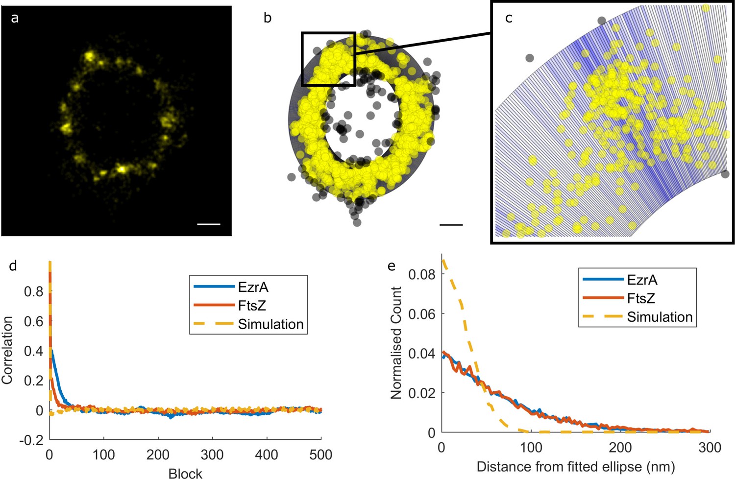
Quantitative analysis of EzrA and FtsZ distributions from localisation microscopy data based on elliptical fits.
(a) Example image of EzrA distribution. Scale bar 200 nm. (b) EzrA distribution represented as a scatter plot overlaid on an elliptical ring. Yellow points are included in the subsequent analyses, grey ones are not. The elliptical ring is derived from an elliptical fit to all of the points. (c) Enlargement of boxed region in b. The elliptical ring is split up into blocks – darker blue blocks contain more localisations than lighter ones. This gives a measure of how the number of localisations varies around the ring. (d) Autocorrelations of localisations around the ring for EzrA, FtsZ and simulated data with a random distribution. EzrA and FtsZ distributions are more self-correlated than a random distribution, but have no periodic order. (e) Distributions of absolute distances of localisations from the fitted ellipse for EzrA, FtsZ and simulated data. Simulated data had a localisation precision from a normal distribution with a mean of 27 nm and a standard deviation of 8.7 nm – representative of our measured values. The spread and magnitude of distances of EzrA and FtsZ localisations from the fitted ellipse cannot be accounted for by localisation uncertainty alone.
Figure 1—figure supplement 4
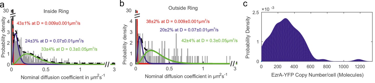
Dynamics of EzrA.
Nominal diffusion coefficient (D) distributions of EzrA-meYFP molecules (a) inside and (b) outside the EzrA ‘ring’ in SH4604 (ezrA-meyfp ΔezrA). The distribution of D values could be fitted using a 1–3 component gamma distribution model, as developed for heterogeneous protein mobility observed previously in bacteria (Stracy et al., 2015), with three components producing the lowest reduced chi2 = 0.05. (c) Distribution of number of EzrA-meYFP molecules per cell.
Figure 2
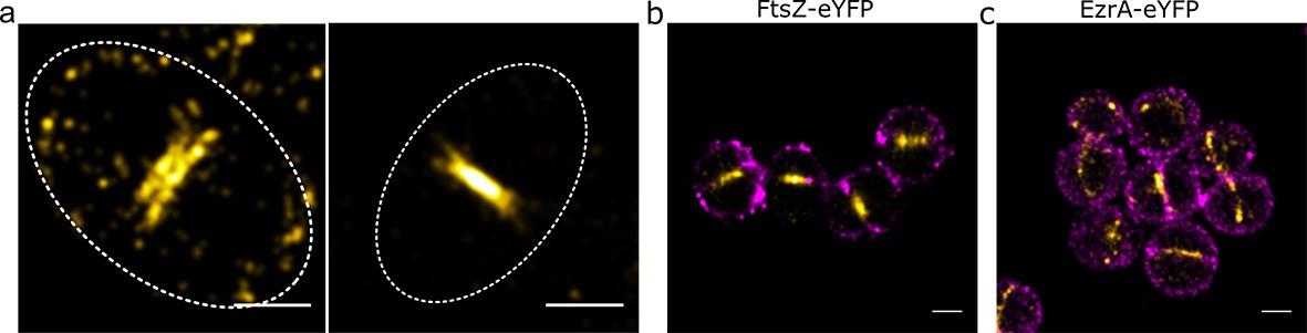
Relative locations of division components.
(a) Localisation microscopy images: of FtsZ-eYFP distributions in bacteria in the late stages of division. Scale bars 500 nm. Ellipses show approximate cell location and orientation. (b) Dual colour localisation microscopy image of FtsZ-eYFP and the cell wall (labelled with Alexa Fluor 647 NHS ester, NHS-647). Scale bars 500 nm. (c) Dual colour localisation microscopy image of EzrA-eYFP and the cell wall (labelled with NHS-647). Scale bars 500 nm.
Figure 3 with 3 supplements
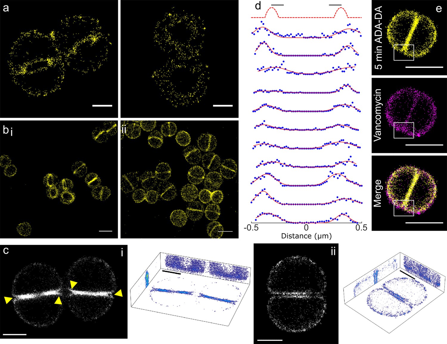
Peptidoglycan insertion.
Localisation microscopy images: (a) 15 s labelling of ADA clicked to Alexa Fluor 647. Scale bars 0.5 µm. (b) 5 min labelling of (i) ADA clicked to Alexa Fluor 647 and (ii) ADA-DA clicked to Alexa Fluor 647. Scale bars 1 µm. (c) 3D projections of S. aureus labelled for 5 min with ADA clicked to Alexa Fluor 647. (i) Cells with incomplete septum (yellow arrows show gaps in labelling), (ii) cell with annulus complete. Images in black boxes are z-projections while 3D representations show projections in all three planes. Scale bar 0.5 µm. (d) Cross sections of incomplete septa. The sketch graph (top row) hypothetically shows labelling exclusively at the leading edge of the septum. This is not the case for the data shown below - labelling is spread throughout the septum. The full width half maximum spread of labelling is ~230 nm. Data are plotted with blue dots, fits in red lines. (e) Two colour STORM, sample labelled for 5 min with ADA-DA clicked to Alexa Fluor 647 (yellow) and vancomycin linked to Amersham Cy3B (magenta). Images are z-projections and in merged images where localisations are in white show labelling by both ADA-DA and vancomycin. Boxed regions show slot in ADA-DA labelling but not vancomycin. Scale bars 1 µm.
Figure 3—figure supplement 1
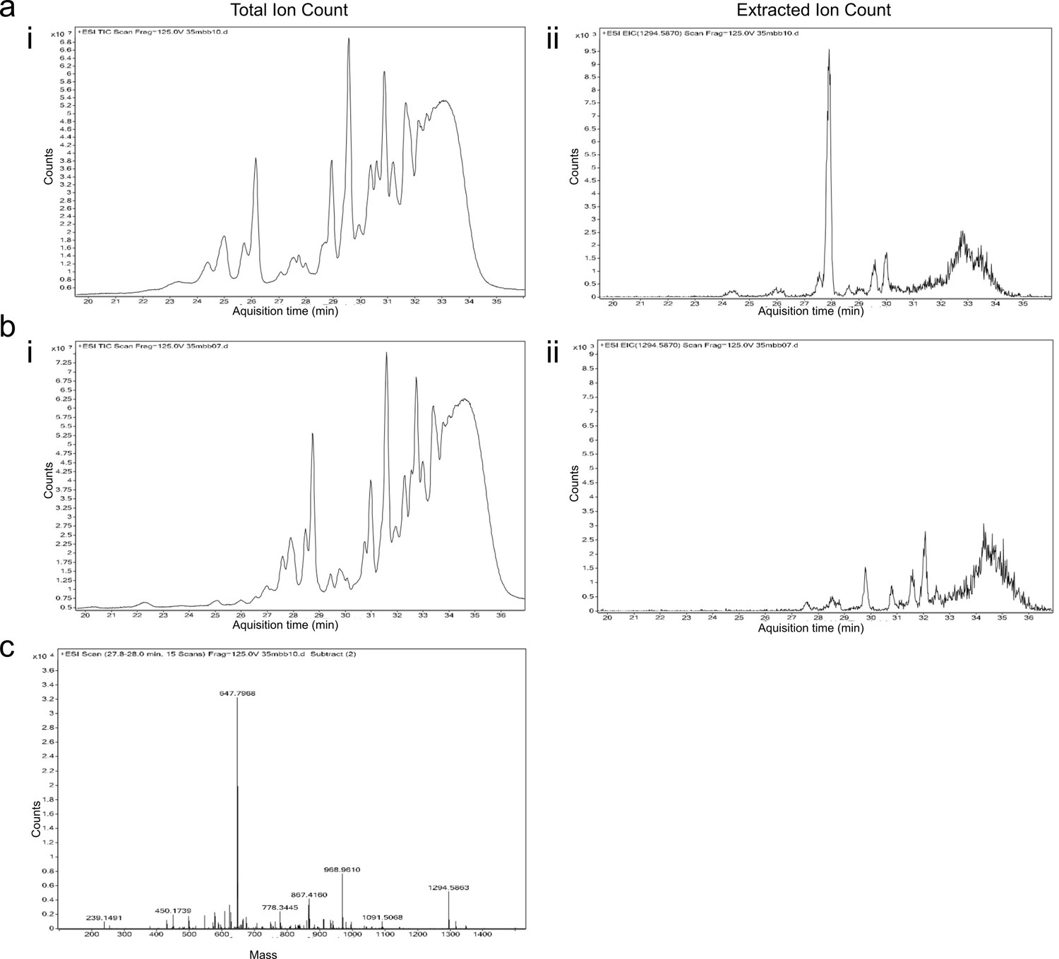
Identification of mechanism of DAA labelling in S. aureus.
Cellosyl digested peptidoglycan from S. aureus SH1000 grown in the presence (a) or absence (b) of ADA for 4 hr were investigated using LC-MS, with total ion chromatogram for acquisition time 20–36 min showing all detected ions (i). (ii) Extracted ion chromatogram for m/z [H+]=1294.5970 shows a clear peak in (a) not present in (b). (c) The mass-spectrum of this peak shows both the monoisotopic mass of the single-charged ion 1294.5863 and the doubly-charged ion at 647.7968, corresponding to disaccharide-pentapeptide-pentaglycine molecule with ADA replacing one of the d-alanine residues.
Figure 3—figure supplement 2
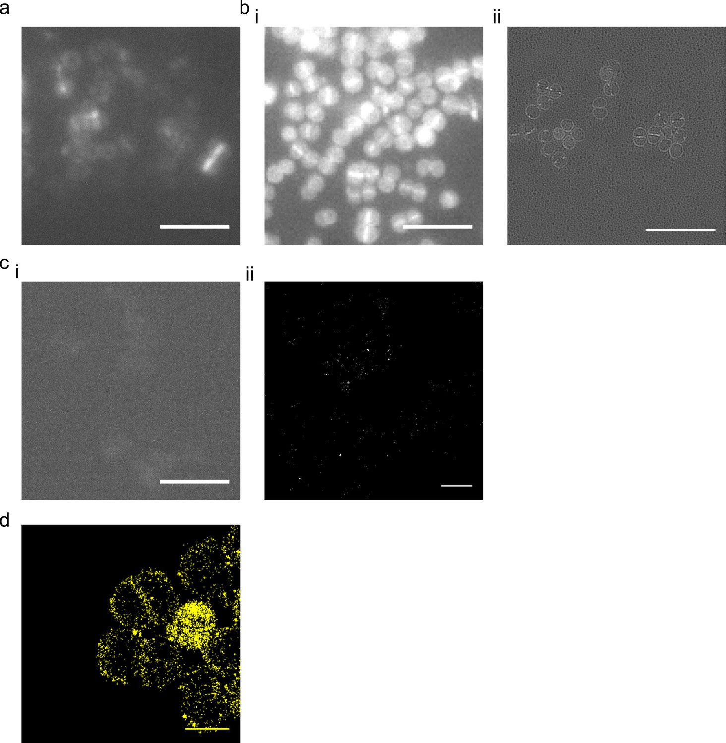
15 s labelling of peptidoglycan insertion with DAAs and controls.
(a) 15 s labelling of ADA clicked to Alexa Fluor 647. Sample imaged by epifluorescence and image is a maximum intensity projection of z stacks. (b) 15 s labelling with HADA imaged by i) epifluorescence and ii) 3D-SIM. (c) Cells labelled with Alexa Fluor 647 by the click reaction in the absence of ADA imaged by (i) epifluorescence and (ii) STORM. (d) localisation microscopy of 15 slabelling of ADA-DA (azido-d-alanyl-d-alanine) clicked to Alexa Fluor 647. Scale bars (a-c) 5 µm (d) 1 µm.
Figure 3—figure supplement 3
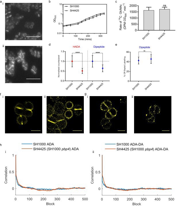
DAA labelling of PBP4 null S. aureus.
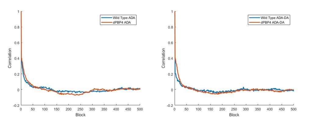
(a) SH4425 (SH1000 pbp4) labelled for 5 min with (i) HADA and (ii) ADA-DA clicked to Alexa Fluor 647. Scale bars 5 µm. (b) Growth rate of SH1000 and SH4425 in CDM, (c) Rate of peptidoglycan synthesis as measured by 14C GlcNAc incorporation. (d) DAA incorporation with 5 min labelling (HADA and ADA-DA) in SH1000 and SH4425. (e) % off-septal labelling in 5 min ADA-DA labelling of SH1000 and SH4425. (f) Localisation microscopy of 5 min FDAA labelling of SH4425 with (i) ADA clicked to Alexa Fluor 647 and (ii) ADA-DA clicked to Alexa Fluor 647. Scale bars 1 µm. (g) Localisation microscopy of 15 s labelling of SH4425 with (i) ADA clicked to Alexa Fluor 647 and (ii) ADA-DA clicked to Alexa Fluor 647. Scale bars 1 µm. (h) Comparison of autocorrelations of localisations around a fitted elliptical ring for SH1000 and SH4425 (SH1000 pbp4) labelled for 15 with (i) ADA or (ii) ADA-DA. n = 10 bacteria per group. There is no substantial difference between autocorrelations in either comparison.
Figure 4 with 1 supplement
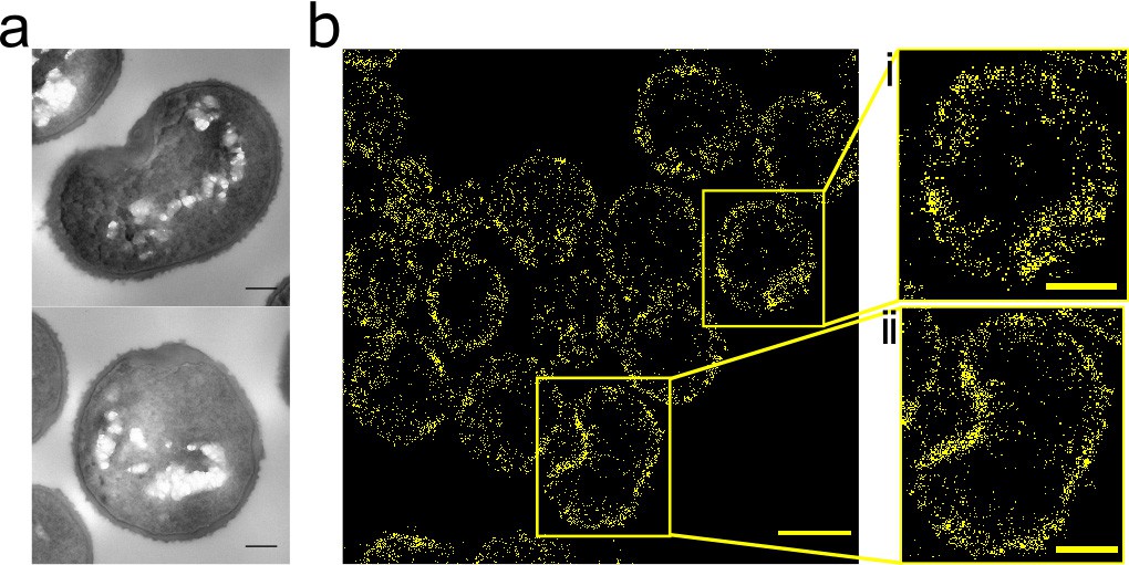
Effect of FtsZ inhibitor PC190723 on S. aureus.
(a) TEM of S. aureus SH1000 grown in the presence of PC190723 (10 µg ml−1) for 60 min. Scale bars 200 nm. (b) STORM image of S. aureus SH1000 pre-treated with PC190723 (10 µg ml−1) for 60 min labelled with ADA clicked to Alexa Fluor 647 for 5 min. Scale bar 1 µm. (i) and (ii) zoomed images of the corresponding area, scale bars 0.25 µm.
Figure 4—figure supplement 1
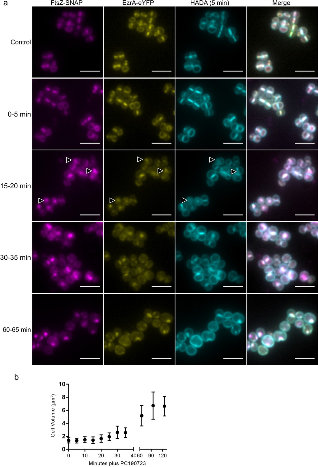
Effect of FtsZ inhibitor PC190723 on S. aureus.
(a) SH4652 (ezrA-eyfp ΔezrA pCQ11-FtsZ-SNAP) grown in the presence of 50 μM IPTG in the absence (control) or presence of PC190723 (10 μg ml−1) for 0, 15, 30 and 60 min, labelled with SNAP-Cell TMR-Star was incubated with HADA for 5 min. Images are average intensity projections of z stacks. Scale bars 3 µm. Arrows indicate localisation defects. (b) Cell volume of S. aureus SH1000 during treatment with PC190723 (10 μg ml−1). Data are expressed as mean ±standard deviation.
Figure 5
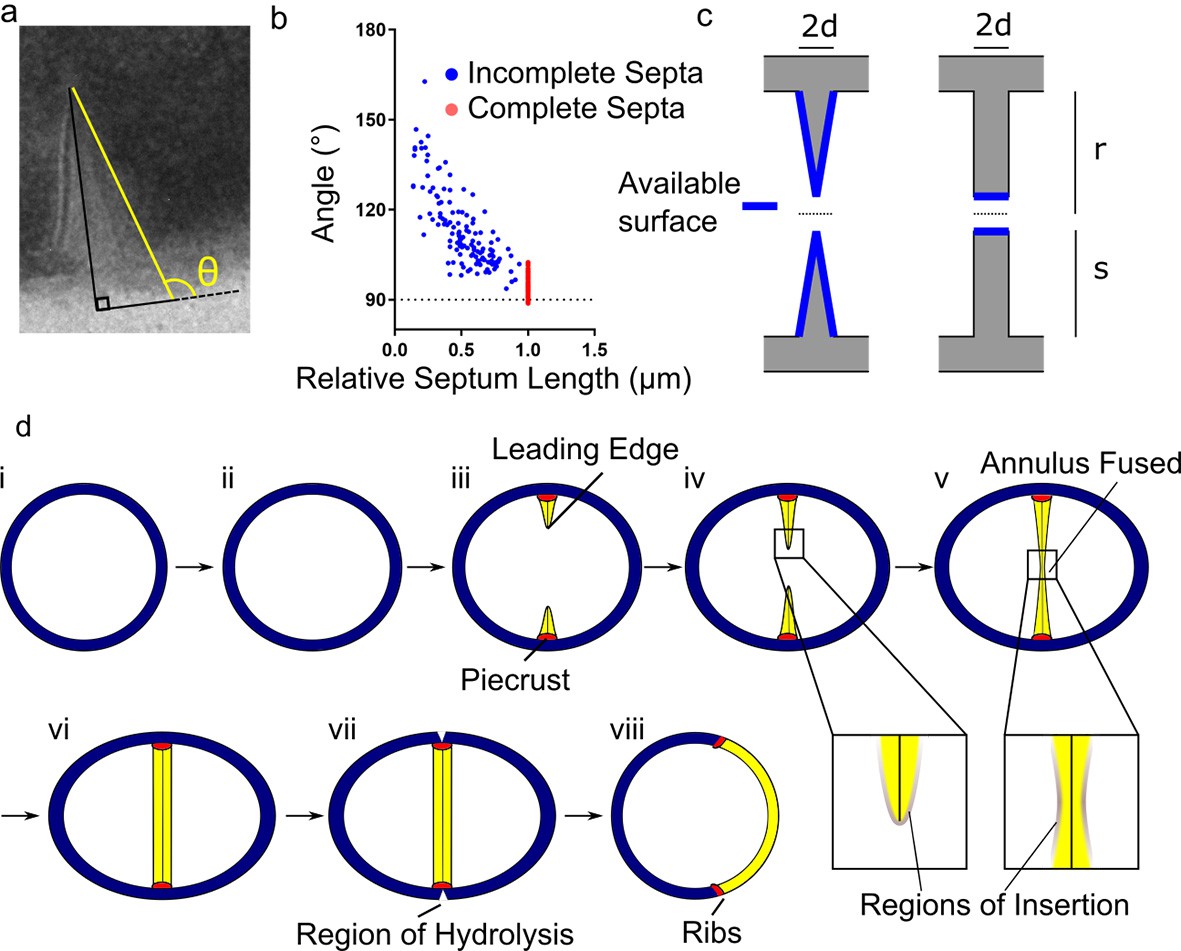
Conceptual model of peptidoglycan insertion during the S. aureus cell cycle.
(a) Schematic of measurement used in (b) measurement of the angle (θ) between a line parallel to the surface of the septum (yellow) and a tangent to the surface of the bacterium in incomplete (blue) and complete (red) septa. (c) Surfaces available for peptidoglycan insertion for different septal geometries where d is half the thickness of the septum, r is the cell radius in the septal plane and s is the distance from the leading to the lagging edge of the septum (measured from the inner surface of the cell wall). (d) Conceptual model of peptidoglycan insertion in S. aureus. (i, ii) Cell size increases and aspect ratio changes prior to observation of the start of septum formation by 3D-SIM (Monteiro et al., 2015). (iii) The septum then starts to form, beginning with the ‘piecrust’ feature (red) observed by AFM (Turner et al., 2010). The septum is thinner at the leading edge (Matias and Beveridge, 2007). (iv) New peptidoglycan is inserted in a zone at the leading edge of the septum, as well as across the rest of the cell surface as visualised here by localisation microscopy. (v, vi) After the annulus has fused, peptidoglycan insertion continues in the septum, executed by cell division components, until it is of uniform thickness. (vii) ATL (a peptidoglycan hydrolase) is present at the outer surface of the cell in the plane of septation(Komatsuzawa et al., 1997). Cracks or splits begin to form at the outer surface in the plane of septation(Touhami et al., 2004), followed by rapid popping apart of the daughter cells (Zhou et al., 2015). (vii) ‘Scars’ or ‘ribs’ remain marking the site of division (Monteiro et al., 2015; Turner et al., 2010) and may provide spatial cues to subsequently enable correct sequentially orthogonal divisions.
Appendix 1—figure 1
Structure of 3-Azido-N-[(1,1-dimethylethoxy)carbonyl]-D-alanine (Boc-D-aza-ala-OH).
https://doi.org/10.7554/eLife.32057.017
Appendix 1—figure 2

1D 1H NMR Spectrum of 3-Azido-N-[(1,1-dimethylethoxy)carbonyl]-D-alanine (Boc-D-aza-ala-OH.
https://doi.org/10.7554/eLife.32057.018
Appendix 1—figure 3
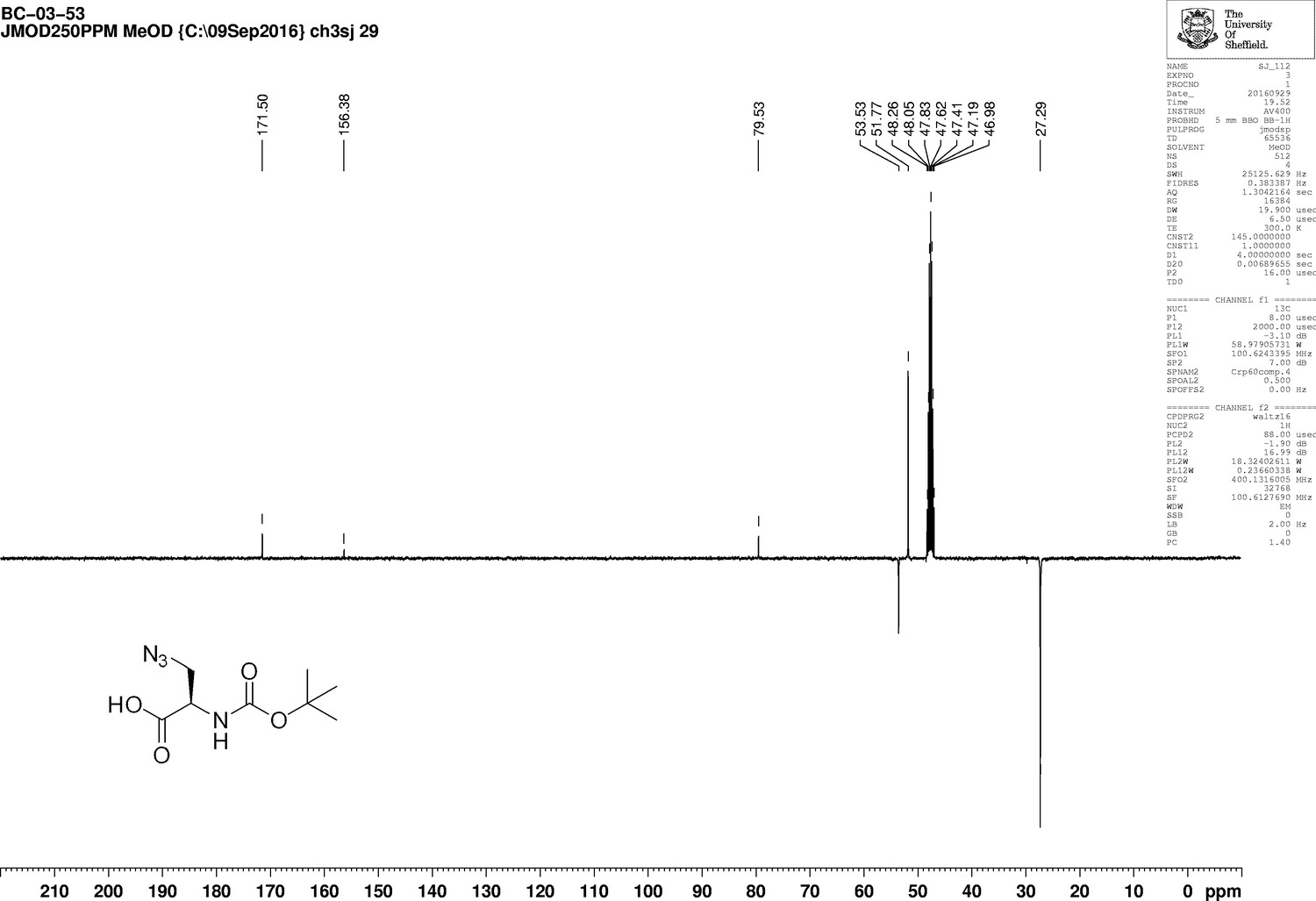
1D 13C NMR Spectrum of 3-Azido-N-[(1,1-dimethylethoxy)carbonyl]-D-alanine (Boc-D-aza-ala-OH).
https://doi.org/10.7554/eLife.32057.019
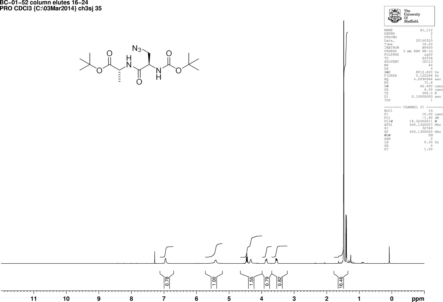
Appendix 1—figure 4

Structure of 3-Azido-N-[(1,1-dimethylethoxy)carbonyl]-D-alanyl-D-alanine 1,1-dimethylethyl ester (Boc-D-aza-ala-D-ala-OtBu).
https://doi.org/10.7554/eLife.32057.020
Appendix 1—figure 5
1D 1H NMR Spectrum of 3-Azido-N-[(1,1-dimethylethoxy)carbonyl]-D-alanyl-D-alanine 1,1-dimethylethyl ester (Boc-D-aza-ala-D-ala-OtBu).
https://doi.org/10.7554/eLife.32057.021
Appendix 1—figure 6
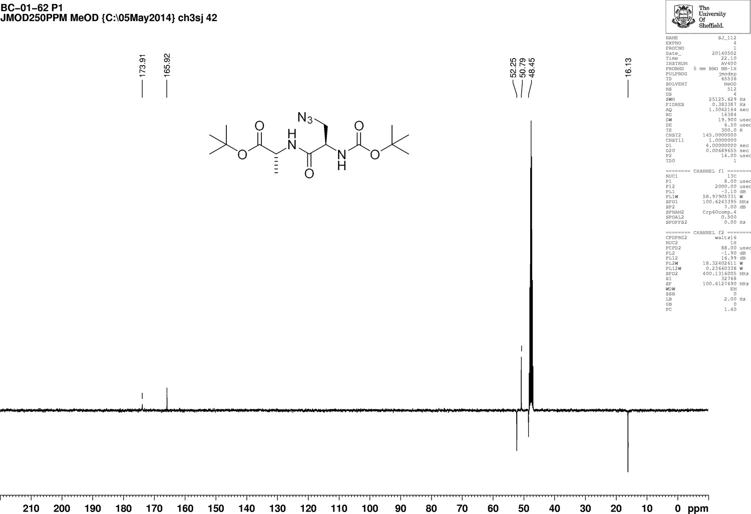
1D 13C NMR Spectrum of 3-Azido-N-[(1,1-dimethylethoxy)carbonyl]-D-alanyl-D-alanine 1,1-dimethylethyl ester (Boc-D-aza-ala-D-ala-OtBu).
https://doi.org/10.7554/eLife.32057.022
Appendix 1—figure 7
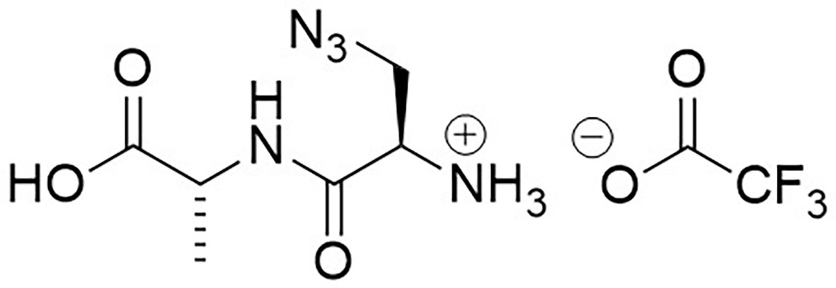
Structure of 3-Azido-D-alanyl-D-alanine, 2,2,2-trifluoroacetate [D-aza-ala-D-ala (ADA-DA) TFA salt].
https://doi.org/10.7554/eLife.32057.023
Appendix 1—figure 8
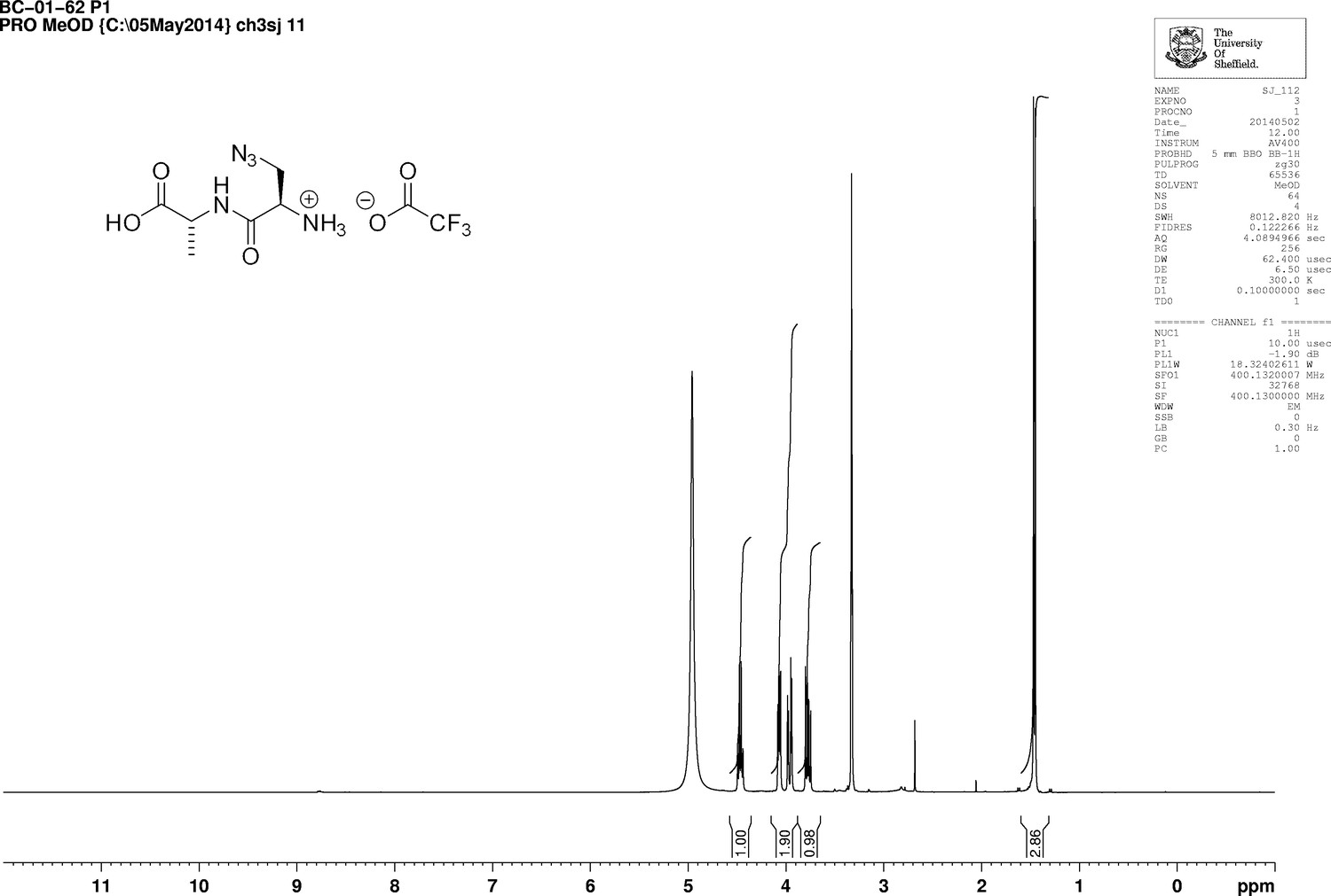
1D 1H NMR Spectrum of 3-Azido-D-alanyl-D-alanine, 2,2,2-trifluoroacetate [D-aza-ala-D-ala (ADA-DA) TFA salt].
https://doi.org/10.7554/eLife.32057.024
Appendix 1—figure 9
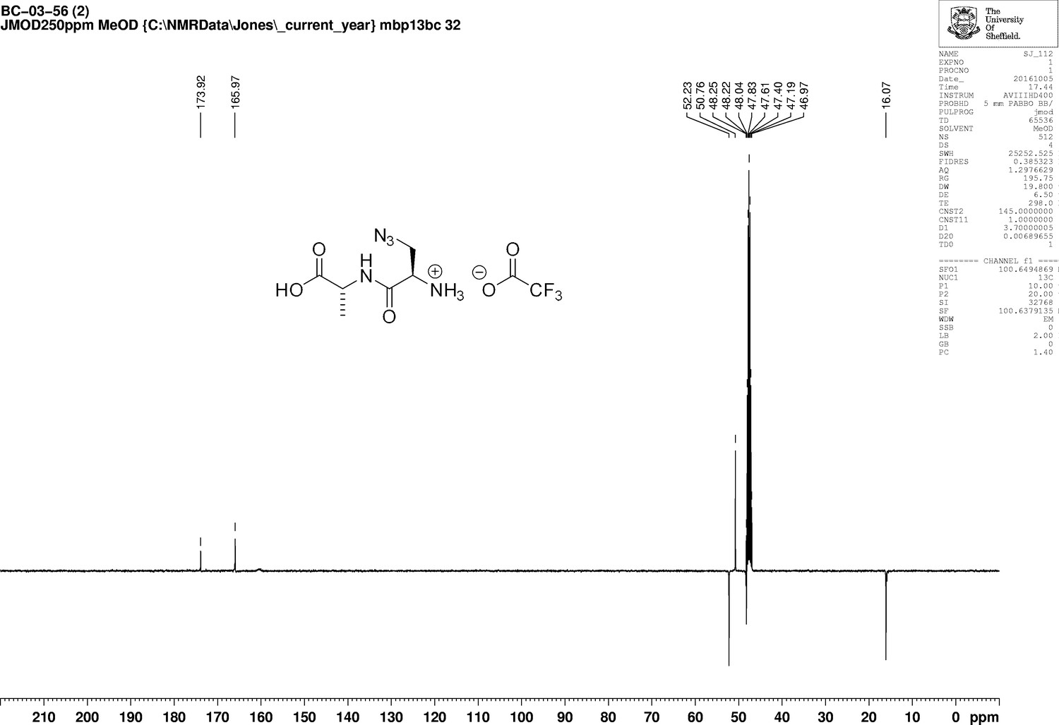
1D 13C NMR Spectrum of 3-Azido-D-alanyl-D-alanine, 2,2,2-trifluoroacetate [D-aza-ala-D-ala (ADA-DA) TFA salt].
https://doi.org/10.7554/eLife.32057.025
Appendix 1—figure 10
Structure of N-[(1,1-Dimethylethoxy)carbonyl]−3-{[(7-hydroxy-2-oxo-2H-1-benzopyran-3-yl) carbonyl] amino} D-alanine (N-Boc-HADA).
https://doi.org/10.7554/eLife.32057.026
Appendix 1—figure 11

1D 1H NMR Spectrum of N-[(1,1-Dimethylethoxy)carbonyl]−3-{[(7-hydroxy-2-oxo-2H-1-benzopyran-3-yl) carbonyl] amino} D-alanine (N-Boc-HADA).
https://doi.org/10.7554/eLife.32057.027
Appendix 1—figure 12
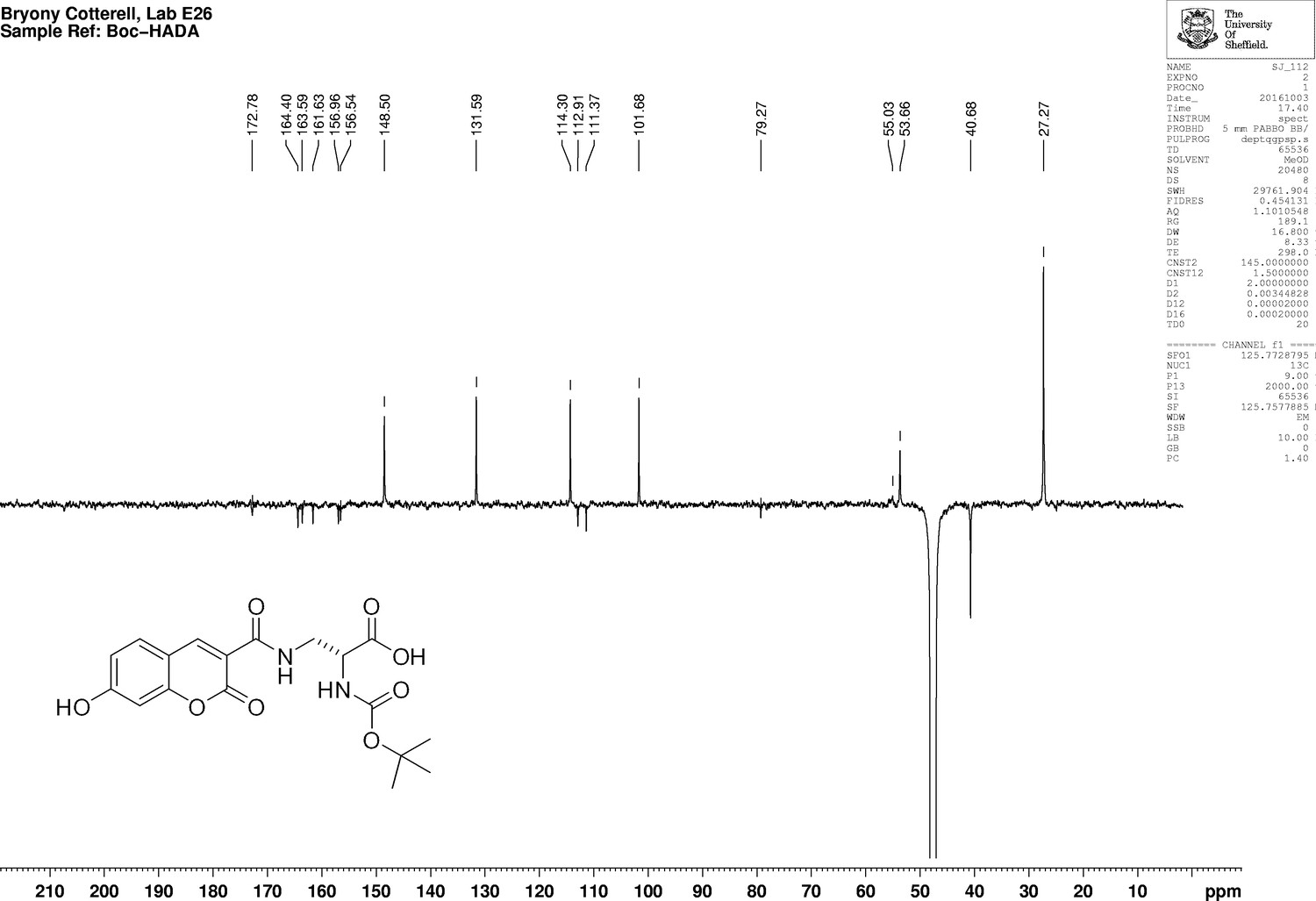
1D 13C NMR Spectrum of N-[(1,1-Dimethylethoxy)carbonyl]−3-{[(7-hydroxy-2-oxo-2H-1-benzopyran-3-yl) carbonyl] amino} D-alanine (N-Boc-HADA).
https://doi.org/10.7554/eLife.32057.028
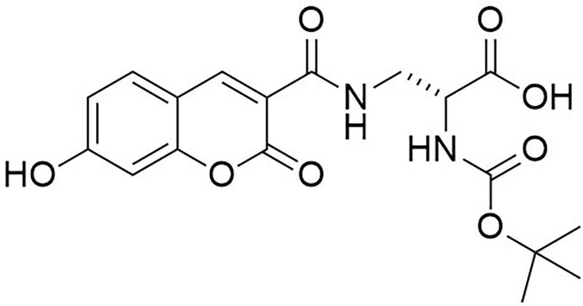
Appendix 1—figure 13
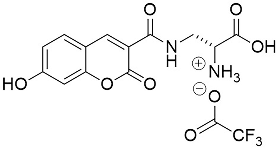
Structure of 3-{[(7-Hydroxy-2-oxo-2H-1-benzopyran-3-yl) carbonyl] amino} D-alanine, 2,2,2-trifluoroacetate (HADA TFA salt).
https://doi.org/10.7554/eLife.32057.029
Appendix 1—figure 14
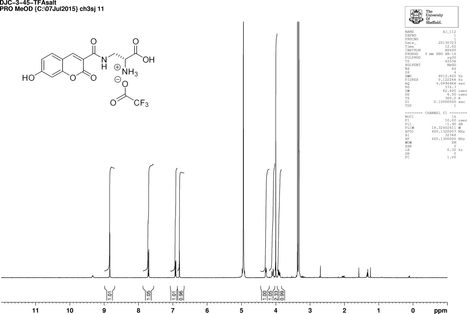
1D 1H NMR Spectrum of 3-{[(7-Hydroxy-2-oxo-2H-1-benzopyran-3-yl) carbonyl] amino} D-alanine, 2,2,2-trifluoroacetate (HADA TFA salt).
https://doi.org/10.7554/eLife.32057.030
Appendix 1—figure 15
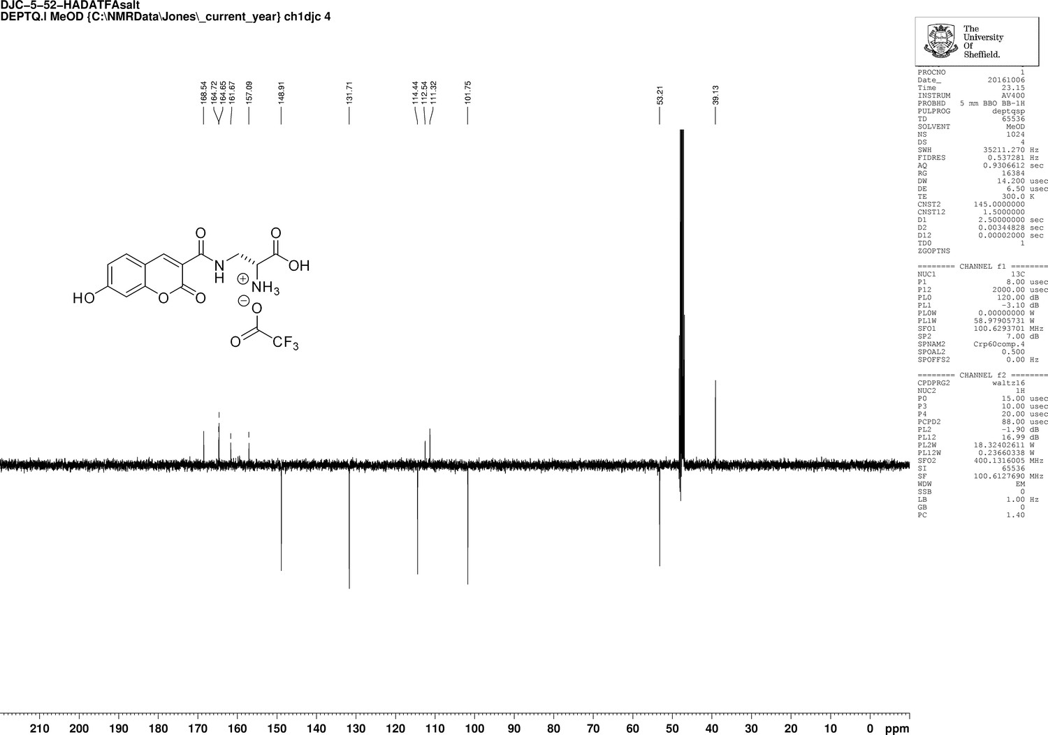
1D 13C NMR Spectrum of 3-{[(7-Hydroxy-2-oxo-2H-1-benzopyran-3-yl) carbonyl] amino} D-alanine, 2,2,2-trifluoroacetate (HADA TFA salt).
https://doi.org/10.7554/eLife.32057.031
Author response image 1
Author response image 2
Author response image 3

Effect of HADA labelling on EzrA-eYFP and FtsZ-SNAP TMR-Star localisation.
S. aureus strain producing EzrA-eYFP grown without (a) and with HADA for 5 min (b). SNAP-Cell TMR-Starlabelled S. aureus strain producing FtsZ-SNAP grow without (c) and with HADA for 5 min (d). Images are average intensity fluorescence projections of z stacks. Scale bars 5 𝝻m.
Author response image 4
Tables
Appendix 1—table 1
Strains used in this study.
https://doi.org/10.7554/eLife.32057.032| Strain | Relevant Genotype/markers | Source |
|---|---|---|
| SH1000 | Functional rsbU+ derivative of 8325–4 | (Horsburgh et al., 2002b) |
| RN4220 | Restriction deficient transformation recipient | (Kreiswirth et al., 1983) |
| CYL316 | S. aureus RN4220 pCL112Δ19 (cm) | (Lee et al., 1991) |
| JGL227 | S. aureus SH1000 ezrA-gfp+ (ery) | (Steele et al., 2011) |
| SH4386 | S. aureus SH1000 ezrA-eyfp (kan) | This study |
| SH4388 | S. aureus SH1000 ezrA-eyfp ΔezrA (kan, tet) | This study |
| SH4603 | S. aureus SH1000 ezrA-meyfp (kan) | This study |
| SH4604 | S. aureus SH1000 ezrA-meyfp ΔezrA (kan, tet) | This study |
| SH4639 | S. aureus SH1000 ezrA-gfp (kan) | This study |
| SH4640 | S. aureus SH1000 ezrA-gfp ΔezrA (kan, tet) | This study |
| SH4641 | S. aureus SH1000 ezrA-snap (kan) | This study |
| SH4642 | S. aureus SH1000 ezrA-snap ΔezrA (kan, tet) | This study |
| SH4652 | S. aureus SH1000 ezrA-eyfp ΔezrA pCQ11-FtsZ-SNAP (kan, tet, ery) | This study |
| SH4665 | S. aureus SH1000 pCQ11-FtsZ-eYFP (ery) | This study |
| NE679 | S. aureus JE2 with transposon insertion in pbp4 (ery) | (Fey et al., 2013) |
| SH4425 | S. aureus SH1000 pbp4 (ery) | This study |
| N315 | Methicillin-resistant S. aureus | (Kuroda et al., 2001) |
| SU492 | B. subtilis SU5 Pxyl-ftsZ-yfp (spec) | (Monahan et al., 2014) |
Appendix 1—table 2
Plasmids used in this study
https://doi.org/10.7554/eLife.32057.033| Plasmid | Relevant genotype/markers | Source |
|---|---|---|
| pGM074 | pKASBAR-kan (Bottomley et al., 2014) carrying ezra-psmorange under the putative ezrA promoter (amp, kan) | G. McVicker |
| pSNAP-tag (T7)−2 | E. coli expression plasmid carrying the snap gene under the control of the T7 promoter (amp) | New England Biolabs |
| pOB | pGEM3Zf(+) cloning vector containing the erythromycin resistance cassette (amp, ery) | (Horsburgh et al., 2002a) |
| pAISH | TetR derivative of pMUTIN4 | (Aish, 2003) |
| pKASBAR-EzrA-eYFP | pKASBAR-kan containing ezrA-eyfp under the putative ezrA promoter (amp, kan) | This study |
| pKASBAR-EzrA-meYFP | pKASBAR-kan containing ezrA-meyfp under the putative ezrA promoter (amp, kan) | This study |
| pKASBAR-EzrA-GFP | pKASBAR-kan containing ezrA-gfp under the putative ezrA promoter (amp, kan) | This study |
| pKASBAR-EzrA-SNAP | pKASBAR-kan containing ezrA-snap under the putative ezrA promoter (amp, kan) | This study |
| pOB-ΔezrA | pOB containing the ezrA deletion cassette consisting of a 1.5 kb fragment of the upstream region of S. aureus ezrA, the tetracycline resistance cassette from pAISH and a 1.5 kb fragment of the downstream region of S. aureus ezrA (amp, ery, tet) | This study |
| pSS26b | pUC19 encoding snap (amp) | Covalys |
| pSS26bFtsZ-C | pSS26b containing ftsZ-snap (amp) | This study |
| pCQ11 | E. coli-S. aureus shuttle vector containing lacI, Pspac and gfp (amp, ery) | (Hardt et al., 2017) |
| pCQ11-FtsZ-SNAP | pCQ11 derivative containing ftsZ-snap under Pspac (amp, ery) | This study |
| pCQ11-FtsZ-eYFP | pCQ11-FtsZ-SNAP with eyfp replacement of snap (amp, ery) | This study |
Appendix 1—table 3
Oligonucleotides used in this study.
https://doi.org/10.7554/eLife.32057.034| Oligonucleotide name | Sequence (5’ to 3’) |
|---|---|
| eYFP-F | CGGCGCGCCTCAGGTTCAGGTTCAGGTATGGTGAGCAAGGGCGAG |
| eYFP-R | CGCGGCCGCTTACTTGTACAGCTCGTCCATGCCGAGAGTGATCCCGGC |
| GFP-F | CGGCGCGCCTCAGGTTCAGGTTCAGGTATGGCTAGCAAAGGAGAAGAA CTTTTCACTGGAGTTGTCCC |
| GFP-R | CGCGGCCGCTTATTTGTAGAGCTCATCCATGCCATGTGTAATCCCAGCAGC |
| SNAP-F | GGGCGCGCCTCAGGTTCAGGTTCAGGTATGGACAAAGACTGCGAAATG AAGCGCAC |
| SNAP-R | CGAATTCTCATTAACCCAGCCCAGGCTTGCCCAGTCTG |
| meYFP-F | CTACCAGTCCAAGCTGAGCAAAGAC |
| meYFP-R | CTCAGGTAGTGGTTGTCG |
| pOB-ezrA-up-F | TTTACGTACACTATCTGCAGATGCTTCTCCTCCTAATTTATCATT |
| pOB-ezrA-up-R | ATTCGAGCTCGGTACCCGGGTTTTAAATTAATAAAAAAAACAC CCACAATT |
| pOB-ezrA-down-F | CACTATAGAATACTCAAGCTTACTCCTTAATTTCCTCATAAATGATGA |
| pOB-ezrA-down-R | GGATCAACTTTGGGAGAGAGAAACTAGTATGTAGTTATACTTAAA TAATATGAGC |
| pOB-TetR-F | TAAATTAGGAGGAGAAGCATCTGCAGATAGTGTACGTAAAAAGA |
| pOB-TetR-R | GTATAACTACATACTAGTTTCTCTCTCCCAAAGTTGATCCC |
| ftsZ-eyfp-F | ACATGGCCATGTCAGGTTCAG |
| ftsZ-eyfp-R | GGCGCGCCTTATTTATATAATTC |
| FGFtsZXhoI-F | CTCGAGATGTTAGAATTTGAACAAGG |
| FGFtsZEcoRI-R | TTAGAATTCACGTCTTGTTCTTCTTGAA |
| FGFtsZNheI-F | GTTGCTAGCATGTTAGAATTTGAACAAGG |
| FGFtsZAscI-R | GTTGGCGCGCCTTATCCCAGACCCGGTTTAC |
Additional files
-
Transparent reporting form
- https://doi.org/10.7554/eLife.32057.015
Download links
A two-part list of links to download the article, or parts of the article, in various formats.
Downloads (link to download the article as PDF)
Open citations (links to open the citations from this article in various online reference manager services)
Cite this article (links to download the citations from this article in formats compatible with various reference manager tools)
Molecular coordination of Staphylococcus aureus cell division
eLife 7:e32057.
https://doi.org/10.7554/eLife.32057


