Proprioceptive and cutaneous sensations in humans elicited by intracortical microstimulation
- California Institute of Technology, United States
- Keck School of Medicine of USC, United States
- UCLA-Caltech Medical Scientist Training Program, United States
- Rancho Los Amigos National Rehabilitation Center, United States
Figures
Figure 1
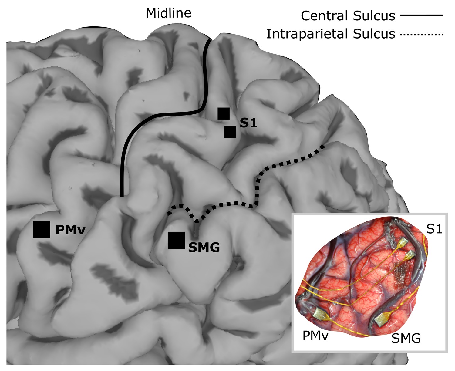
Array implant locations on rendered MRI image of the left hemisphere of FG.
96-channel microelectrode arrays were implanted into ventral premotor cortex (PMv) and supramarginal gyrus (SMG), and two 48-channel stimulating arrays were implanted into primary somatosensory cortex (S1). The insert shows the in situ array locations.
Figure 2
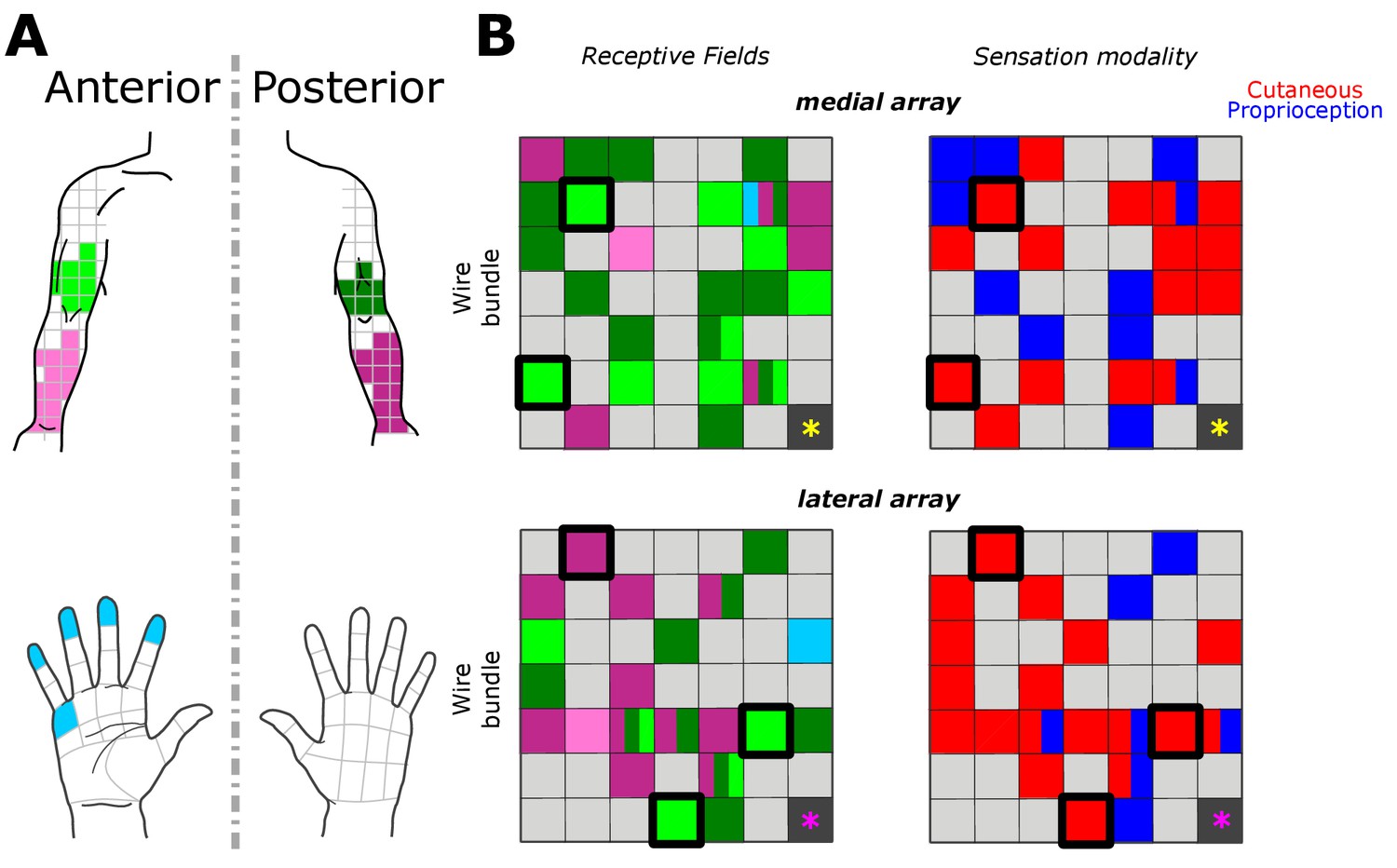
Receptive fields and sensation modality across all amplitude mapping experiments.
(A) Receptive field location on anterior (lighter shades) and posterior (darker shades) planes of the right upper arm (green), forearm (pink), and hand (cyan). Grid is the same that the subject referenced during the experiment. (B) Schematic of the two electrode arrays implanted over S1 (Figure 1). Left side panels display the reported receptive fields at each electrode location, and right side panels display the sensation modality (cutaneous - red, proprioceptive - blue). Light gray boxes show electrodes with no reported sensation, while dark gray boxes represent reference channels which are not used in recording. The five electrodes with a thick black outline represent the subset tested in the additional parameter-wide mapping task. Yellow and magenta asterisks mark the inferior-posterior corner of the implants, for the medial and lateral arrays respectively.
Figure 3
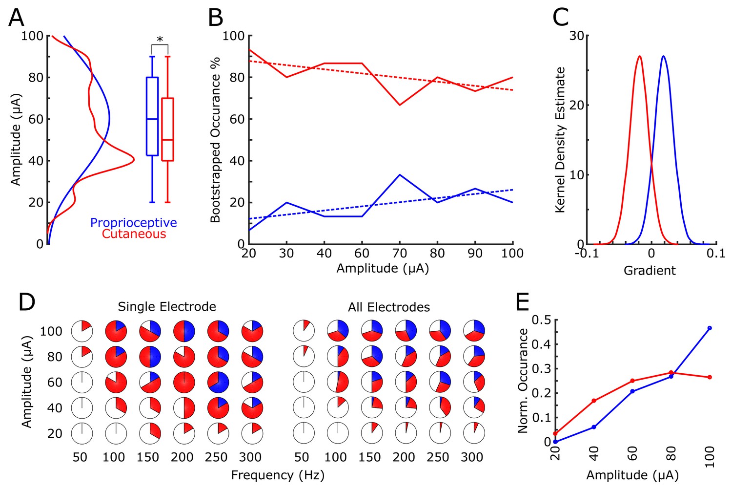
Proprioceptive and cutaneous responses.
(A) Kernel density estimate and box plot showing the difference in the distribution of amplitudes associated with each report of proprioceptive (blue) or cutaneous (red) responses. (B) The median percentage of responses in the bootstrapped sample (solid line) for proprioceptive and cutaneous responses at each amplitude tested. Dashed line shows 1st-order polynomial fit. (C) Kernel density estimates of the distribution of slopes from 1st-order polynomial fits in each bootstrap iteration. (D) Pie charts show the percentage of total stimulations of responses for the subset of electrodes tested over a range of both current amplitudes and pulse frequencies. The left panel shows an individual example electrode (six trials per combination of amplitude and frequency) and the right panel shows data pooled over all five electrodes (30 total stimulations per combination). The percentage of no response (white), proprioceptive (blue) or cutaneous (red) are shown. (E) A normalized histogram of proprioceptive (blue) and cutaneous (red) responses at each of the amplitudes tested in experiment 2.
Tables
Table 1
Descriptions of the most prevalent sensations by percentage of total responses.
Entries cover 90% of 381 reported sensations, with the final 10% comprising a mixture of other naturalistic cutaneous and proprioceptive descriptors. Each sensation is accompanied by the mode and 25th-75th percentiles in the distribution of amplitudes that elicited each sensation, and by the same quantities for the perceived reported intensities (on a scale of 1 [weak] to 10 [strong]).
| Description | % Total Sensations (381 total) | Amplitude μA (mode) | Amplitude μA (25th, 75th percentile) | Intensity (mode) | Intensity (25th, 75th percentile) |
|---|---|---|---|---|---|
| Squeeze | 24.9 | 40 | 40, 87.5 | 7 | 4, 7 |
| Tap | 17.3 | 70 | 40, 80 | 1 | 1, 4 |
| Right movement | 9.7 | 90 | 55, 90 | 1 | 1, 3 |
| Vibration | 8.1 | 40 | 40, 90 | 2 | 2, 3 |
| Blowing | 6.6 | 60 | 30, 80 | 1 | 1, 2 |
| Forward Movement | 5.8 | 70 | 40, 80 | 1 | 1, 4 |
| Pinch | 5.5 | 40 | 40, 90 | 3 | 3, 6 |
| Press | 5.0 | 40 | 40, 70 | 7 | 4, 7 |
| Upward Movement | 3.9 | 70 | 70, 85 | 1 | 1.25, 4 |
| Goosebumps | 3.1 | 100 | 60, 90 | 5 | 2, 5 |
Additional files
-
Source code 1
Stimulation commands.
- https://doi.org/10.7554/eLife.32904.007
-
Transparent reporting form
- https://doi.org/10.7554/eLife.32904.008
Download links
A two-part list of links to download the article, or parts of the article, in various formats.
Downloads (link to download the article as PDF)
Open citations (links to open the citations from this article in various online reference manager services)
Cite this article (links to download the citations from this article in formats compatible with various reference manager tools)
Proprioceptive and cutaneous sensations in humans elicited by intracortical microstimulation
eLife 7:e32904.
https://doi.org/10.7554/eLife.32904


