FGF mediated MAPK and PI3K/Akt Signals make distinct contributions to pluripotency and the establishment of Neural Crest
- Northwestern University, United States
Figures
Figure 1 with 1 supplement

FGF signaling is required for proper blastula stage gene expression.
(A) In situ hybridization examining FGFR4 expression in wildtype Xenopus embryos collected at blastula (stage 9, lateral view, animal pole up), late gastrula (stage 12, dorsal view, anterior up), and mid-neurula (stage 15, dorsal view, anterior up) stages. Expression is seen in the pluripotent cells of the animal hemisphere at blastula stages and in the neural plate and neural crest forming regions at gastrula and neurula states. (B) Animal pole explant assay examining FGFR4 expression. Explants were cultured alongside sibling embryos and collected at blastula (stage 9), late gastrula (stage 12), and mid-neurula (stage 15) stages. (C) In situ hybridization examining Vent2, Id3, Myc, and FoxD3 expression in blastula stage (stage 9) embryos injected with dominant-negative FGFR4 (dnFGFR4). Asterisk denotes injected side, marked by staining of the lineage tracer β-galactosidase (red). Dominant-negative FGFR4 blocks expression of Vent2 and Id3.
Figure 1—figure supplement 1
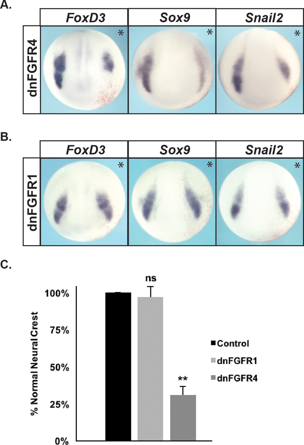
Blocking FGF signaling using dnFGFR4, but not dnFGFR1, leads to a loss in neural crest gene expression at mid-neurula stages.
(A–B). In situ hybridization examining FoxD3, Sox9, and Snail2 expression in mid-neurula stage (stage 16) embryos injected with dnFGFR4 (A) or dnFGFR1 (B). Asterisk denotes injected side, marked by staining of the lineage tracer β-galactosidase (red). Blocking FGF signaling using dnFGFR4 causes a loss of FoxD3, Sox9, and Snail2 expression. (C) Quantification of effects on FoxD3 expression in embryos injected with dnFGFR4 or dnFGFR1 scored for loss of neural crest/exclusion of injected cells from the neural crest, normalized to control injections. (ns, not significant; **p<0.01).
Figure 2
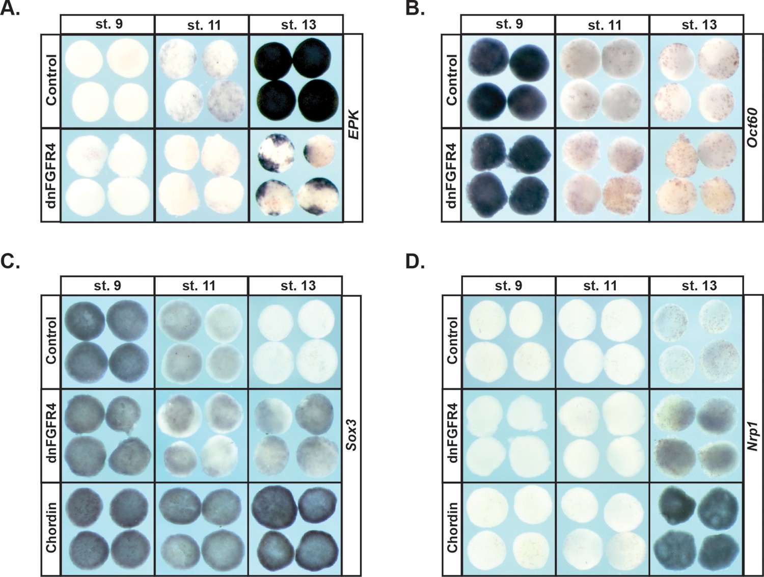
Blocking FGF signaling in pluripotent blastula cells interferes with adoption of an epidermal state and neuralizes cells.
(A–D) In situ hybridization examining expression of Epidermal Keratin (EPK) (A) Oct60 (B) Sox3 (C) or Nrp1 (D) in animal pole explants injected with dnFGFR4 or chordin for phenotypic comparison. Explants were cultured alongside sibling embryos and collected at blastula (stage 9), midgastrula (stage 11), and early neurula (stage 13) stages. Blocking FGF signaling interferes with EPK expression and mildly induces Nrp1 expression.
Figure 3 with 1 supplement
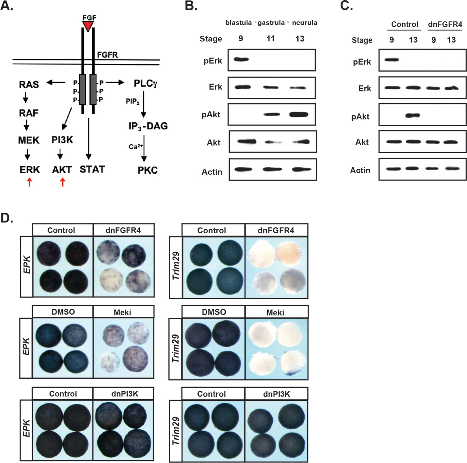
FGF signaling directs the transit from a pluripotent to a lineage restricted state through regulation of Erk and Akt activation.
(A) Schematic representation of the FGF receptor and select signaling cascades activated downstream, highlighting the Ras/MAPK (Erk) and PI3K/Akt cascades. (B) Western blot of lysates from animal pole explants cultured alongside sibling embryos and collected at blastula (stage 9), midgastrula (stage 11), and early neurula (stage 13) stages to examine levels of phosphorylated and unphosphorylated Erk1/2 and Akt. Pluripotent cells show high pErk while lineage restricted cells display high pAkt. (C) Western blot of lysates from animal pole explants injected with dnFGFR4. Explants were cultured alongside sibling embryos and collected at blastula (stage 9) and early neurula (stage 13) stages to examine levels of phosphorylated and unphosphorylated Erk1/2 and Akt. Both pErk and pAkt are blocked by dnFGFR4. (D) Animal pole explant assay examining Epidermal Keratin (EPK) and Trim29 expression in explants injected with either dnFGFR4 or dominant- negative PI3K (dnPI3K) or treated with Meki (RDEA119) and collected alongside sibling embryos at early neurula stages (stage 13–14). Meki treatment phenocopies dnFGFR4.
Figure 3—figure supplement 1
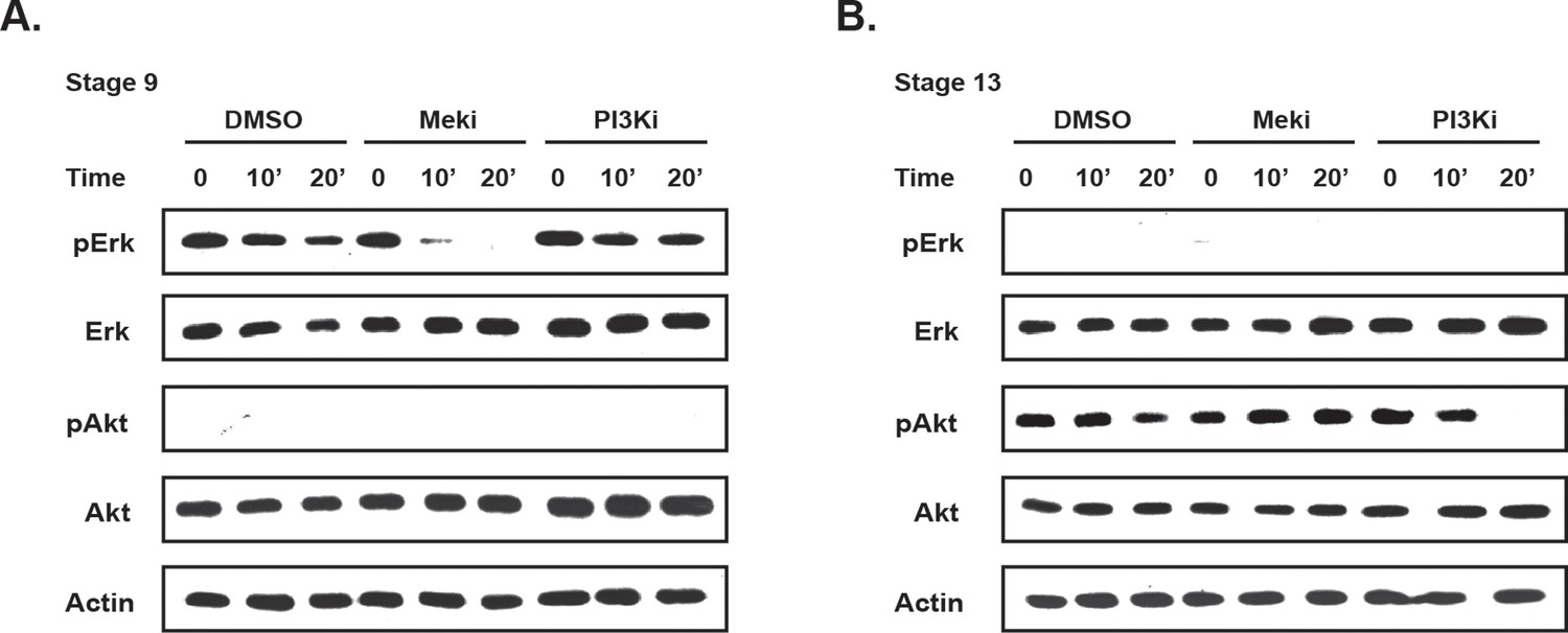
Meki (RDEA119) and PI3Ki (LY294) block activation of the MAPK and Akt cascades respectively.
(A–B) Western blot of lysates from animal pole explants treated with DMSO, Meki, or PI3Ki. Stage 9 (A) or Stage 13 (B) explants were cultured in vehicle or inhibitor treated media and collected after 0, 10, or 20 min to examine levels of phosphorylated and unphosphorylated Erk1/2 and Akt. Meki blocks pErk but not pAkt, while PI3Ki blocks pAkt but not pErk.
Figure 4 with 1 supplement
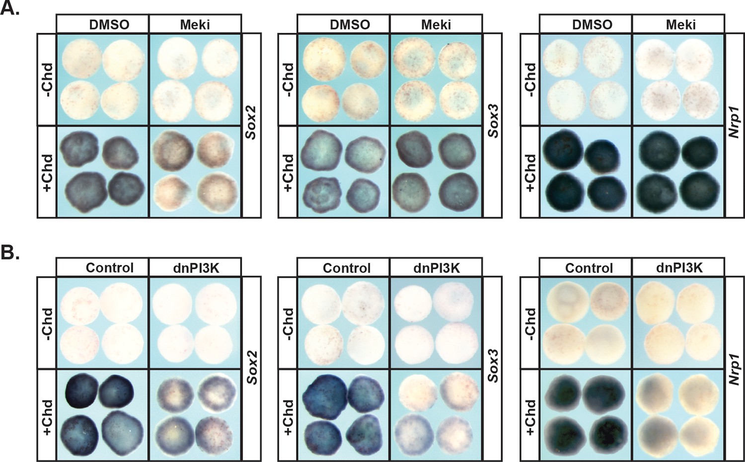
PI3K/Akt signaling but not MAPK signaling is required for pluripotent blastula cells to transit to a neural progenitor state.
(A–B) Animal pole explant assay examining Sox2, Sox3, and Nrp1 expression in Chordin (Chd) induced animal cap explants treated with Meki (RDEA119) (A) or injected with dnPI3K (B). Explants were cultured alongside sibling embryos and collected at early neurula stages (stage 13) for Sox2/3 or late neurula stages (stage 18) for Nrp1. Meki treatment does not affect Chordin-mediated neural induction whereas dnPI3K blocks induction of all three neural markers.
Figure 4—figure supplement 1
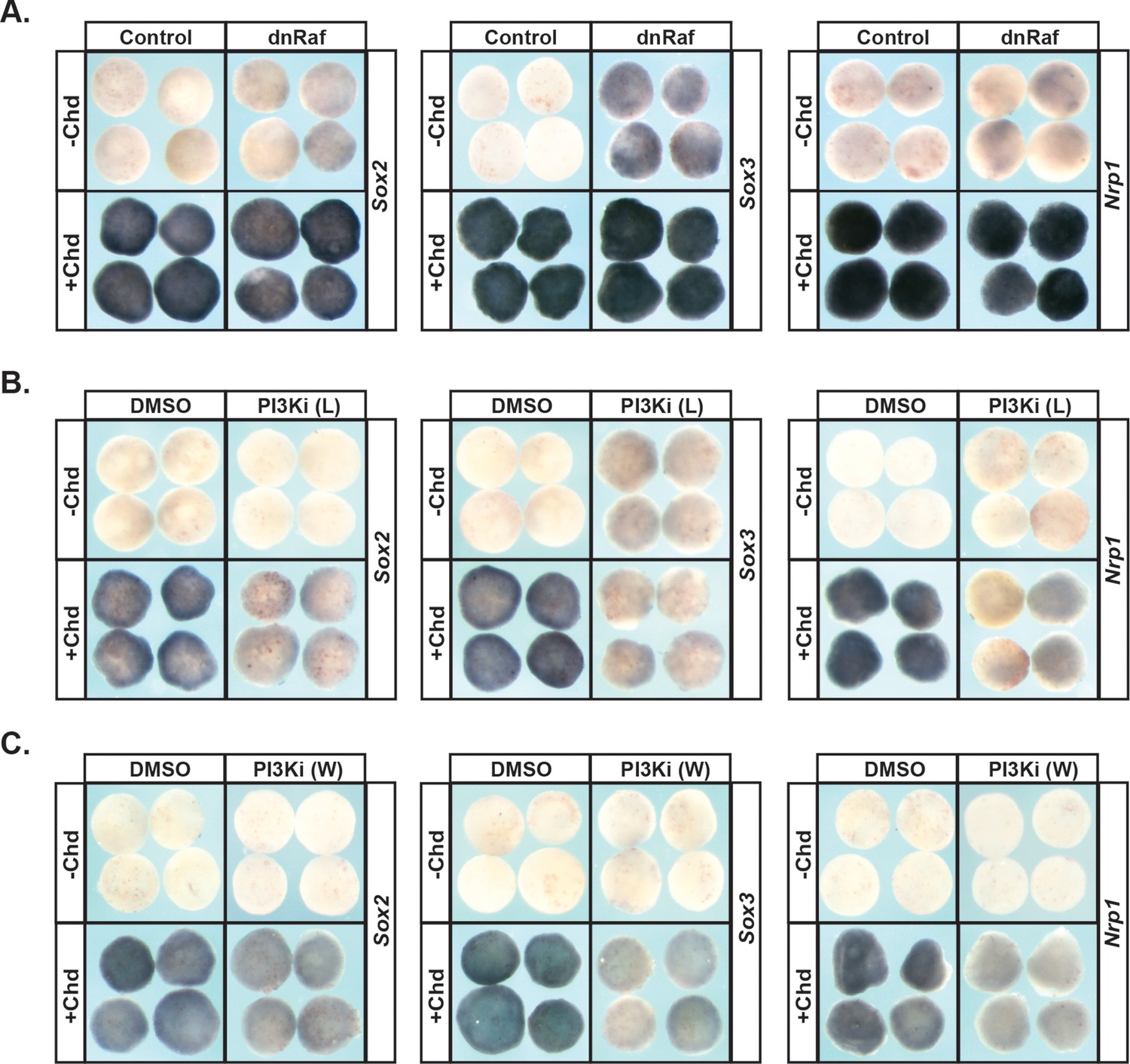
Blocking PI3K/Akt activation using PI3Ki (L) (LY294) or PI3Ki (W) (Wortmannin) phenocopies the effects of dnPI3K on Chordin-mediated neural induction.
(A–C) Animal pole explant assay examining Sox2, Sox3, and Nrp1 expression in Chordin (Chd) induced animal cap explants injected with dnRaf (A) or treated with PI3Ki (L) (B) or PI3Ki (W) (C). Explants were cultured alongside sibling embryos and collected at early neurula stages (stage 13) for Sox2/3 or late neurula stages (stage 18) for Nrp1. PI3Ki (L) and PI3Ki (W) phenocopy the effects of dnPI3K on Chordin-mediated neural induction.
Figure 5 with 2 supplements
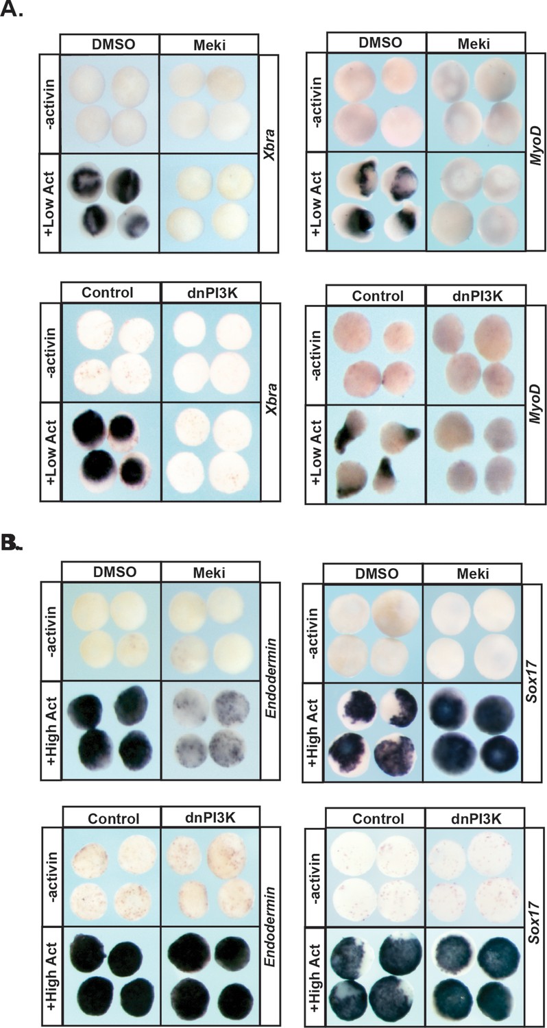
MAPK and PI3K/Akt differentially alter the transit of pluripotent cells to restricted cell states.
(A–B) Animal pole explant assay examining expression of Xbra and MyoD (A) or Endodermin and Sox17 (B) in explants cultured with or without activin after treatment with Meki (RDEA119) or injection with dnPI3K. Explants were cultured alongside sibling embryos and collected at midgastrula stages (stage 11.5) for Xbra, Endodermin, and Sox17 expression and midneurula stages (stage 15/16) for MyoD expression. Blocking either cascade interferes with mesoderm formation whereas only MAPK signaling is required for Endodermin induction.
Figure 5—figure supplement 1
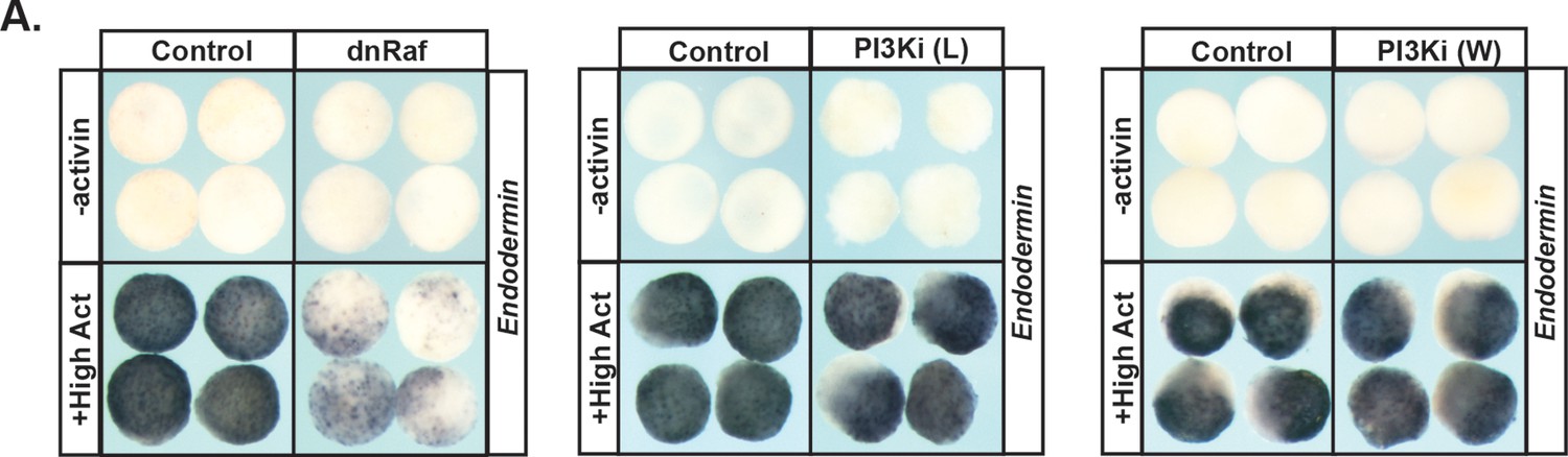
MAPK and PI3K/Akt signaling are differentially required for Endodermin expression.
(A) Animal pole explant assay examining expression of Endodermin in explants cultured with or without activin after injection with dnRaf or treatment with PI3Ki (L) (LY294) or PI3Ki (W) (Wortmannin). Explants were cultured alongside sibling embryos and collected at midgastrula stages (stage 11.5). MAPK signaling is required for Endodermin induction.
Figure 5—figure supplement 2
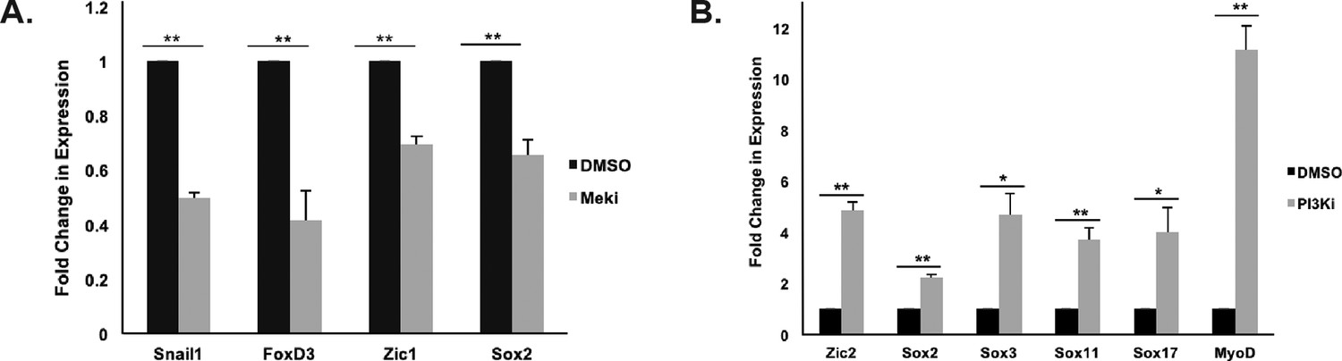
Blocking MAPK or PI3K activation differentially alters animal pole explant gene expression (A–B) qRT-PCR analysis of animal cap explants treated with Meki (RDEA119) and cultured alongside sibling embryos collected at blastula stages (stage 9) (A) or treated with PI3Ki (LY294) and cultured alongside sibling embryos collected at early neurula stages (stage 13) (B).
Blocking MAPK activation inhibits Snail1, FoxD3, Zic1, and Sox2 expression while blocking PI3K activation increases Zic2, Sox2, Sox3, Sox11, Sox17, and MyoD expression. (*p<0.05; **p<0.01).
Figure 6
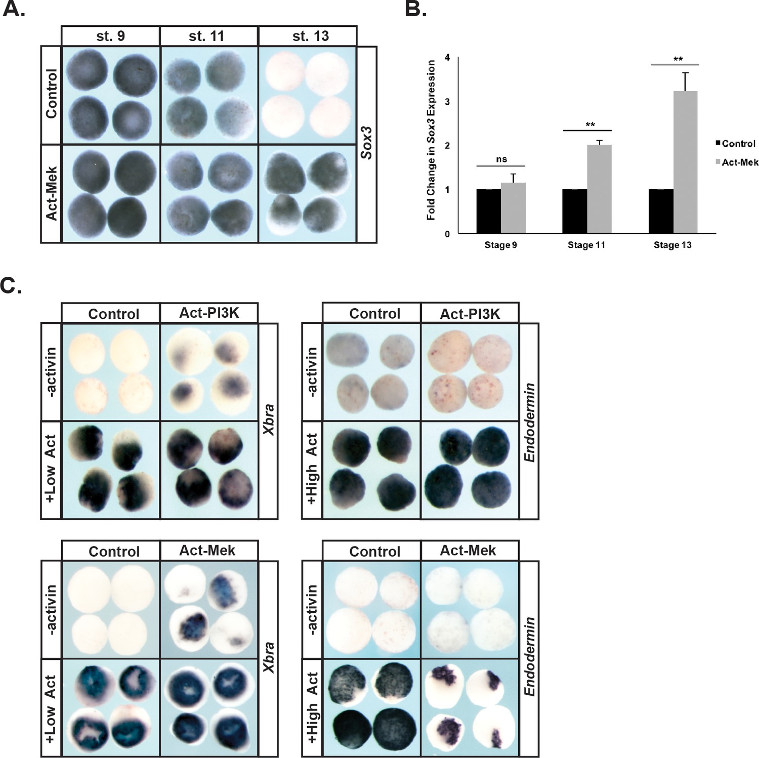
Prolonged MAPK activation alters the timing of pluripotency gene expression.
(A–B) Animal pole explant assays examining Sox3 expression in animal cap explants injected with constitutively active Mek (Act-Mek). Explants were cultured alongside sibling embryos and collected at blastula (stage 9), midgastrula (stage 11), and early neurula (stage 13) stages for in situ hybridization (A) or qRT-PCR (B). Activating MAPK leads to retained Sox3 expression. (C) Animal cap explant assay examining Xbra and Endodermin expression in explants cultured with or without activin after injection with constitutively active PI3K (Act-PI3K) or Act-Mek. Explants were cultured alongside sibling embryos and collected at midgastrula stages (stage 11.5). Sustained MAPK activity interferes with Endodermin induction. (ns, not significant; **p<0.01).
Figure 7 with 2 supplements
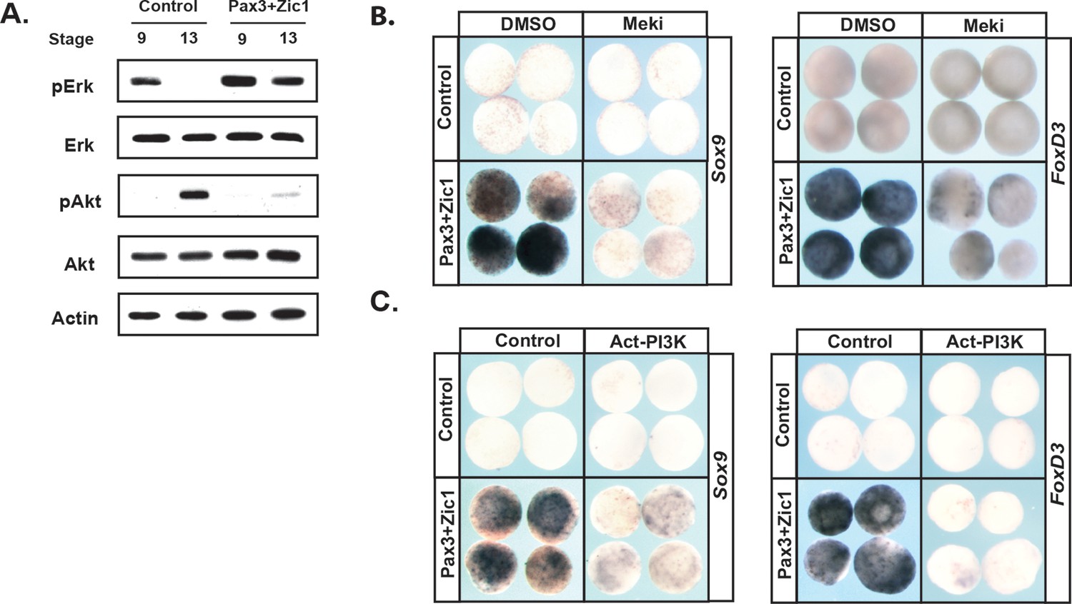
Reprograming cells to a Neural Crest state establishes and requires high MAPK and low PI3K/Akt activity.
(A) Western blot of lysates from Pax3-GR/Zic1-GR injected animal pole explants. Explants were cultured alongside sibling embryos and collected at blastula (stage 9) and early neurula (stage 13) stages to examine levels of phosphorylated and unphosphorylated Erk1/2 and Akt. Reprograming to a neural crest state retains the activities of these pathways characteristic of pluripotent blastula cells. (B–C) Animal cap explant assay examining Sox9 and FoxD3 expression in Pax3GR/Zic1-GR injected explants treated with Meki (RDEA119) (B) or co-injected with Act-PI3K (C). Explants were cultured alongside sibling embryos and collected at late neurula stages (stage 18). Blocking MAPK activation or activating PI3K/Akt blocks expression of neural crest markers.
Figure 7—figure supplement 1
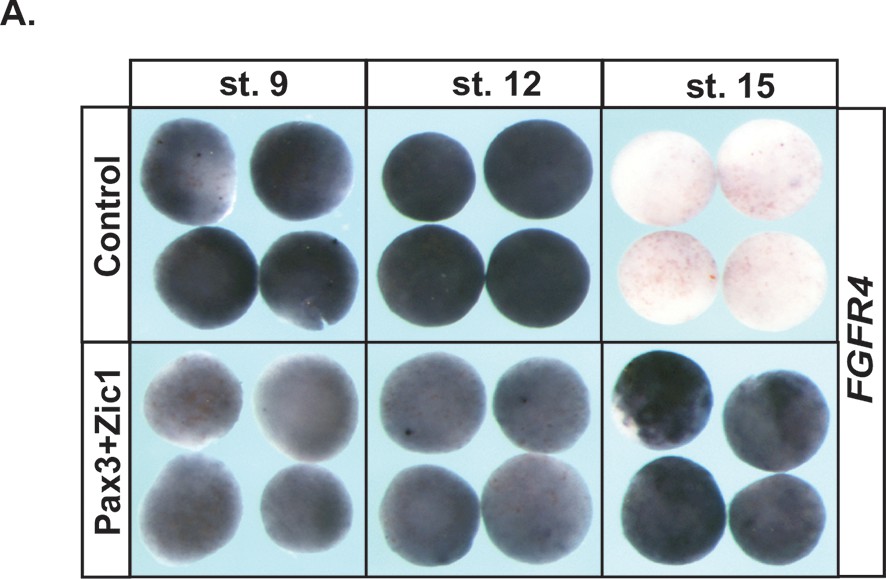
Reprograming to a Neural Crest state sustains FGFR4 expression.
(A) Animal pole explant assay examining FGFR4 expression in animal cap explants injected with Pax3-GR/Zic1-GR. Explants were cultured alongside sibling embryos and collected at blastula (stage 9), midgastrula (stage 12), and midneurula (stage 15) stages. Reprogramming to a neural crest state causes sustained FGFR4 expression.
Figure 7—figure supplement 2
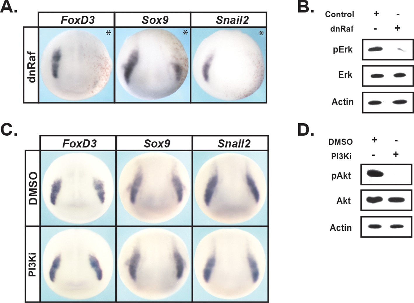
MAPK activation is required for neural crest factor expression at mid-neurula stages.
(A–D). In situ hybridization examining FoxD3, Sox9, and Snail2 expression in mid-neurula stage (stage 15) embryos injected with dnRaf (A) or treated with the PI3Ki (Wortmannin) (C). Western Blot analyses were conducted with stage nine animal cap explants dissected from dnRaf injected sibling embryos (B) or with mid-neurula stage (stage 15) whole embryos treated with PI3Ki (D). Asterisk denotes injected side, marked by staining of the lineage tracer β-galactosidase (red). Blocking MAPK activation causes a loss of FoxD3, Sox9, and Snail2 expression.
Figure 8
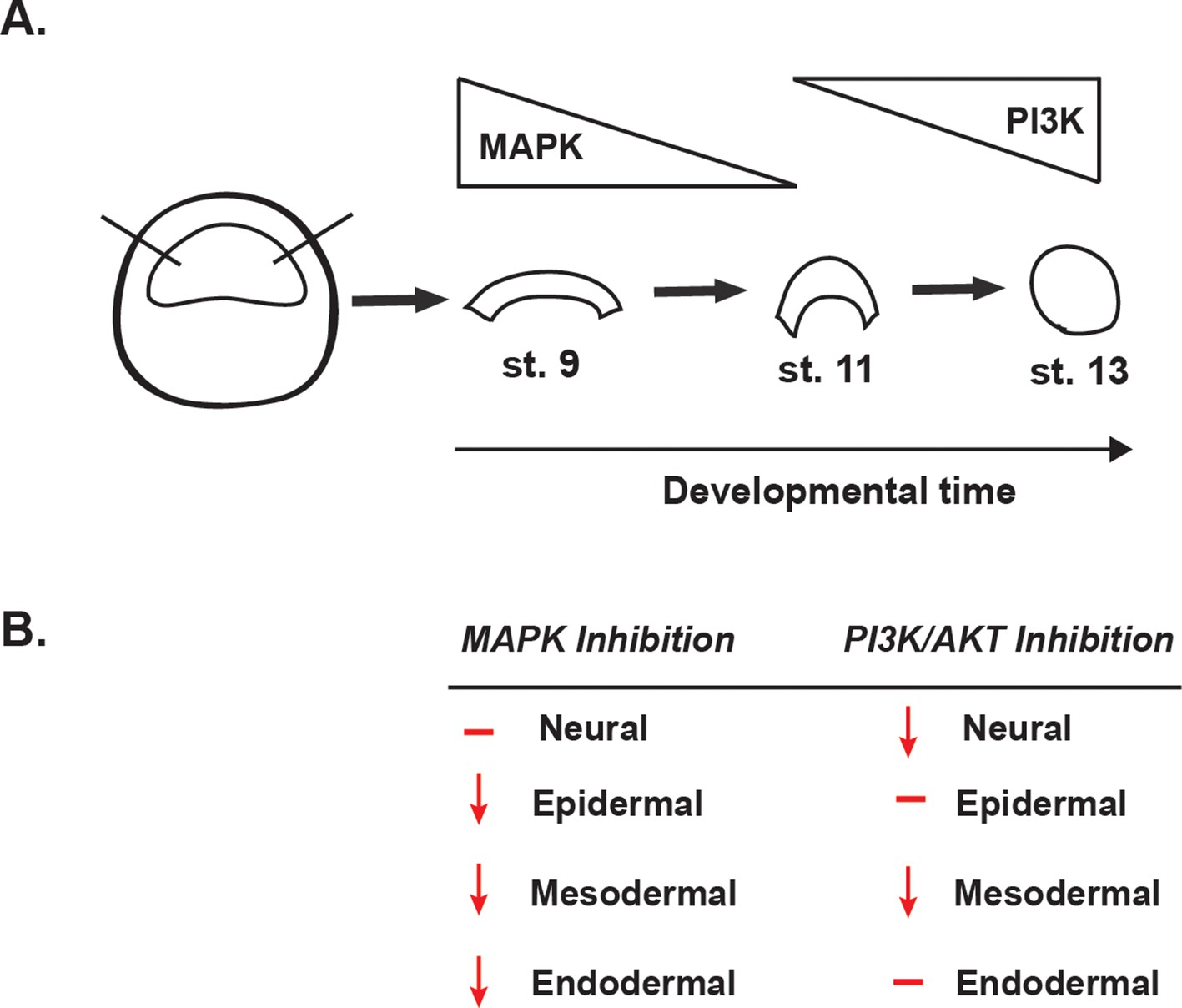
Summary of the effects of MAPK or PI3K/Akt inhibition.
(A) Schematic representation of MAPK and PI3K/Akt cascade activation in animal cap explants (staged by sibling embryos) at the blastula stage (stage 9), midgastrula stage (stage 11), and early neurula stage (stage 13). (B) Diagram summarizing the effects of MAPK or PI3K/Akt inhibition on the adoption of neural, epidermal, mesodermal, and endodermal states.
Additional files
-
Transparent reporting form
- https://doi.org/10.7554/eLife.33845.017
Download links
A two-part list of links to download the article, or parts of the article, in various formats.
Downloads (link to download the article as PDF)
Open citations (links to open the citations from this article in various online reference manager services)
Cite this article (links to download the citations from this article in formats compatible with various reference manager tools)
FGF mediated MAPK and PI3K/Akt Signals make distinct contributions to pluripotency and the establishment of Neural Crest
eLife 7:e33845.
https://doi.org/10.7554/eLife.33845


