Neurocranial anatomy of an enigmatic Early Devonian fish sheds light on early osteichthyan evolution
- Flinders University, Australia
- Evolutionary Biology Centre, Uppsala University, Sweden
- Museum Victoria, Australia
- Naturalis Biodiversity Center, Netherlands
- University of Oxford, United Kingdom
- Research School of Physics & Engineering, Australian National University, Australia
- Australian Museum Research Institute, Australia
Figures
Figure 1 with 1 supplement
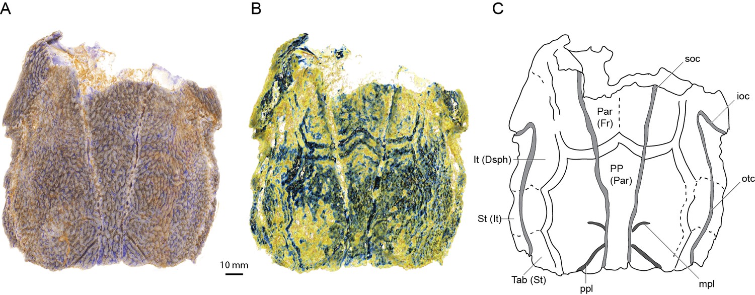
Skull roof of ‘Ligulalepis’ AM-F101607 in dorsal view.
Artificial colouration added in Drishti to highlight (A) sensory canals; and (B) bone sutures. (C) Interpretive diagram showing skull roof pattern; patterns of sensory canals inferred from both specimens. Bone names use sarcopterygian conventions, with actinopterygian conventions in brackets.
Figure 1—figure supplement 1
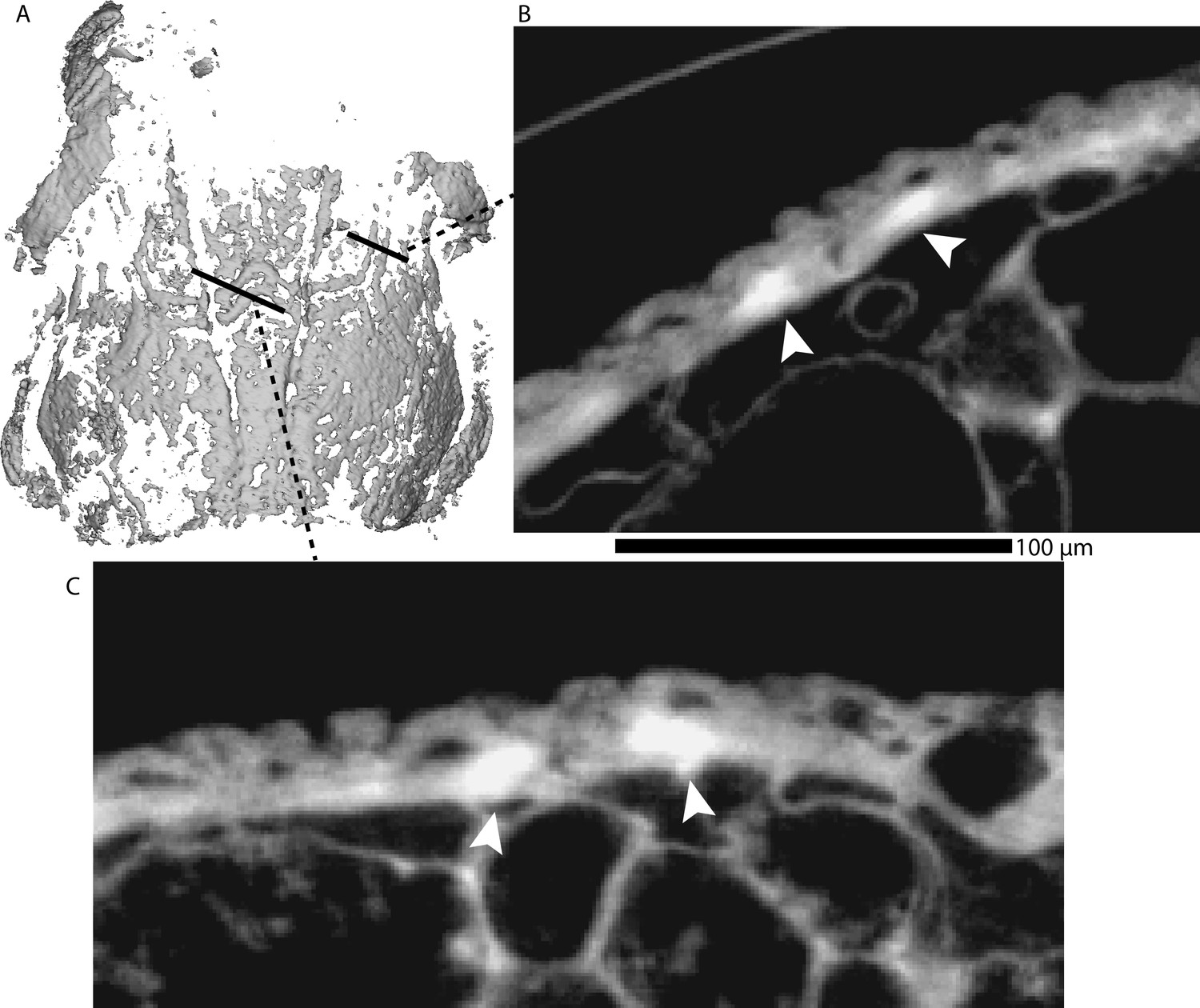
Bands of high-density bone in the basal layer of the skull roof dermal bone are assumed to follow sutures in AM-F101607.
(A) Mimics rendering of a mask with a high minimum threshold, thereby showing only the highest density bone, including the double bands that we presume occurred either side of sutures. (B-C) Cross sections, with these high-density bands marked with arrows. Note that they appear to be confined to the basal part of the bone.
Figure 2
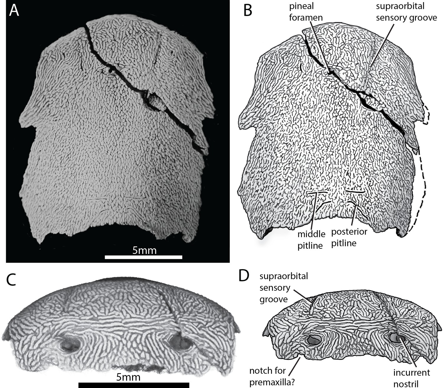
Skull of ‘Ligulalepis’ ANU V3628.
(A) Dorsal view, photograph of specimen whitened with ammonium chloride. (B) Line drawing of A. (C) Anterior view, imaged using Drishti to reveal parts embedded in resin. (D) Line drawing of C.
Figure 3

Skull and sensory canals of ‘Ligulalepis’ ANU V3628.
(A) Segmented model of dermal and perichondral bone of the left orbit, showing the posterior nostril within the orbit and endochondral bone in the eyestalk. (B) Position of supraorbital canal (soc) and infraorbital canal (ioc) on the skull. (C) Left supraorbital canal in left lateral view. Arrow indicates point where anterior and posterior canal sections overlap. (D) Right infraorbital and postotic canal in anterior view. Arrows indicate tubules that connect the canal to the surface.
Figure 4
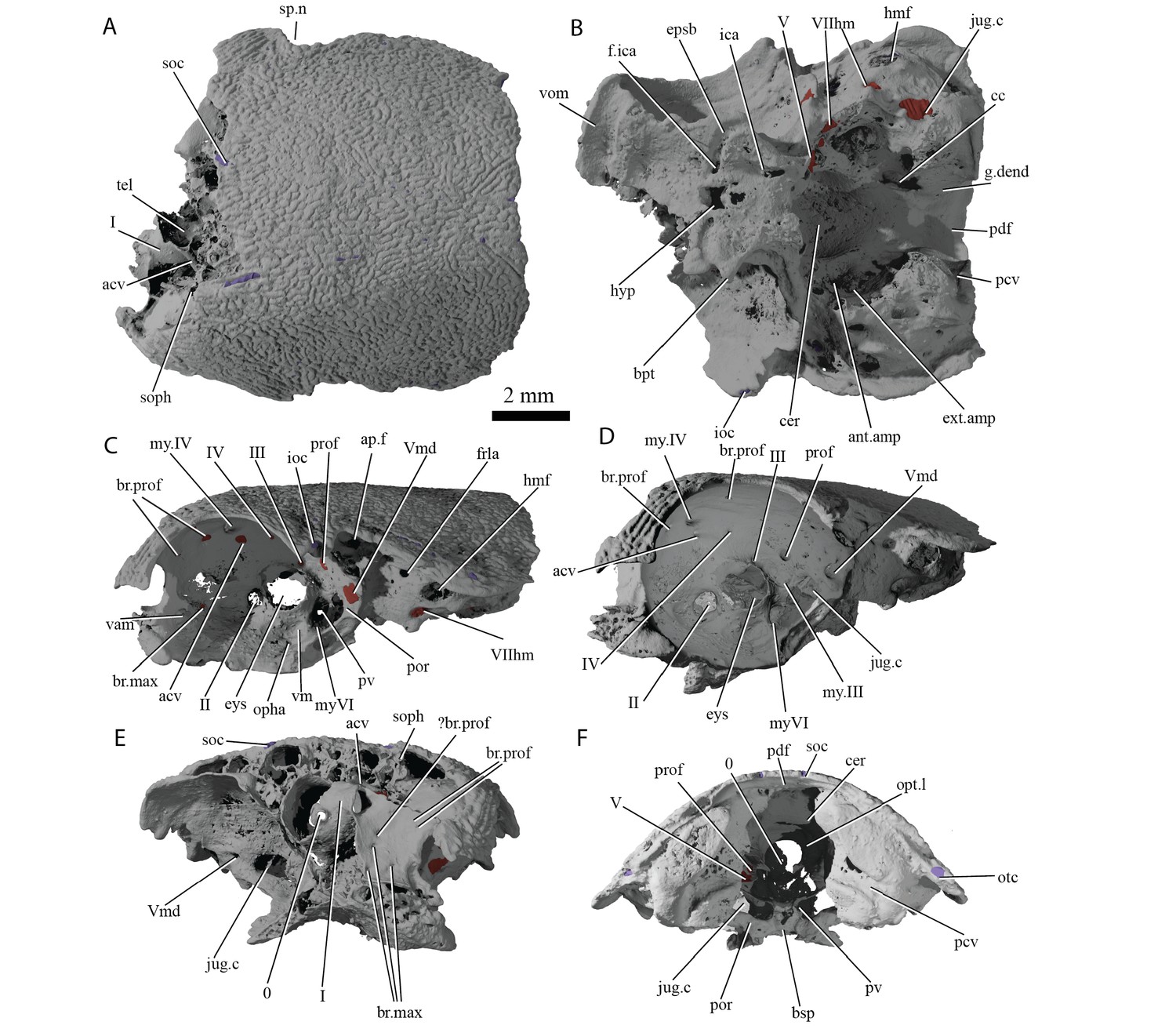
Cranium of ‘Ligulalepis’ AM-F101607.
(A) dorsal; (B) ventral; (C) left lateral; (D) left anterolateral showing details of orbit; (E) anterior; and (F) posterior view.
Figure 5
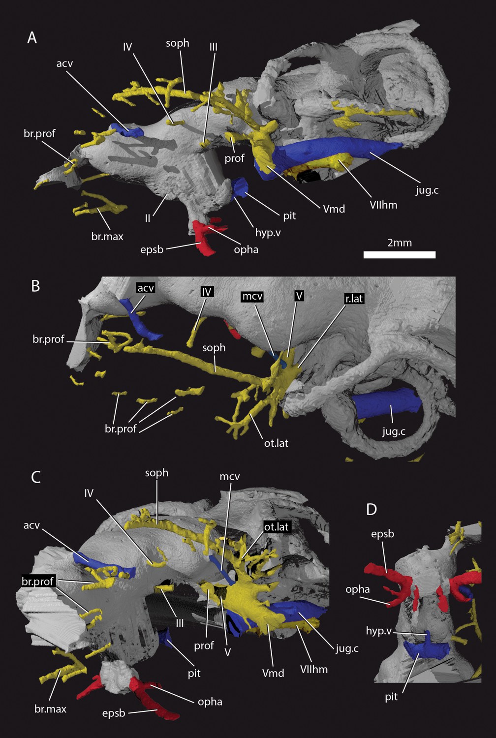
Cranial nerves and vessels of ‘Ligulalepis’ AM-F101607.
(A) left lateral; (B) dorsal; (C) left anterolateral and (D) ventral view of anterior section only. Cranial endocast in grey, nerves in yellow, veins in blue and arteries in red.
Figure 6
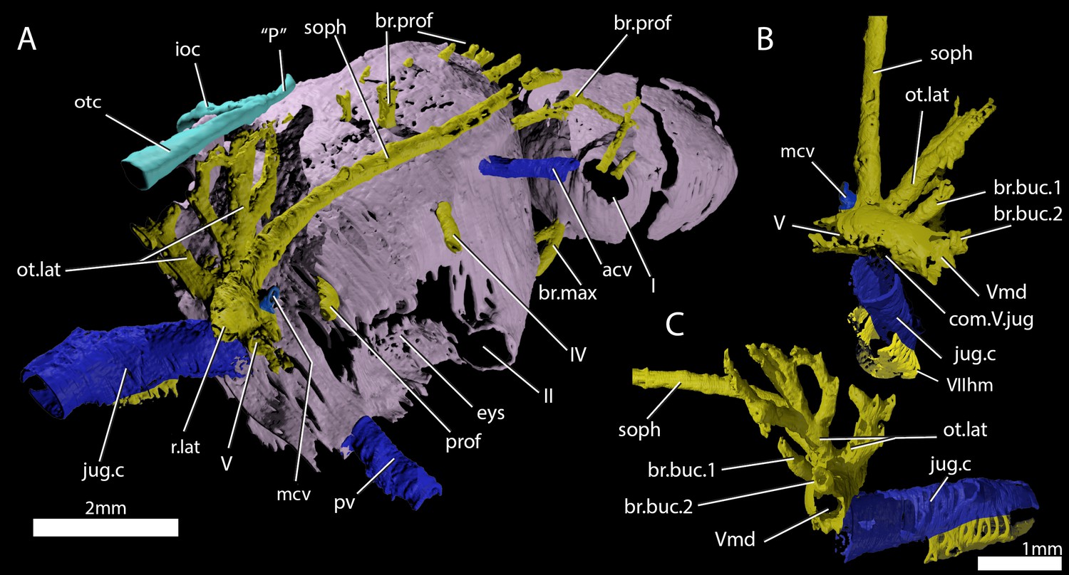
Cranial nerves and vessels of ‘Ligulalepis’ ANU V3628.
(A) ANU V3628, segmentation of the interior of the left orbital region, viewed from a postero-dorsal-medial viewpoint. The cranial endocast is not shown. Perichondral bone lining the orbit and nasal capsules is in lilac. Nerves are yellow, veins blue and sensory canals are in turquoise. The trigeminal, lateralis and facial nerves and their branches and the jugular vein, viewed from an anterior-ventral (B) and left lateral (C) viewpoints.
Figure 7
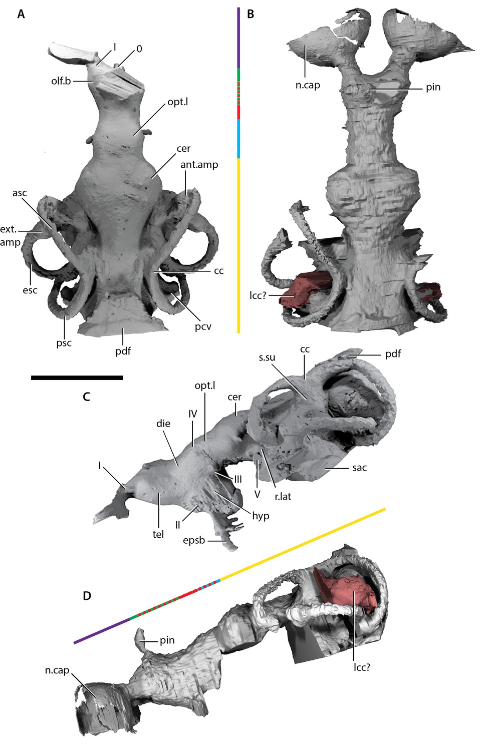
Endocast of ‘Ligulalepis’.
(A) AM-F101607, dorsal view. (B) ANU V3628, dorsal view. (C) AM-F101607 lateral view. (D) ANU V3628 lateral view with possible lateral cranial canal in red. Major brain regions indicated by coloured bars: nasal capsules (purple), telencephalon (green), diencephalon (red), mesencephalon (blue), metencephalon and myelencephalon (yellow).
Figure 8
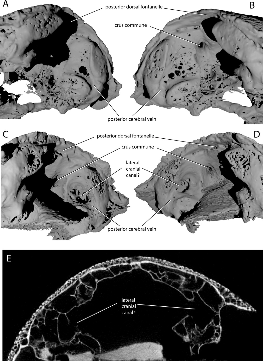
Variability in the development of a lateral cranial canal in ‘Ligulalepis’.
(A–B) Ventrolateral view of AM-F101607, showing internal view of the otic region on the right hand side (A) and the left hand side (B). (C–D) Ventrolateral view of ANU V3628, showing internal view of the otic region on the right hand side (C) and the left hand side (D). (E) CT scan cross-section of ANU V3628 showing diverticula that may represent lateral cranial canals.
Figure 9
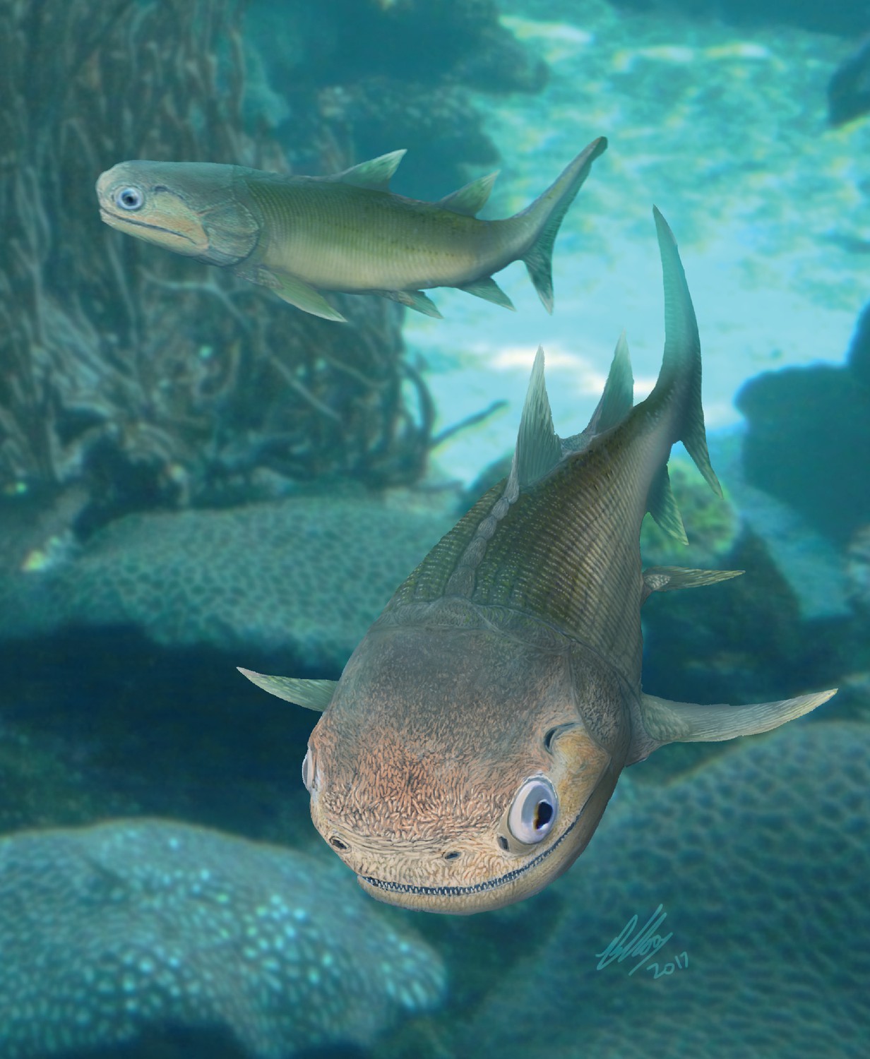
Life reconstruction of ‘Ligulalepis’.
Based on the skull roof morphology of AM-F101607 and ANU V3628, other features remain hypothetical.
Figure 10
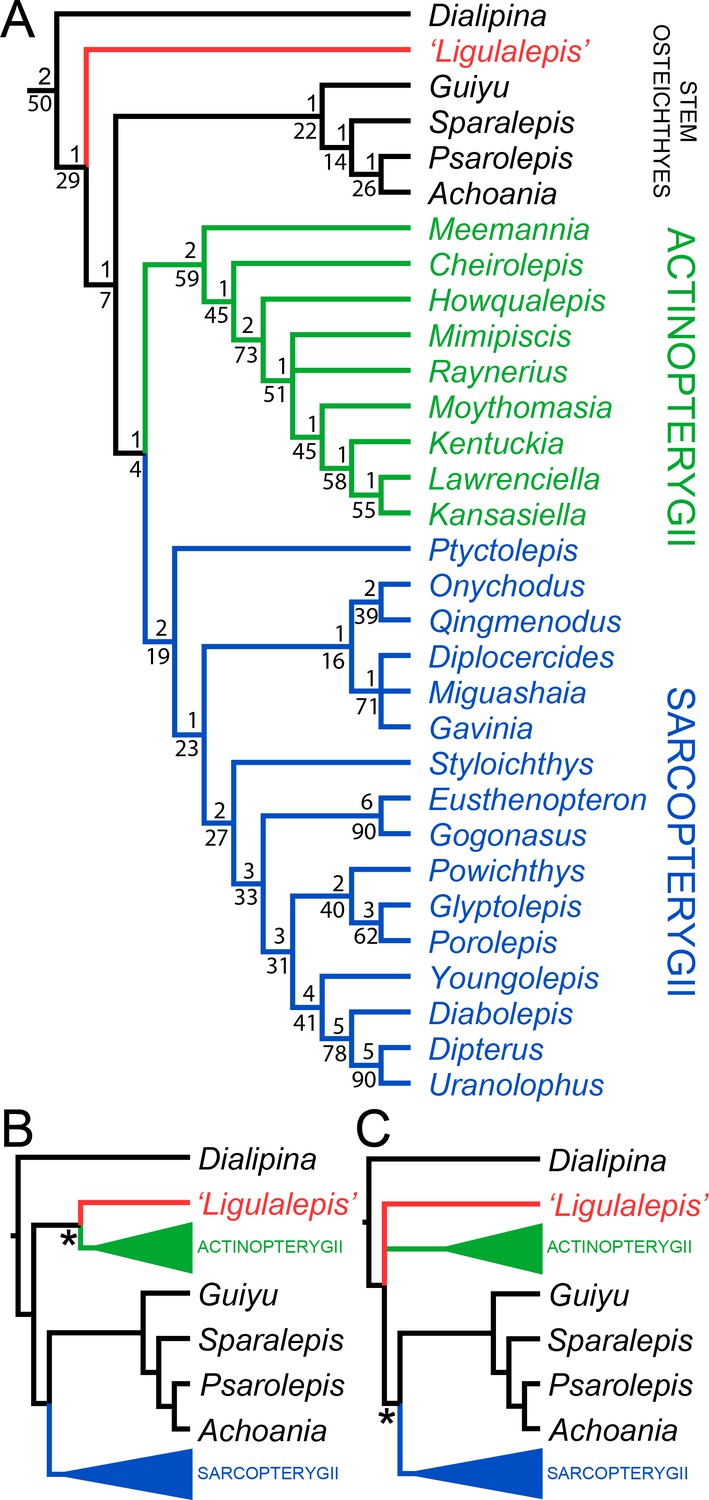
Results of parsimony phylogenetic analysis.
(A) Strict consensus tree. Numbers above nodes refer to bremer support, numbers below nodes represent bootstrap support. (B) Strict consensus tree after enforcing ‘Ligulalepis’ as a stem actinopterygian. (C) Strict consensus tree after constraining ‘psarolepids’ (Guiyu, Sparalepis, Psarolepis, Achoania) as stem sarcopterygians. Asterisks indicate constrained nodes.
Figure 11
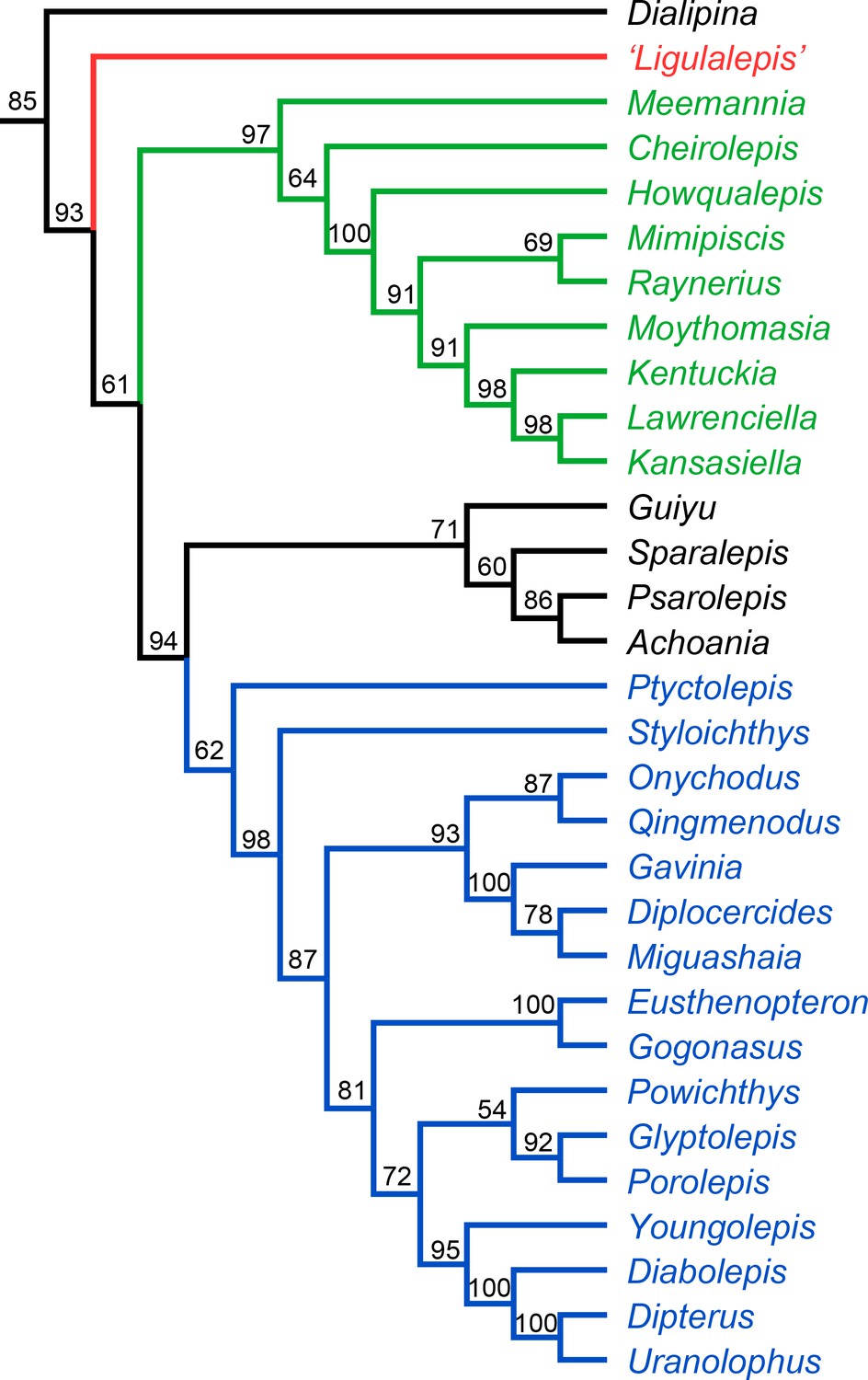
Results of Bayesian phylogenetic analysis.
Maximum clade credibility tree. Numbers represent posterior probabilities, displayed as percentages for presentation purposes.
Additional files
-
Transparent reporting form
- https://doi.org/10.7554/eLife.34349.015
Download links
A two-part list of links to download the article, or parts of the article, in various formats.
Downloads (link to download the article as PDF)
Open citations (links to open the citations from this article in various online reference manager services)
Cite this article (links to download the citations from this article in formats compatible with various reference manager tools)
Neurocranial anatomy of an enigmatic Early Devonian fish sheds light on early osteichthyan evolution
eLife 7:e34349.
https://doi.org/10.7554/eLife.34349


