Dynamics of venom composition across a complex life cycle
- The Hebrew University of Jerusalem, Israel
- University of North Carolina at Charlotte, United States
- Indian Institute of Science, India
Figures
Figure 1
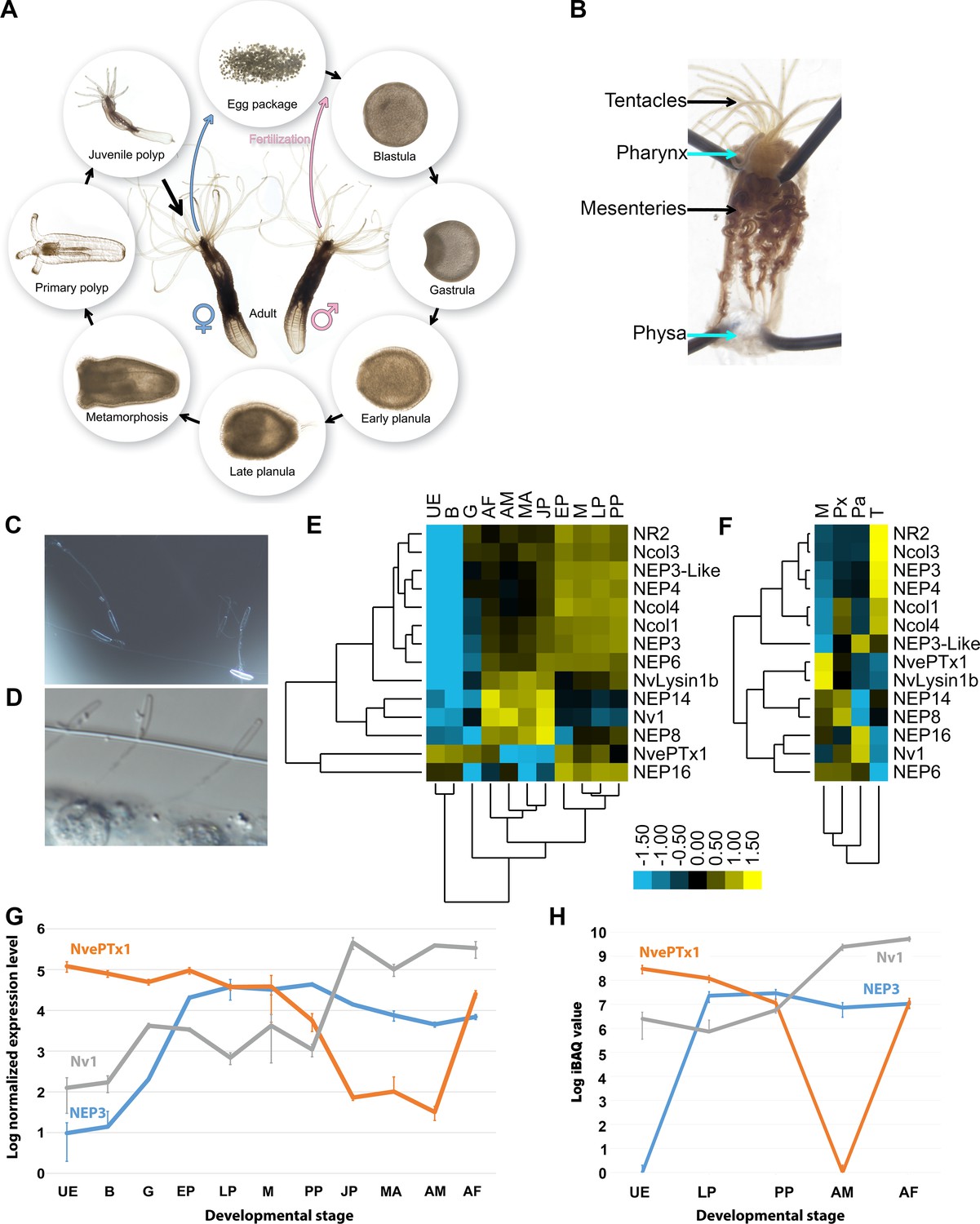
Expression of toxins across Nematostella development and tissues.
(A) The life cycle of N. vectensis. (B) Dissected Nematostella female polyp. (C) Discharged planula nematocysts found in the medium after an encounter with A. salina nauplii. (D) Nematocysts of Nematostella planula pinned in the cuticle of an A. salina nauplii. (E–F) Heat maps of the nCounter differential expression levels of genes encoding toxins and other nematocyst proteins in various developmental stages and adult female tissues. (G) A graph at a logarithmic scale of the nCounter normalized expression levels of the genes encoding NEP3, NvePTx1 and Nv1 at each developmental stage. Each point is the average of three biological replicates and the error bars represent standard deviation. (H) A graph at a logarithmic scale of the mass spectrometry-measured iBAQ (Intensity-Based Absolute Quantification) values of the peptides NEP3, NvePTx1 and Nv1 at five developmental stages. Each point is the average of four technical replicates and the error bars represent standard deviation. Key for panels E and G: UE = Unfertilized Egg; B = Blastula; G = Gastrula; EP = Early Planula; LP = Late Planula; M = metamorphosis; PP = Primary Polyp; JP = Juvenile Polyp; MA = Mixed Adults; AM = Adult Male; AF = Adult Female. Key for panel F: M = Mesenteries; Px = Pharynx; Pa = Physa; T = Tentacles.
Figure 2
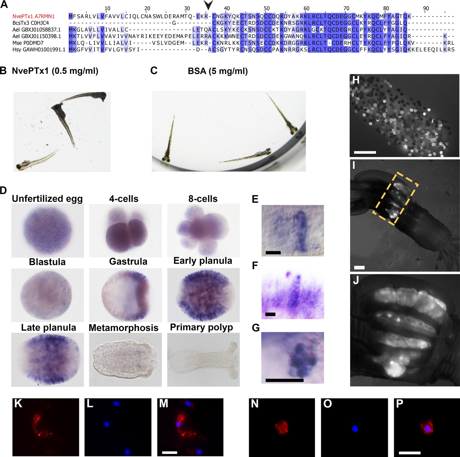
NvePTx1 is a toxin that is maternally deposited in Nematostella eggs.
(A) Sequence alignment of NvePTx1 and its homologs from other cnidarian species. The arrowhead represents the proteolysis site that separates the mature toxin from the signal peptide. Accession numbers appear near each sequence. Nve = Nematostella vectensis; Bcs = Bunodosoma caissarum; Ael = Anthopleura elegantissima; Mse = Metridium senile; Hsy = Hydractinia symbiolongicurpus. (B–C) Zebrafish larvae 16 hr after incubation in NvePTx1 and BSA control. (D) Detection of NvePTx1 expression by ISH. From the blastula stage on, the oral pole is to the right. Close up on planula large elongated (E–F) and small round (G) gland cells stained by ISH for NvePTx1. Gland cells, which are discernible by their round vesicles are free of stain, scale bar is 10 µm. (H–I) An adult F0 female polyp that as a zygote was injected by an NvePTx1::mOrange2 construct. mOrange2 expression is noticeable in the gonads. Panel G is a close up of the region indicated in panel H by the yellow dotted box. Scale bar is 1000 µm (J) An egg package laid by the female from panel H. Many of eggs express mOrange2. Scale bar is 1000 µm. (K–M) Large elongated and (N–P) small round gland cells stained by anti mCherry. Scale bar is 10 µm.
Figure 3 with 1 supplement
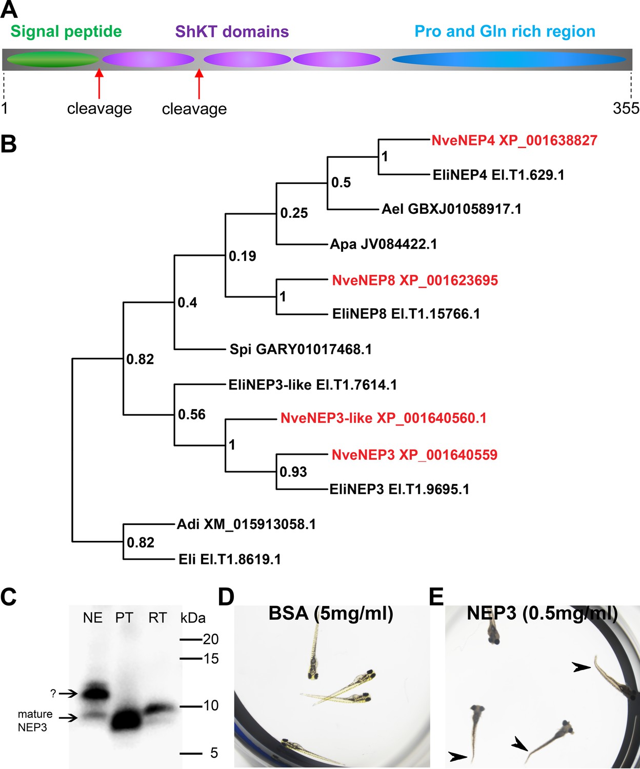
NEP3 is a toxin that is processed from a long precursor protein and is part of a large family.
(A) The primary structure of the NEP3 precursor. (B) A maximum-likelihood tree of the NEP3 family. Accession numbers appear near each sequence and bootstrap values (fraction of 1000 bootstraps) appear near each node. Nve = Nematostella vectensis; Eli: Edwardsiella lineata; Ael = Anthopleura elegans; Apa = Aiptasia pallida; Spi = Stylophora pistillata; Adi = Acropora digitifera. (C) Western blot with rat Anti-NEP3 antibody. Samples are discharged nematocyst extract (NE), native purified toxin (PT) and recombinant toxin (RT). (D–E) Zebrafish larvae after 16 hr incubation in NEP3 and BSA control. Arrowheads are pointing at twitched tails.
Figure 3—figure supplement 1
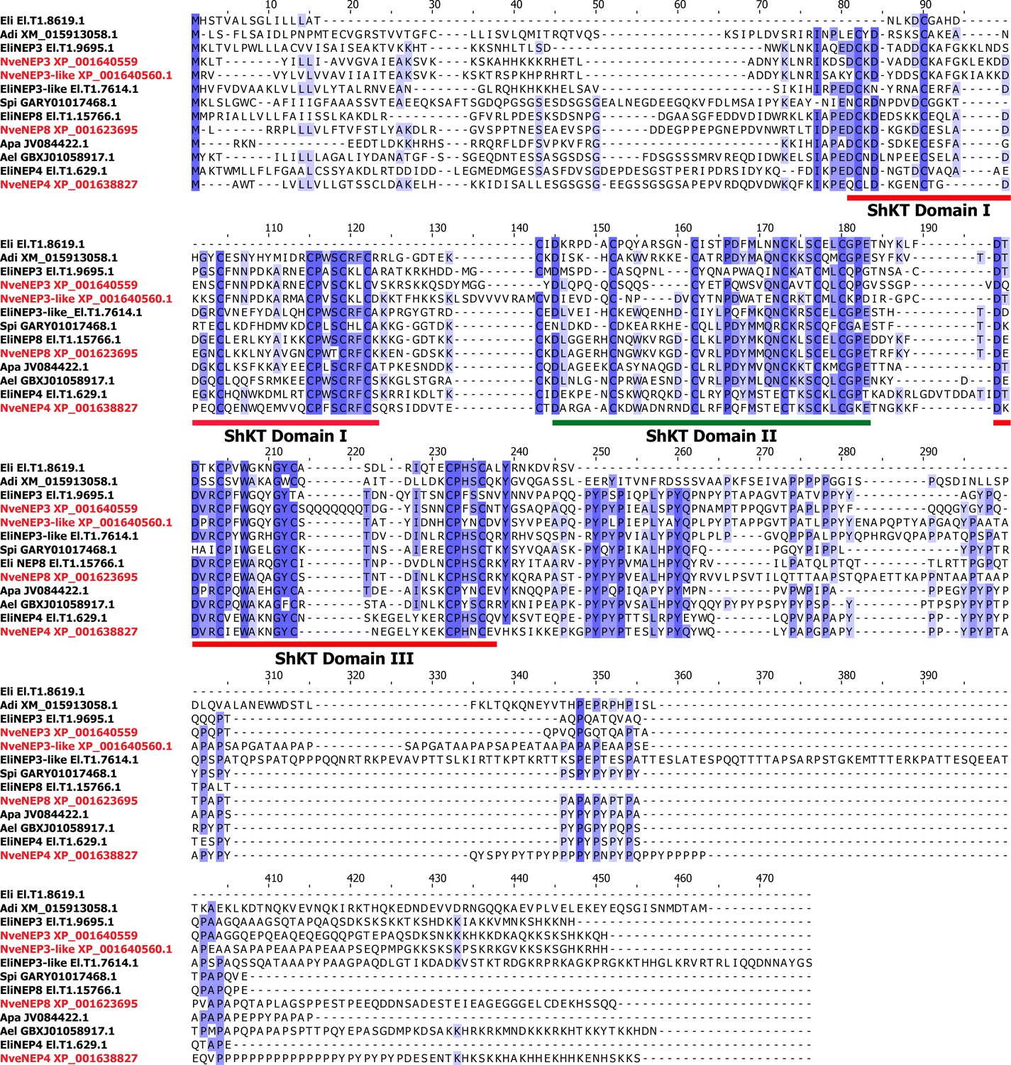
Multiple sequence alignment of the NEP3 family members from Figure 3B.
Accession numbers appear near each sequence. Nematostella sequences are in red.
Figure 4 with 1 supplement
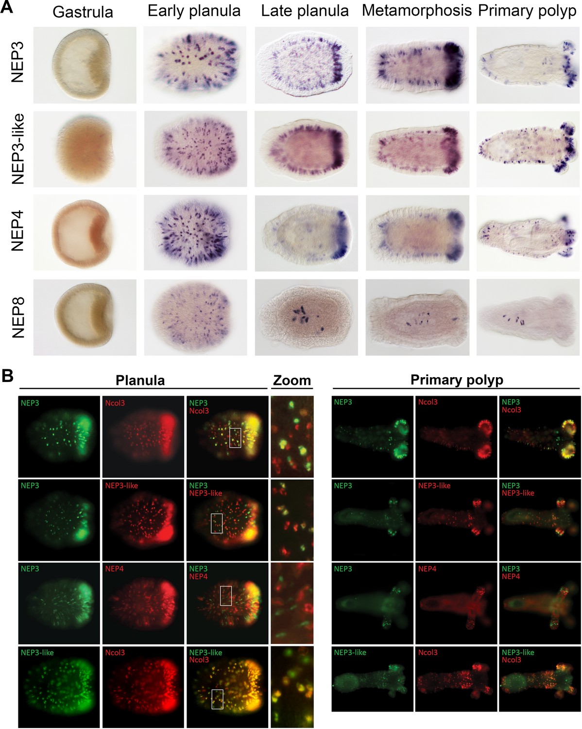
Partially overlapping and distinct expression patterns of NEP3 family members.
(A) Expression of the NEP3 family members in five developmental stages of Nematostella as determined by ISH. (B) Expression of the NEP3 family members in planulae and polyps of Nematostella as determined by dFISH. In all panels, the oral pole is to the right.
Figure 4—figure supplement 1

Close up on ISH staining of NEP3, NEP3-like, NEP4 and NEP8 reveals that they are expressed in nematocytes.
Scale bars are 20 µm.
Figure 5
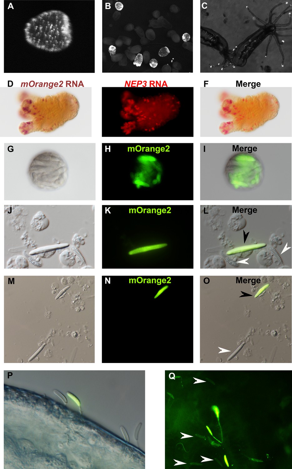
NEP3 is expressed only in a subset of nematocytes.
(A) Three days of planula of an F1 transgenic line expressing mOrange2 under a NEP3 promoter. (B) 6 days old metamorphosing larva of the same transgenic line. (C) Two months old juvenile polyps of the same line. (D–F) Double ISH of an F2 primary polyp of the transgenic line with probes against NEP3 and mOrange2. mOrange2 transcripts were stained with an NBT (nitro-blue tetrazolium chloride) and BCIP (5-bromo-4-chloro-3'-indolyphosphate p-toluidine) solution that forms purple crystals, whereas NEP3 transcripts were stained with FastRed that provides fluorescent red signal. (G–I) A nematosome of the transgenic line exhibiting mOrange2 fluorescent signal within its nematocytes. (J–L) A spread of dissociated cells of the tentacles of an F2 transgenic NEP3 polyp. An mOrange2-positive nematocyte is indicated by a black arrow and negative spirocytes are indicated by white arrows. (M–O) A picture of another field of the cell spread from previous panels. An mOrange2-positive nematocyte is indicated by a black arrow and a negative nematocyte is indicated by a white arrow. Scale bar is 10 µm in panels G-O. (P) Nematocytes of a transgenic F2 polyp pinned in the cuticle of A. salina. Only one nematocyte in the picture is mOrange2-positive. (Q) Nematocytes of an F2 polyp of the same transgenic line are pinned in the skin of a zebrafish larva. Only some of them are mOrange2 positive. White arrows indicate examples for negative nematocytes.
Figure 6

Time (seconds) each fish spent when exposed to Artemia (red), silica beads (black), or Nematostella primary polyps (blue) in the top (open symbols) or bottom (closed symbols) third of the aquarium.
Statistically significant differences between treatments identified using separate one way ANOVAs and the Tukey post hoc test are noted with an asterisk.
Figure 7
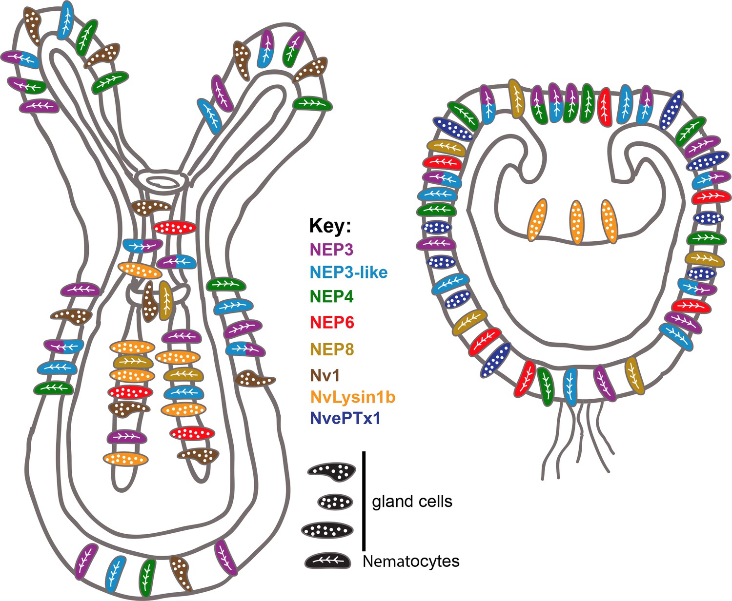
Summary of the current knowledge on spatiotemporal toxin expression in early planula and primary polyp of Nematostella.
This illustration summarizes the current work as well as previous works (Moran et al., 2012a; Moran et al., 2012b; Moran et al., 2013). The degree of expression overlap of NEP8 with other toxins is currently unknown.
Author response image 1
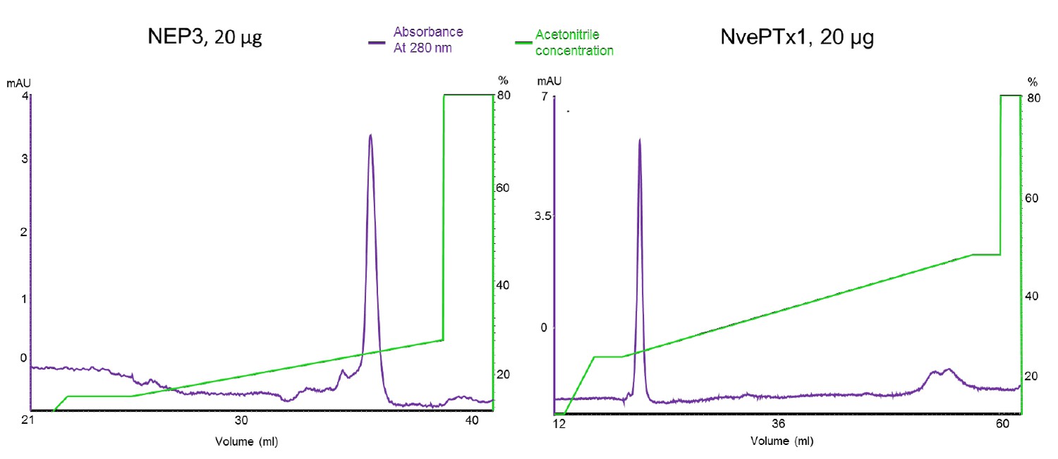
Chromatograms of the recombinant sea anemone toxins used in the zebrafish experiments.
The purified toxins were re-run on a Resource RPC 3 ml reverse phase column (GE Healthcare).
Author response image 2

Double Fluorescent in Situ Hybridization experiment demonstrating that the expression patterns of NEP3 and NEP3-like only partially overlap (yellow signal demonstrates overlapping expression of the two genes).
https://doi.org/10.7554/eLife.35014.022Videos
Video 1
The interaction between Nematostella planulae and Artemia nauplii.
The panulae are rapidly paralyzing the nauplii after physical interaction. This movie is related to Figure 1.
Video 2
The interaction between a burrowed adult Nematostella with grass shrimps.
Only the Nematostella tentacles are exposed and can be encountered by the shrimp. The shrimp readily jumps away once it comes into contact with the tentacles.
Video 3
The interaction of Fundulus larvae with Nematostella egg package.
Upon contact with the egg package, Fundulus reacts swiftly and escapes.
Video 4
The interaction of Fundulus larvae with Nematostella planulae.
Fundulus actively expels planulae from their mouths, if they were erroneously perceived as food.
Tables
Table 1
Interaction of Nematostella vectensis with potential predators, Palaemonetes sp. and Fundulus heteroclitus, at different life stages. ++ readily eats/eats when food is restricted for at least 2 days,+eats when food is restricted for at least 7 days, - does not consume or consider a food source, -- attempted to consume, but exhibited an adverse reaction in all treatments, NA not available. *Only when the eggs are treated with cysteine.
https://doi.org/10.7554/eLife.35014.011| Egg package | Individual eggs | Planulae | Primary polyps | Adults (body exposed) | Adults burrowed in the substrate | |
|---|---|---|---|---|---|---|
| Grass shrimp | ++ | ++ | - | + | + | -- |
| Fish larvae | -- | ++* | -- | -- | -- | - |
| Fish adults | - | - | - | + | - | NA |
Additional files
-
Supplementary file 1
Information of nCounter probe sequences, nCounter raw and normalized data and entropy list for Nematostella transcripts.
This file is related to Figure 1E-G.
- https://doi.org/10.7554/eLife.35014.017
-
Supplementary file 2
Results of MaxQuant analysis of tandem mass spectrometry on Nematostella lysates from different developmental stages.
- https://doi.org/10.7554/eLife.35014.018
-
Transparent reporting form
- https://doi.org/10.7554/eLife.35014.019
Download links
A two-part list of links to download the article, or parts of the article, in various formats.
Downloads (link to download the article as PDF)
Open citations (links to open the citations from this article in various online reference manager services)
Cite this article (links to download the citations from this article in formats compatible with various reference manager tools)
Dynamics of venom composition across a complex life cycle
eLife 7:e35014.
https://doi.org/10.7554/eLife.35014


