Occurrence of long-term depression in the cerebellar flocculus during adaptation of optokinetic response
- Kyoto University, Japan
Figures
Figure 1
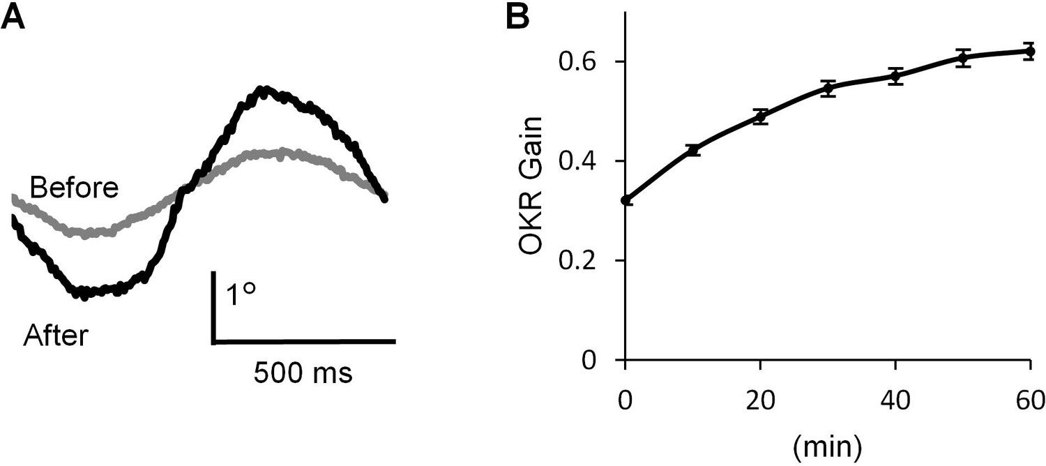
OKR adaptation.
(A) Representative eye position traces before (grey) and after (black) 60 min continuous optokinetic stimulation (OKR training). Ten traces were averaged for each. (B) OKR gain changes during the OKR training (n = 31). Mean ± SEM.
-
Figure 1—source data 1
Time course of OKR gain change during OKR training.
- https://doi.org/10.7554/eLife.36209.003
Figure 2
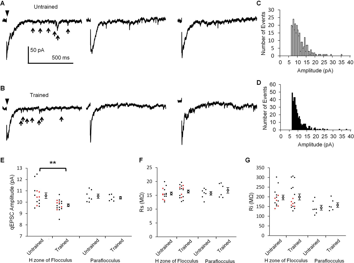
OKR training decreased qEPSC amplitudes.
(A, B) Representative current traces showing qEPSCs (arrows) induced by parallel fiber stimulation (arrowheads) in the H zone of untrained (A) or trained mice (B). (C, D) Histograms of qEPSC amplitudes, which were obtained from the same PC as shown in (A) or (B). (E) Distribution of mean qEPSC amplitudes. Each small circle represents the mean amplitude in a PC (red, blind experiment; black, non-blind experiment), and large open circles and bars represent Mean ± SEM of all data. (F, G) Series (F) and input (G) resistances of PCs. Data shown in (E, F, G) were recorded with or without OKR training in both the flocculus and paraflocculus. **p<0.01, Student’s t-test.
-
Figure 2—source data 1
qEPSC amplitudes with or without OKR training in the H-zone of flocculus or in the paraflocculus.
- https://doi.org/10.7554/eLife.36209.005
Figure 3
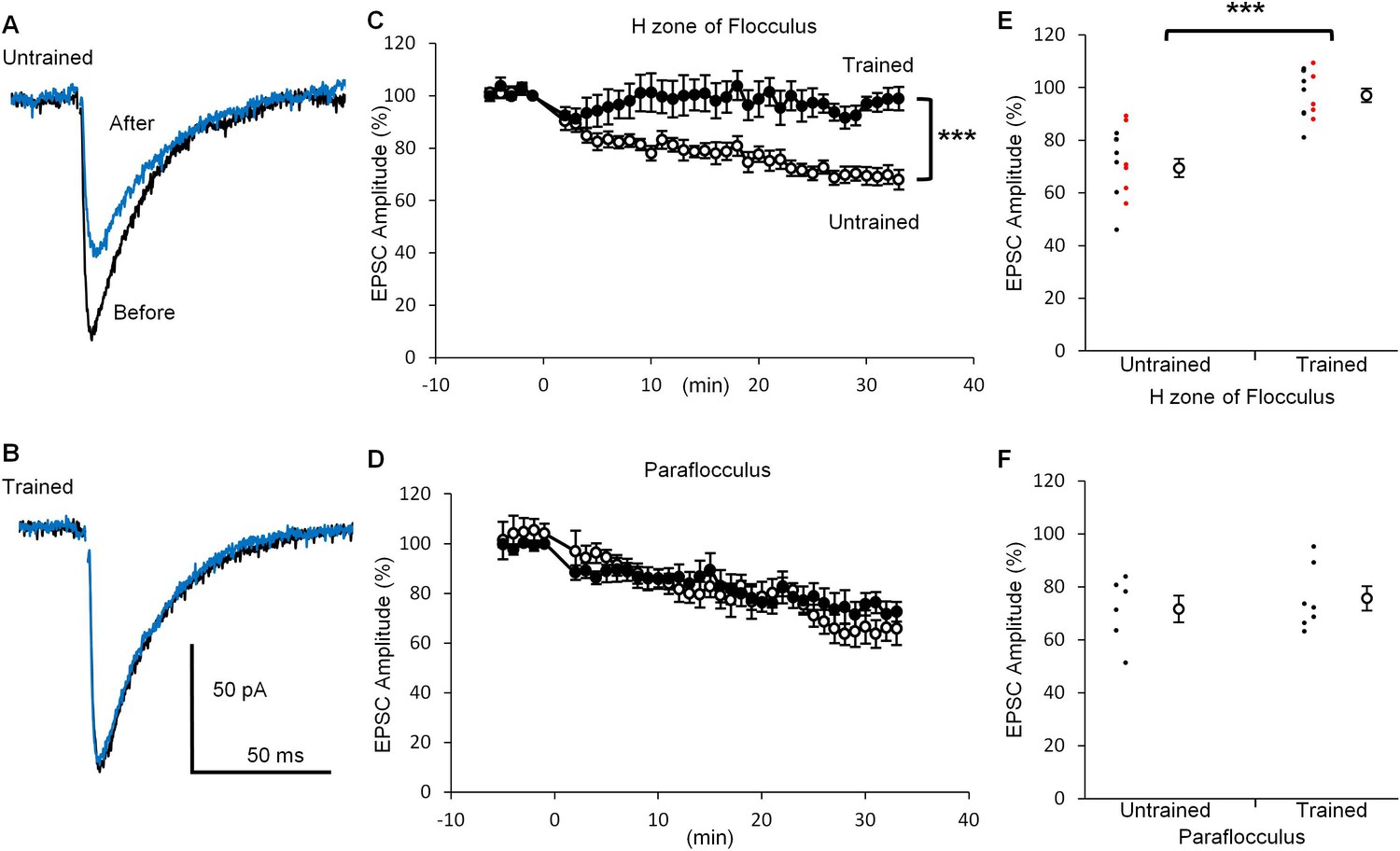
OKR training suppressed LTD induction.
(A, B) Representative EPSC traces recorded before (black) and 30 min after (blue) the conditioning stimulation in the H zone of untrained (A) or trained (B) mice. (C, D) Time courses of normalized EPSC amplitudes before and after the conditioning stimulation at PF-PC synapse in the H zone (untrained, open circles, n = 14; trained, filled circles, n = 12) (C) or the paraflocculus (untrained, open circles, n = 6; trained, filled circles, n = 7) (D). EPSC amplitudes were normalized by setting the amplitude between −1 min and 0 min at 100%. At 0 min, the conditioning depolarizations coupled with the PF stimulation were applied to a PC. (E, F) Distribution of EPSC amplitudes at 30 min after the start of conditioning relative to those before in the H zone (E) and in the paraflocculus (F). Each small circle represents a result obtained from a PC (red, blind experiment; black, non-blind experiment), and large circles and bars represent Mean ± SEM of all data. ***p<0.001, Student’s t-test at 30 min.
-
Figure 3—source data 1
EPSC amplitudes before and after the conditioning stimulation with or without OKR training in the H zone of flocculus or in the paraflocculus.
- https://doi.org/10.7554/eLife.36209.007
Figure 4
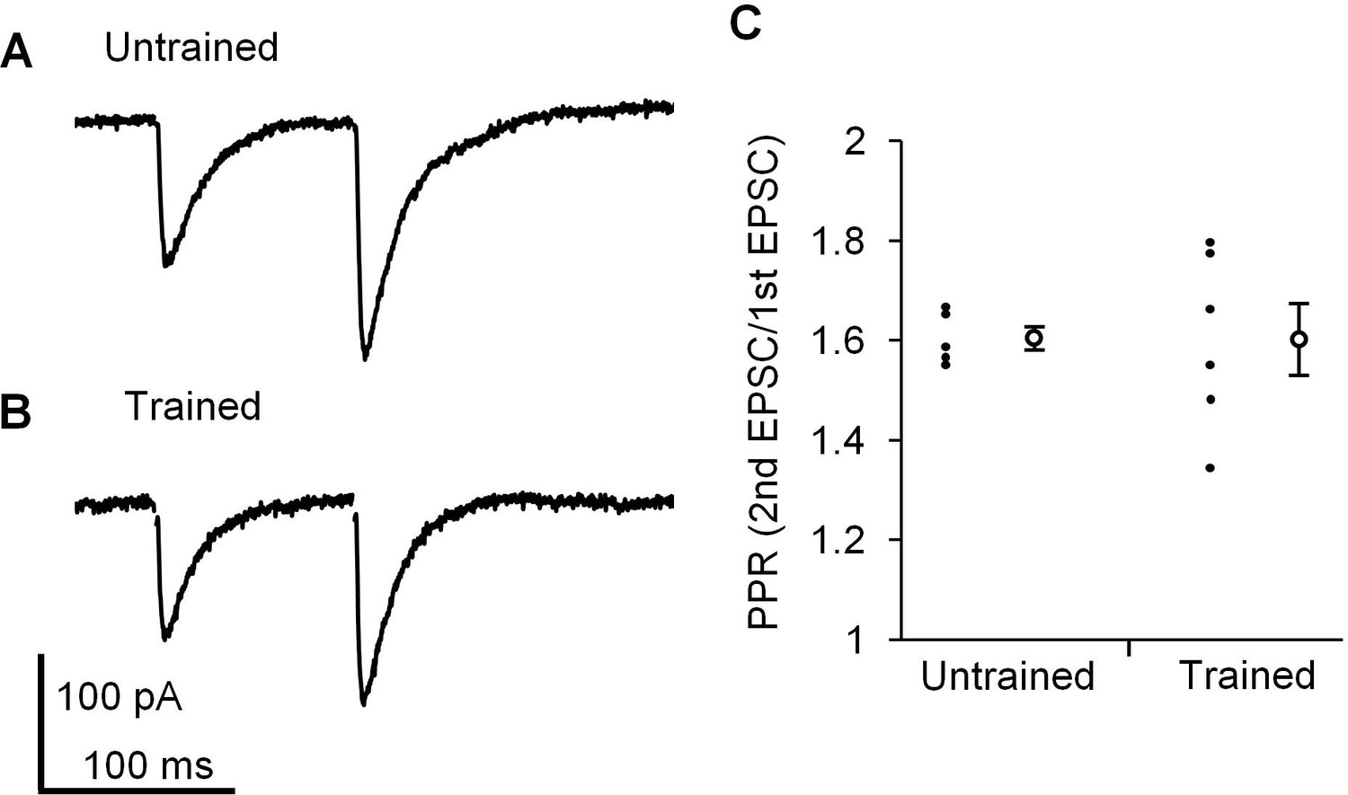
Paired pulse facilitation at PF-PC synapse in the H zone.
(A, B) Representative EPSCs induced by paired-pulse stimulation in the H zone of untrained (A) or trained mice (B). (C) Distribution of paired-pulse ratio of EPSCs with 100 ms interval. Each small circle represents a result obtained from a PC, and large circles and bars represent Mean ± SEM of all data.
-
Figure 4—source data 1
Paired pulse ratio of EPSC amplitudes with or without training in the H zone of flocculus.
- https://doi.org/10.7554/eLife.36209.009
Additional files
-
Transparent reporting form
- https://doi.org/10.7554/eLife.36209.010
Download links
A two-part list of links to download the article, or parts of the article, in various formats.
Downloads (link to download the article as PDF)
Open citations (links to open the citations from this article in various online reference manager services)
Cite this article (links to download the citations from this article in formats compatible with various reference manager tools)
Occurrence of long-term depression in the cerebellar flocculus during adaptation of optokinetic response
eLife 7:e36209.
https://doi.org/10.7554/eLife.36209


