Resting-state gamma-band power alterations in schizophrenia reveal E/I-balance abnormalities across illness-stages
- University of Glasgow, United Kingdom
- Institute for Biomagnetism and Biosignalanalysis, University of Muenster, Germany
- Goethe University, Germany
- Institute of Health and Wellbeing, University of Glasgow, United Kingdom
- University of Edinburgh, United Kingdom
- University Edinburgh, United Kingdom
- University Hospital of Child and Adolescent Psychiatry and Psychotherapy, University of Bern, Switzerland
- Medical Faculty, Heinrich-Heine-University, Germany
- Norwegian University of Science and Technology, Norway
- Central Institute of Mental health, Medical Faculty Mannheim, Heidelberg University, Germany
- University of Sydney, Australia
- Medical Faculty Mannheim, Heidelberg University, Germany
- Max Planck Institute for Brain Research, Germany
- Ernst Strüngmann Institute for Neuroscience and the Max Planck Society, Germany
- Frankfurt Institute for Advanced Studies, Germany
Figures
Figure 1
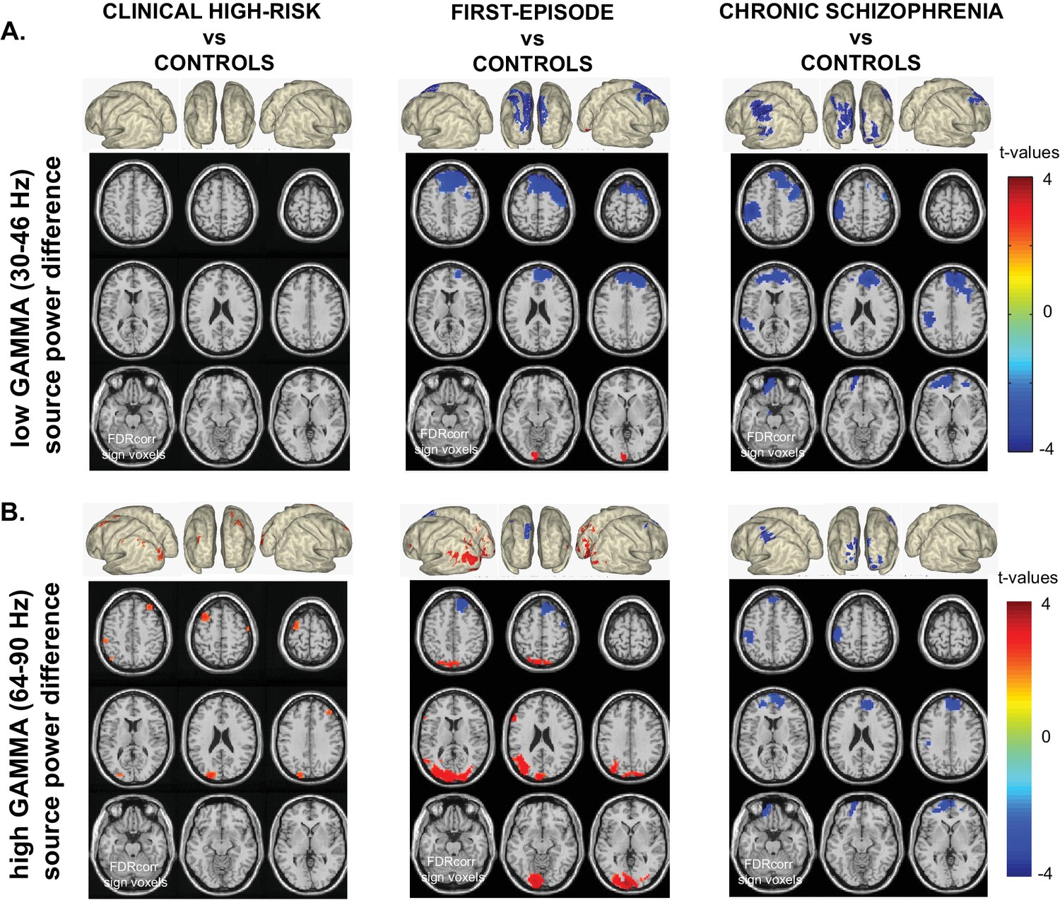
Whole-Brain Gamma-Band Power Group Differences Across Illness-Stages.
(A) Low gamma (30–46 Hz) source-power differences for the three main group contrasts: CHR vs.CON1 (left panel), FEP vs.CON2 (middle panel), ScZ vs.CON2 (right panel). Sources were estimated using a DICS beamformer method. Slice- and surface plot representations are shown with t-values corresponding to significant voxels (non-parametric, Monte-Carlo permutation based independent t-tests, FDR corrected at p<0.05, two-sided). Red colors (positive t-values) indicate an increase in gamma-band power compared to controls, whereas blue colors (negative t-values) reflect decreased gamma-band power in the clinical groups. (B) As panel A, but for high gamma (64 – 90 Hz) band activity.
Figure 2 with 1 supplement
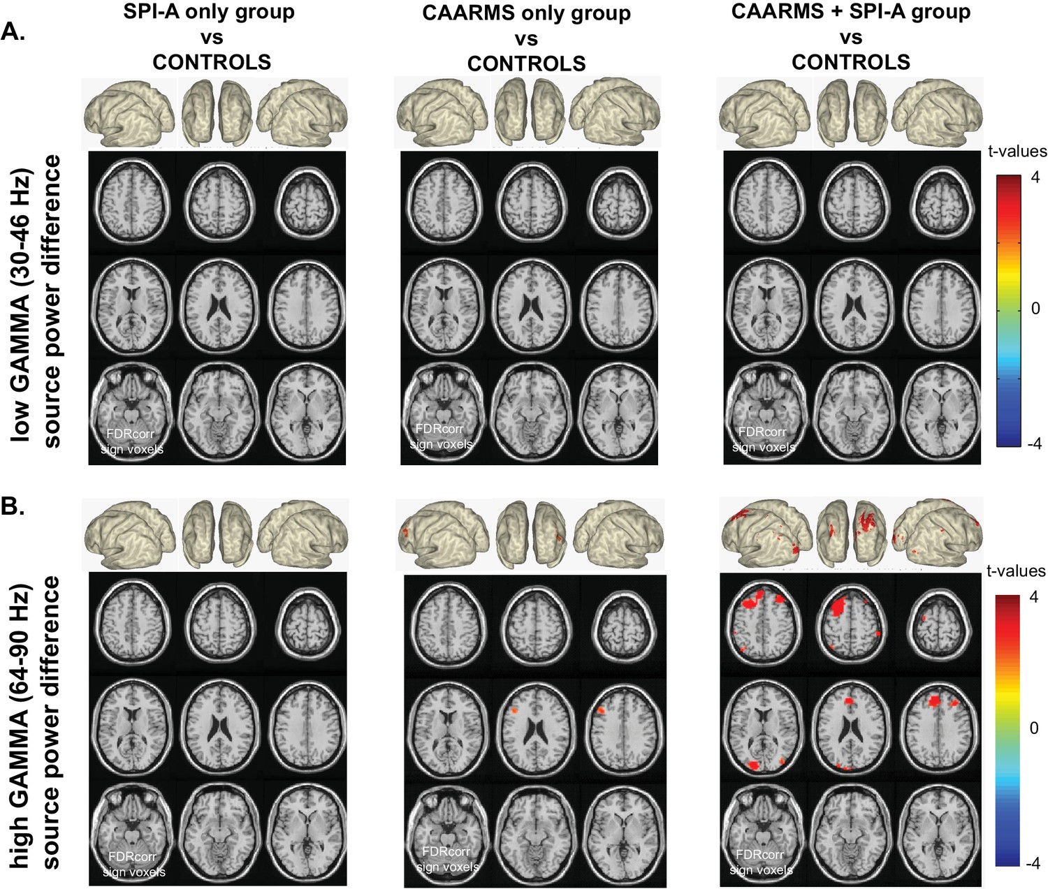
Whole-Brain Gamma-Band Power for CHR-Groups.
(A) Low gamma-band (30–46 Hz) source-power differences for the three CHR-group contrasts: SPI-A vs.CON1 (left panel), CAARMS vs.CON1 (middle panel), CAARMS + SPI-A vs.CON1 (right panel). Sources were estimated using a DICS beamformer method. Slice- and surface plot representations are shown with t-values corresponding to significant voxels (non-parametric, Monte-Carlo permutation based independent t-tests, FDR corrected at p<0.05, two-sided). Red colors (positive t-values) indicate an increase in gamma-band power compared to controls, whereas blue colors (negative t-values) reflect decreased gamma-band power in the clinical groups. (B) As panel A, but for high gamma (64 – 90 Hz) band activity.
Figure 2—figure supplement 1
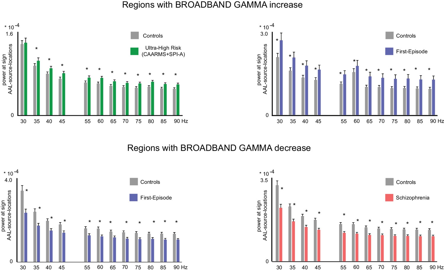
Broadband nature of gamma band effects.
For each group, virtual channel data was reconstructed from central AAL-atlas nodes within the brain regions of significant group effect (Table 2, Manuscript). These data were then submitted to FFT analyses, focusing on 5 Hz bins between 30–90 Hz. Non-parametric, Monte-Carlo based permutation statistics, FDR corrected, were then used to find significant group differences within each 5 Hz bin, averaged across all significant regions. The results showed that spectral changes were broadband in nature, with increased gamma activity in CHR (CAARMS + SPI A group) participants between 35–90 Hz (0.006 < p < 0.031) and FEP group between 30–90 Hz (0.004 < p < 0.015), and decreased gamma-band activity in FEP patients and chronic SCZ patients between 30–90 Hz (FEP: 0.0001 < p < 0.006; SCZ: 0.0001 < p < 0.002). Significant bins are indicated with an asterisk in the Figure.
Figure 3
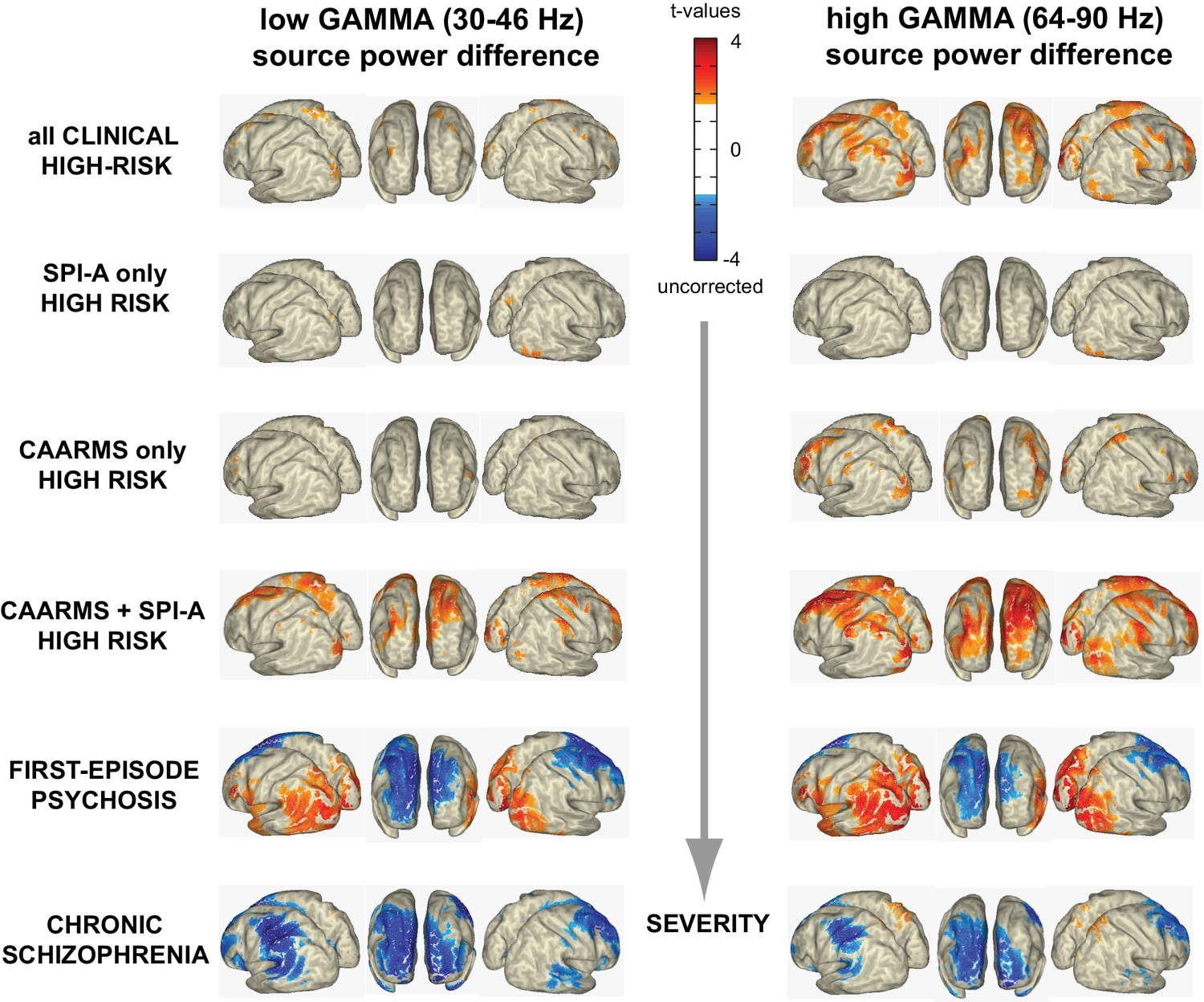
Illness Severity and Aberrant Gamma Activity.
Surface-projected statistical group differences in low gamma (30–46 Hz; left column) and high gamma-band (64–90 Hz; right column) for all main and the three CHR-subgroups contrasts. Values represent t-values corresponding to significant voxels (p<0.05; uncorrected, masked at critical t-values of non-parametric, Monte-Carlo permutation independent t-tests).
Figure 4 with 1 supplement

Clinical and Demographical Variables and Gamma-band Effects.
Overview of the influence of AGE, SEX, total CAARMS, total PANSS, and composite BACS scores on low g and high gamma-band power GROUP differences. As with the main effects of GROUP, non-parametric, Monte-Carlo permutation-based independent t-test were used to test for GROUP differences, but data was permutated over the control variable data rather than the actual gamma-band source power data. The resulting remaining significant activity then represents the interaction between the main group effect and the variation in the control variable. Surface-projected interaction-effects are shown between control groups and CHR group: (top panel), FEP group (mid panel) and chronic SciZ group (lower panel).
Figure 4—figure supplement 1

Influence of Control Variable AGE on main GROUP effect and interaction effect.
Overview of results of control analyses including a subset of 25 chronic SCZ patients and 25 age-matched controls (from CON2 group) to investigate whether AGE is a confounding or a contributing factor to the main group effects on low-Gamma (30–46 Hz; left panels) and high gamma-band (64–90 Hz; right panels) RS power changes.
Compared to the reported results in the main manuscript (including non-age-matched samples of 34 SCZ and 37 CON2 participants), the main effects are very similar, and the interaction effect with AGE is still significant.
Figure 5
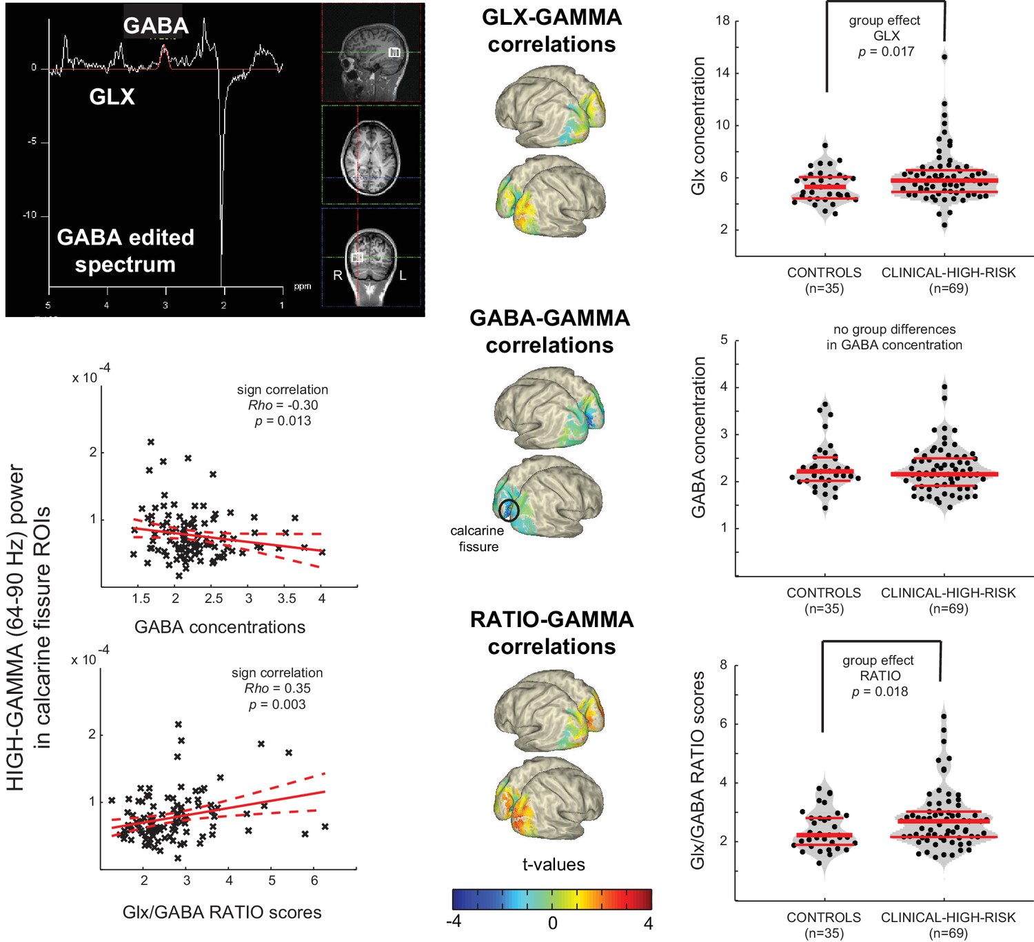
Aberrant Gamma band activity is linked to changes in E/I balance.
Upper Left Panel: Data from a 2 × 2 × 2 cm voxel placed in the right Middle Occiptal Gyrus (RMOG) during 1H-MRS of GABA and Glx (Glutamate/Glutamine) concentrations (MEGAPRESS GABA editing sequence). Right Column: dot-violin distribution plots showing concentration of each metabolite (or ratio between them) for each individual participant (black dots), separately forControls (n = 35) and CHR (n = 69) participants. Red lines indicate median concentration (middle line) and 1st and 3rd quartiles of the distribution. Data was tested for statistical group differences, using one-way repeated-measures ANOVAs, followed up by post-hoc Welch t-tests (bootstrapping: n = 1000, LSD corrected for multiple comparisons). Significant increases were found for CHRs, compared to CONs, in both Glx concentration and Glx/GABA ratio scores. Middle Column: Surface-projected t-values representing linear-regression based correlations between MRS variables and high gamma-band (64– 90 Hz) power from all 104 participants (35 CON plus 69 UHR). Both Glx and ratio scores correlate positively with increased occipital gamma-band power (uncorrected), whereas GABA concentrations correlate negatively with increased gamma-band power in calcarine areas (FDR corrected), resulting in a significantly increased ratio score in the same regions. Lower Left Panel: Correlation plots for the two strongest effects in calcarine regions.
Author response image 1
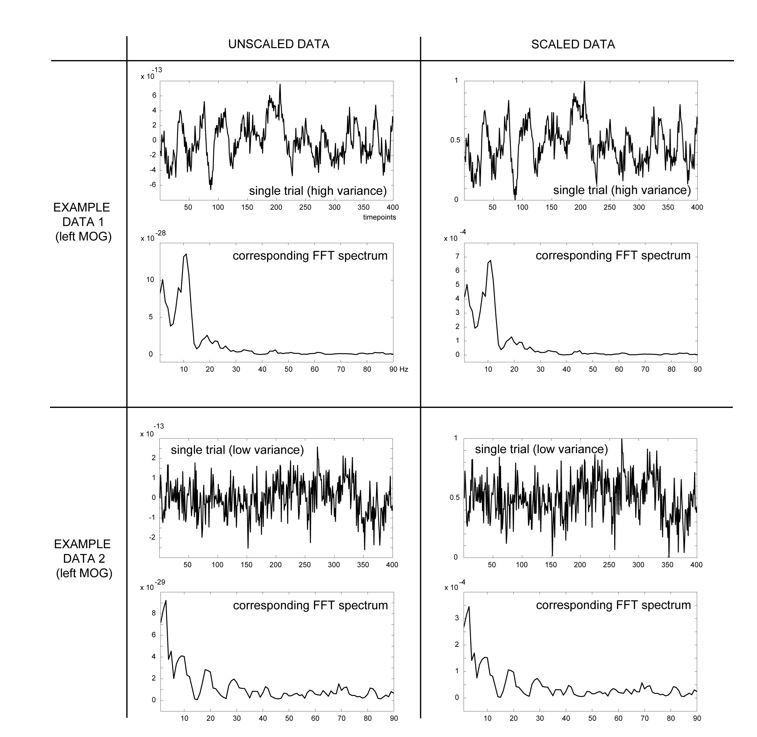
Overview of effects of the scaling procedure on shifts in the FFT spectrum: shown are two example trials from the same virtual channel data (LMOG), but from different participants, with either high (four top panel figures) or low variance across trials (four bottom panel figures).
Left column shows the unscaled data prior to (top) and after (bottom) Fast-Fourier transformation. Right column shows the same data, but rescaled, using the minimum and maximum values within that trial across time (formula: X(t) – min/ (max-min), with X(t) representing the amplitude at time t). These comparisons show that the rescaling procedure does not change the shape of the powerspectrum.
Tables
Table 1
Demographical and clinical data.
https://doi.org/10.7554/eLife.37799.002| CHR (n = 88) | CON1 (n = 48) | FEP (n = 21) | SCZ (n = 34) | CON2 (n = 37) | GROUP effect* | Pairwise comparisons* | H/p -values | |
|---|---|---|---|---|---|---|---|---|
| Age (mean/SEM) | ||||||||
| 22.0/0.5 | 22.7/0.5 | 27.0/1.5 | 37.1/2.0 | 28.6/1.2 | H(4)=80.8 p<0.0001 | CHR vs. FEP CHR vs. SCZ | −54.6/0.006 −104.5/0.000 | |
| Sex (mean/SEM) | ||||||||
| female/male | 67/21 | 33/15 | 5/16 | 12/22 | 13/24 | H(4)=38.9 p<0.0001 | FEP vs. CHR CON2 vs.CON1 | 59.6/0.000 38.2/0.020 |
| Education (mean/SEM) | ||||||||
| Years | 15.5/0.5 | 16.6/0.4 | 14.1/0.7 | 14.2/0.6 | 16.6/0.6 | H(4)=16.7 p=0.002 | CON1 vs. SCZ | 41.8/0.027 |
| BACS†(mean/SEM) | CHR (n = 88) | CON1 (n = 48) | FEP (n = 18) | SCZ (n = 28) | CON2 (n = 37) | GROUP effect | Pairwise comparisons | H/p -values |
| Verbal Memory | −0.36/0.17 | 0.23/0.17 | −0.41/0.38 | −0.93/0.24 | 0.79/0.14 | H(4)=26.5 p<0.0001 | SCZ vs. CON2 | −76.1/0.000 |
| Digit Sequencing | −0.39/0.12 | −0.07/0.11 | 0.26/0.36 | −1.07/0.20 | 0.62/0.17 | H(4)=35.5 p<0.0001 | SCZ vs. FEP SCZ vs. CHR SCZ vs. CON2 | 66.9/0.003 38.6/0.036 −90.1/0.000 |
| Token Motor Task | −0.64/0.15 | 0.28/0.16 | 0.60/0.27 | 0.47/0.21 | 1.39/0.15 | H(4)=56.9 p<0.0001 | SCZ vs. CHR CHR vs. CON1 CHR vs. FEP SCZ vs. CON2 | 46.9/0.004 −37.8/0.005 −54.5/0.006 −45.3/0.050 |
| Verbal Fluency | 0.15/0.12 | 0.38/0.19 | −0.85/0.49 | −0.90/0.20 | 0.64/0.21 | H(4)=27.1 p<0.0001 | SCZ vs. CHR FEP vs. CON2 SCZ vs. CON2 | 52.0/0.001 −51.7/0.000 −73.3/0.000 |
| Symbol Coding | −0.04/0.14 | 0.62/0.16 | −0.96/0.27 | −1.19/0.23 | −0.26/0.15 | H(4)=46.6 p<0.0001 | SCZ vs. CHR FEP vs. CHR SCZ vs. CON2 CHR vs. CON1 | 57.0/0.000 44.5/0.049 −48.0/0.030 −32.4/0.031 |
| Tower of London | 0.18/0.12 | 0.28/0.10 | 0.51/0.24 | −0.19/0.21 | 0.85/0.13 | H(4)=15.0 p<0.0001 | SCZ vs. CON2 | −76.1/0.000 |
| COMPOSITE score | −0.31/0.14 | 0.46/0.10 | −0.22/0.35 | −1.03/0.21 | 1.11/0.11 | H(4)=61.0 p<0.0001 | SCZ vs. CON2 FEP vs. CON2 CHR vs. CON1 | −111.3/0.000 −72.1/0.001 −38.5/0.004 |
| PANSS (mean/SEM) | FEP (n = 16) | SCZ (n = 30) | GROUP effect | |||||
| Negative | 18.0/1.3 | 16.6/1.1 | not sign diff | |||||
| Excitation | 9.4/0.8 | 7.2/0.7 | H(1)=6.1, p=0.013 | |||||
| Cognitive | 12.3/1.1 | 10.5/0.7 | not sign diff | |||||
| Positive | 12.5/0.7 | 9.8/0.7 | H(1)=5.1, p=0.024 | |||||
| Depression | 14.8/1.1 | 12.2/0.6 | H(1)=3.9, p=0.047 | |||||
| TOTAL | 66.9/3.2 | 56.3/3.0 | H(1)=5.4, p=0.020 | |||||
| CAARMS (mean/SEM) *frequency | CHR (n = 88) | SPI-A (n = 25) | CAARMS (n = 29) | BOTH‡(n = 34) | GROUP effect | Pairwise comparisons | H/p -values | |
| Unusual Thought Content | 5.2/0.8 | 3.6/1.4 | 3.9/1.1 | 7.6/1.3 | H(2)=6.8 p=0.033 | not sign diff | ||
| Non-Bizarre Ideas | 9.9/0.8 | 5.6/1.1 | 9.7/1.4 | 13.3/1.3 | H(2)=14.3 p=0.001 | SPI-A vs. SPI-A+CAARMS | −25.2/0.000 | |
| Perceptual Abnormalities | 8.1/0.7 | 3.9/0.7 | 9.4/1.3 | 10.2/1.1 | H(2)=15.7 p<0.0001 | SPI-A vs. SPI-A+CAARMS SPI-A vs. SPI-A+CAARMS | −21.5/0.006 −25.2/0.000 | |
| Disorganized Speech | 4.3/0.6 | 3.8/0.9 | 2.1/0.8 | 6.5/0.9 | H(2)=11.9 p=0.003 | CAARMS vs. SPI-A+CAARMS | −20.8/0.002 | |
| TOTAL | 27.6/1.8 | 16.8/2.9 | 25.0/2.4 | 37.6/2.8 | H(2)=22.2 p<0.0001 | SPI-A vs. SPI-A+CAARMS CAARMS vs. SPI-A+CAARMS | −31.4/0.000 −17.4/0.021 | |
| Global Functioning (GAF: mean/SEM) | CHR (n = 88) | CON1 (n = 48) | GROUP effect | |||||
| 59.8/1.2 | 87.4/1.0 | H(1)=81.0, p<0.0001 | ||||||
| MEDICATION | CHR (n = 88) | CON1 (n = 48) | ||||||
| None | 39 | 46 | ||||||
| Anti-psychotic | 1 | 0 | ||||||
| Mood-stabilizer | 1 | 0 | ||||||
| Anti-depressant | 20 | 0 | ||||||
| Anti-convulsant | 0 | 0 | ||||||
| Other | 11 | 0 | ||||||
| Multiple | 16 | 2 | ||||||
-
*Kruskal-Wallis independent-sample test. Alpha-level 0.05, two-sided with p-values adjusted for ties.
†Kruskal-Wallis independent-sample test performed on z-standardized data (Keefe et al., 2008). Alpha-level 0.05, two-sided, p-values adjusted for ties.
Table 2
Overview of AAL regions of significantly modulated resting-state low and high gamma-band power.
https://doi.org/10.7554/eLife.37799.004| Group contrast | Labels of significant AAL regions* | t-values (range) | p-values (range) |
|---|---|---|---|
| Low GAMMA (30–46 Hz) | |||
| FEP vs CON2 | left Calcarine Fissure, left Inferior Occipital Gyrus | 2.82 to 3.80 | 0.002–0.006 |
| right and left Superior Medial Frontal Gyrus, right Middle Frontal Gyrus | −2.15 to −3.79 | 0.002–0.006 | |
| SCZ vs CON2 | right and left Superior Medial Frontal Gyrus, right Middle Frontal Gyrus, left Inferior Parietal Lobule, left Superior Orbital Frontal Gyrus, left Superior Temporal Gyrus, left PostCentral Gyrus, right PreCentral Gyrus | −2.35 to −4.24 | 0.002–0.006 |
| High GAMMA (64–90 Hz) | |||
| CHR vs CON1 | left Middle Occipital Gyrus, right and left Middle Frontal Gyrus, left Angular Gyrus, left Inferior Parietal Lobule | 2.40 to 2.74 | 0.002–0.006 |
| SPI-A only vs CON1 | No significant voxels | − | − |
| CAARMS only vs CON1 | left Middle Frontal Gyrus, left Middle Occipital Gyrus | 2.67 | 0.006 |
| CAARMS + SPI A vs CON1 | right and left Middle Occipital Gyrus, right and left Middle Frontal Gyrus. left Angular Gyrus, right Inferior Parietal Lobule, left Superior Medial Frontal Gyrus | 2.16 to 3.43 | 0.002–0.006 |
| FEP vs CON2 | right and left Calcarine Fissure, right and left Inferior Occipital Gyrus, right and left Middle Occipital Gyrus,right and left PreCuneus, left Inferior Frontal Gyrus,left Angular Gyrus | 2.48 to 4.08 | 0.002–0.006 |
| right Middle Frontal Gyrus | −2.42 to −3.26 | 0.002–0.006 | |
| SCZ vs CON2 | right and left Superior Medial Frontal Gyrus, left Superior Orbital Frontal Gyrus, left Middle Orbital Frontal Gyrus, left PostCentral Gyrus | −2.40 to −3.56 | 0.002–0.006 |
-
*Non-parametric Monte-Carlo permutation based independent-sample tests, alpha-level 0.05, two-sided, FDR corrected voxels.
Additional files
-
Transparent reporting form
- https://doi.org/10.7554/eLife.37799.011
Download links
A two-part list of links to download the article, or parts of the article, in various formats.
Downloads (link to download the article as PDF)
Open citations (links to open the citations from this article in various online reference manager services)
Cite this article (links to download the citations from this article in formats compatible with various reference manager tools)
Resting-state gamma-band power alterations in schizophrenia reveal E/I-balance abnormalities across illness-stages
eLife 7:e37799.
https://doi.org/10.7554/eLife.37799


