The Natural History of Model Organisms: From molecular manipulation of domesticated Chlamydomonas reinhardtii to survival in nature
- Friedrich Schiller University, Germany
- Ludwig Maximilian University, Germany
- Carnegie Instiution for Science, United States
Figures
Figure 1
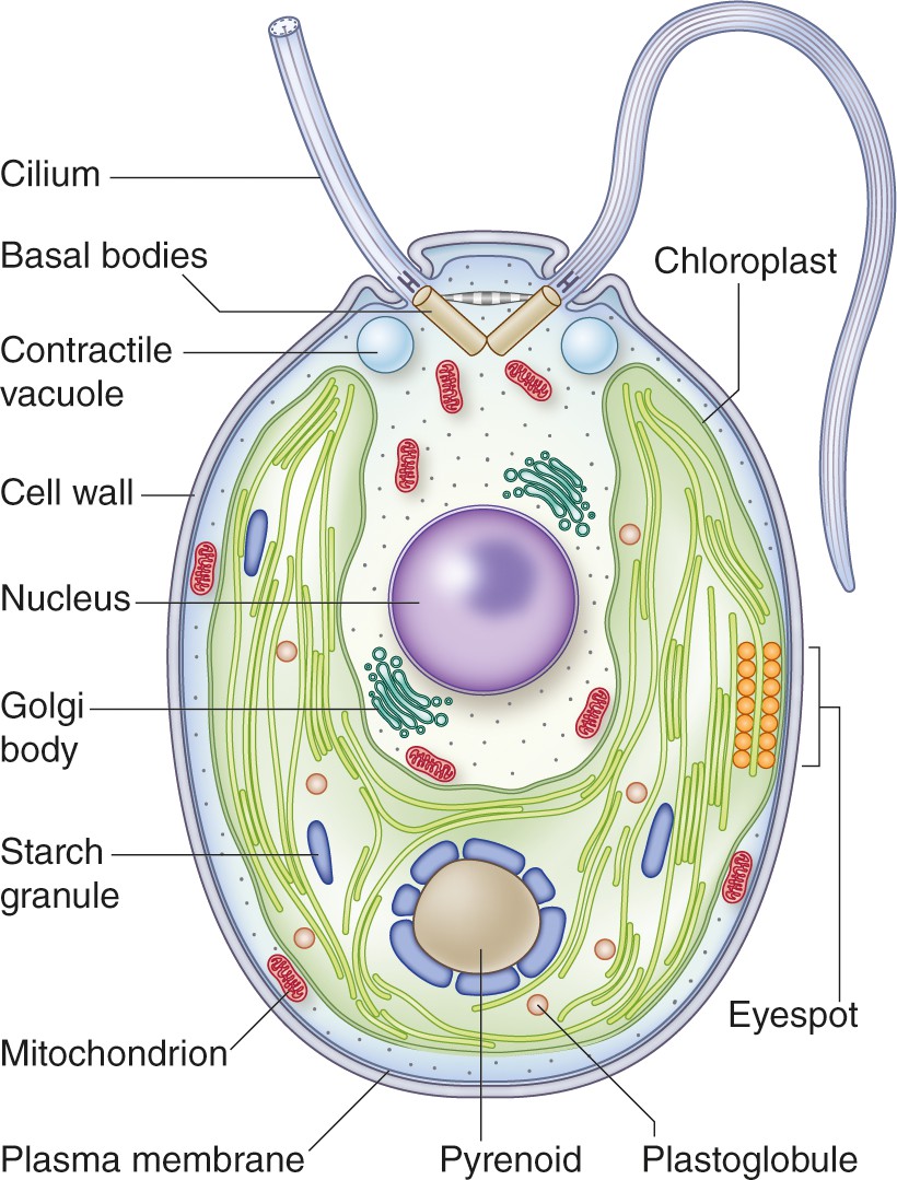
Structure of a vegetative Chlamydomonas reinhardtii cell.
This cell has a 5-10 µm diameter (Gallaher et al., 2015). The two anterior cilia possess a 9+2 microtubule structure characteristic of motile cilia of eukaryotes. The cilia are critical for mating processes and confer motility to the cell (Harris, 2001). A single cup-shaped chloroplast occupies a large proportion of the cell's volume. This organelle houses the machinery for oxygenic photosynthesis and contains the pyrenoid, a structure in which Rubisco is concentrated; the pyrenoid is a component of the carbon concentrating mechanism (CCM) which functions to concentrate inorganic carbon in the cell against a concentration gradient (Mackinder et al., 2016). Close to the cell equator, at the edge of the chloroplast, is the eyespot. This primordial visual system allows the cells to orient their swimming toward or away from the light (phototaxis). Under hypoosmotic conditions, the cytoplasmic water content is maintained by pumping water out of the cell through contractile vacuoles positioned at the cell’s anterior (Komsic-Buchmann et al., 2014). At the base of the cilia are the basal bodies, which are responsible for ciliary assembly (Dutcher and O'Toole, 2016). Other features of the cell include a centrally located nucleus, a proteinaceous cell wall, Golgi bodies within the cup-shaped region formed by the chloroplast, and mitochondria. Image credit: Debbie Maizels.
Figure 2
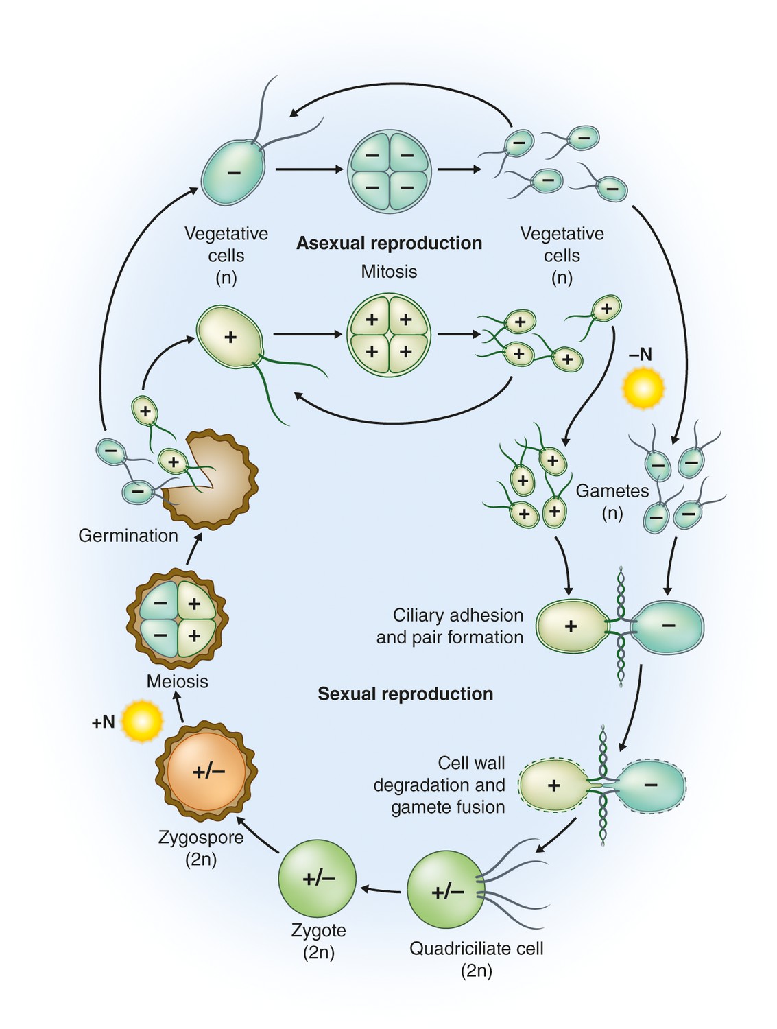
Life cycle of C.reinhardtii.
Haploid (n) vegetative cells occur as two mating types, mt+ and mt-, that divide by mitosis (“Asexual reproduction”; Harris, 2001, Goodenough et al., 2007). Gametogenesis can be induced by nitrogen starvation (-N) in the presence of light, and gametes of opposite mating types can fuse to form diploid (2n) zygotes (“Sexual reproduction”). Within a few hours of fertilization, zygotes resorb their four cilia to become immotile. Over the course of several days these zygotes are remodeled into highly resistant, dormant zygospores. In this process, a strong, multilayered cell wall is formed, and chlorophyll is degraded (Harris, 2001; Goodenough et al., 2007). As a result, mature zygospores appear orange, which reflects their carotenoid content (Lohr, 2009). When environmental conditions improve, the zygote undergoes meiosis to release four haploid cells (sometimes 8 and 16 when mitosis also occurs within the zygote wall; “Germination”). The haploid cells then resume vegetative growth. In the laboratory, zygote germination is induced by the addition of nitrogen (+N) to the medium in the light (Harris, 2001); nitrogen also causes reprogramming of gametes to vegetative cells (Pozuelo et al., 2000). Image credit: Debbie Maizels.
Figure 3

C. reinhardtii ingested by the predatory protist Peranema trichophorum.
Image credit: Santosh Sathe and Pierre Durand.
Videos
Video 1
C. reinhardtii surrounded by the harmful bacteria Pseudomonas protegens (Aiyar et al., 2017)
Video credit: Prasad Aiyar, Severin Sasso and Maria Mittag.
Download links
A two-part list of links to download the article, or parts of the article, in various formats.
Downloads (link to download the article as PDF)
Open citations (links to open the citations from this article in various online reference manager services)
Cite this article (links to download the citations from this article in formats compatible with various reference manager tools)
The Natural History of Model Organisms: From molecular manipulation of domesticated Chlamydomonas reinhardtii to survival in nature
eLife 7:e39233.
https://doi.org/10.7554/eLife.39233


