Odor-evoked category reactivation in human ventromedial prefrontal cortex during sleep promotes memory consolidation
- Northwestern University, United States
- University of Pennsylvania, United States
Figures
Figure 1 with 2 supplements
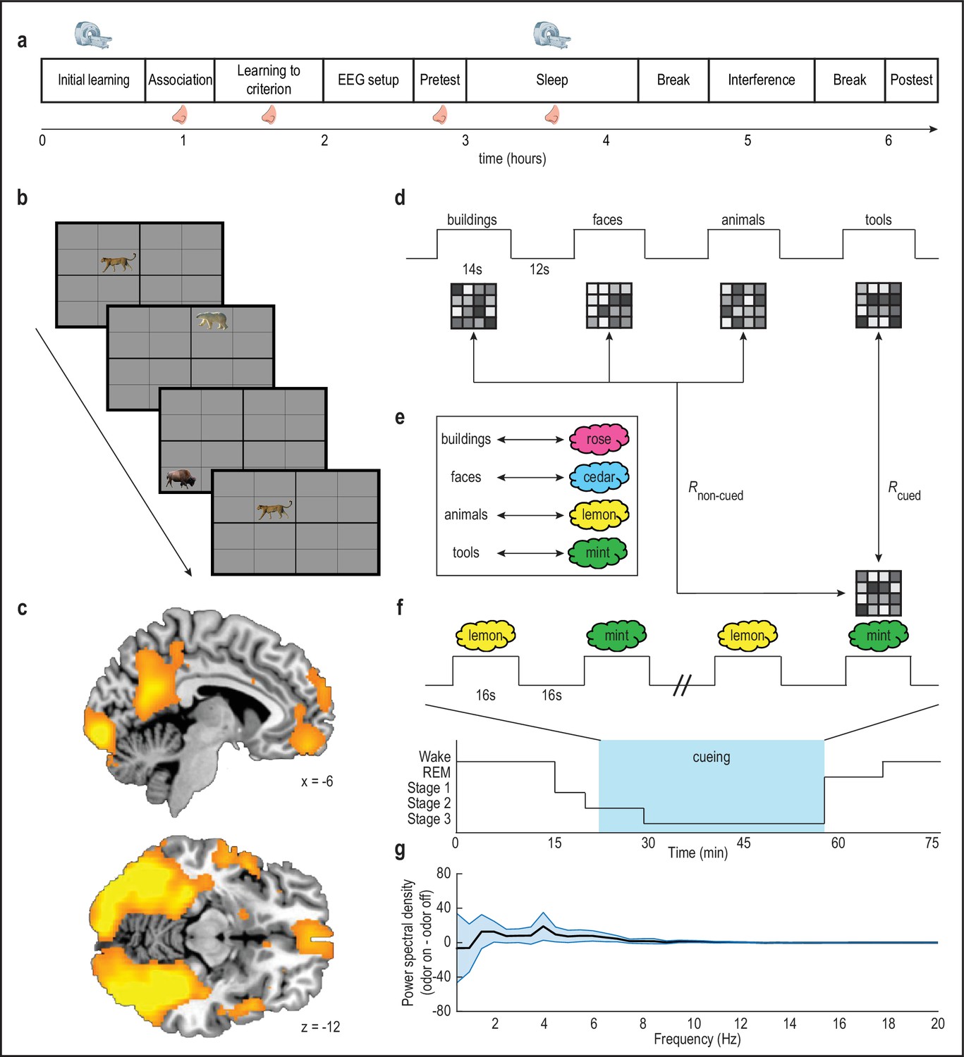
Olfactory cueing paradigm.
(a) Experimental timeline. MRI scanner symbols indicate scanned study phases, and nose symbols indicate study phases during which odors were presented. (b) Initial learning task. Subjects learned spatial locations of objects from four categories (animals, buildings, faces, tools; 32 objects per category) during blocked-design fMRI scanning. Category-sensitive voxels were then identified for each subject by comparing visually evoked fMRI ensemble patterns of activity across runs, and calculating within-category versus between-category pattern correlations. This multivoxel pattern analysis was implemented using a whole-brain searchlight approach. (c) Category-sensitive brain regions included visual processing areas, and parts of parietal cortex and ventromedial prefrontal cortex. fMRI activity is displayed at p < 0.001 uncorrected, and images are overlaid on a canonical single-subject T1-weighted MRI scan. Voxels from this analysis were retained as an image mask for the main analysis. (d-e) During initial learning, category templates (depicted as 4-x-4 greyscale grids of voxels) were defined for each subject. Next, subjects learned to associate each object category with a unique odor (e.g. mint odor + tool images). (f) Subjects were placed into the fMRI scanner, and during NREM sleep stages 2 and 3, two of the four odor cues were presented in 16 s on/16s off blocks during fMRI scanning (i.e. during cueing, blue box) to cue content from their associated object categories. In this example, presentation of the mint odor in sleep would induce reactivation, such that its fMRI ensemble pattern would more closely resemble the tool category ensemble pattern defined in the wake state during initial learning. (g) There was no significant effect of odor presentation on power spectral density (µV2/Hz) between 0.5 and 20 Hz (repeated-measures ANOVA, condition by frequency interaction: F(39,17) = 0.31, p = 1.00). Error bars depict mean ±SEM across subjects.
-
Figure 1—source data 1
Relates to panel (g).
- https://doi.org/10.7554/eLife.39681.007
Figure 1—figure supplement 1
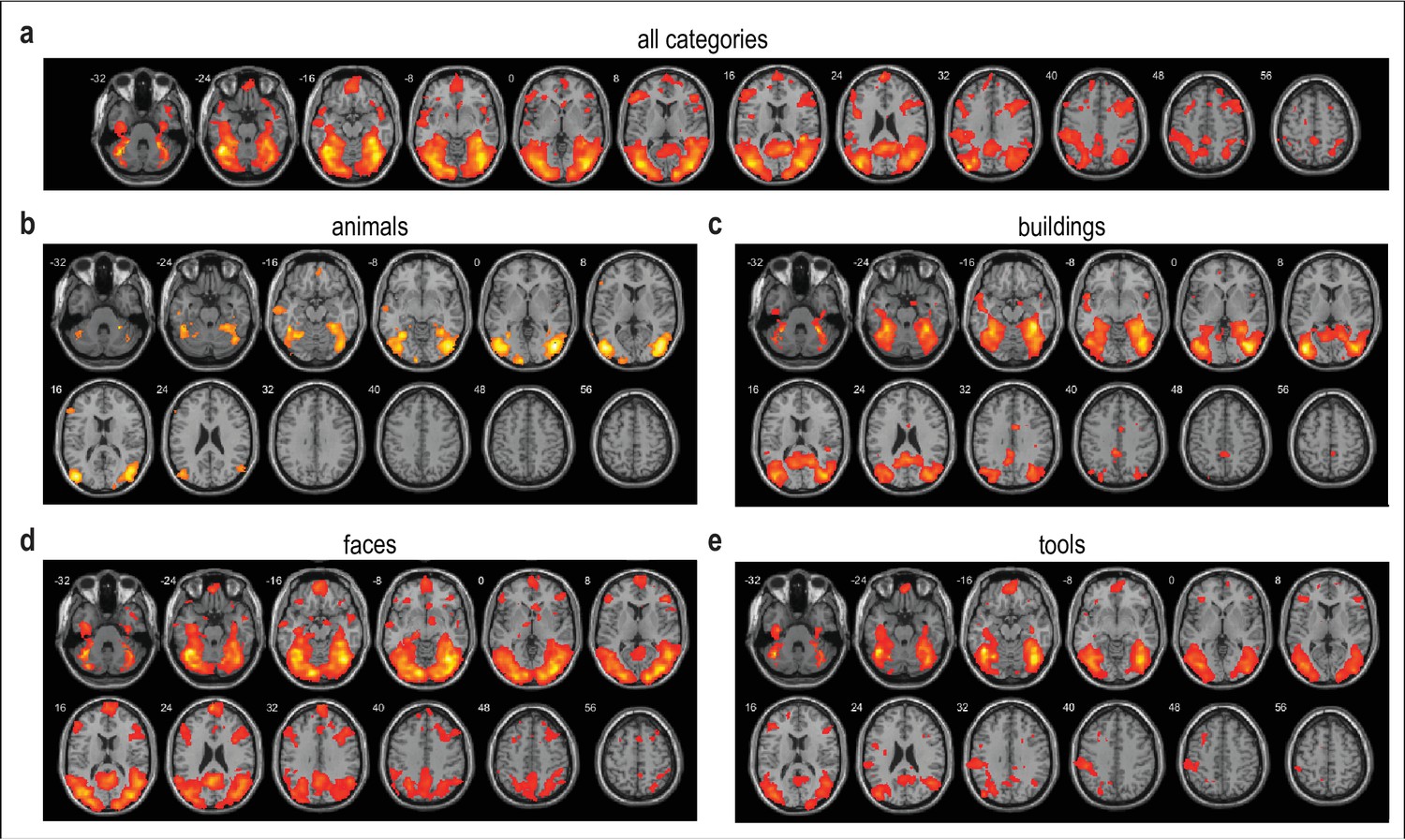
Brain regions demonstrating category selectivity during initial learning.
(a–e) Multivoxel pattern analysis of initial learning data reveals widespread category specificity across the four object categories, and for each of the four object categories individually. fMRI activity is shown at p < 0.001 uncorrected, and images are overlaid on a canonical single-subject T1-weighted MRI scan. This result was visualized using the xjView toolbox (http://www.alivelearn.net/xjview).
Figure 1—figure supplement 2
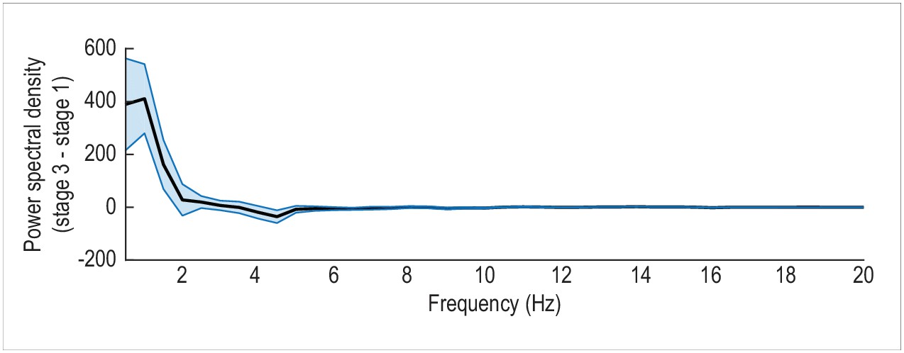
Differences in EEG spectral power between stage 3 and stage 1 sleep.
As predicted, an analysis of EEG power spectral density (µV2/Hz) from 0.5 Hz to 20 Hz reveals enhanced slow-wave activity (0.5 to 2 Hz) during stage 3 sleep, as compared to stage 1 sleep (repeated-measures ANOVA, condition by frequency interaction: F(39,17) = 5.90, p < 0.001). Error bars depict mean ±SEM across subjects.
-
Figure 1—figure supplement 2—source data 1
Relates to entire figure.
- https://doi.org/10.7554/eLife.39681.006
Figure 2 with 1 supplement
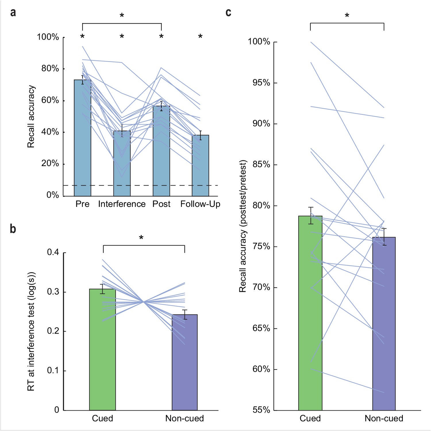
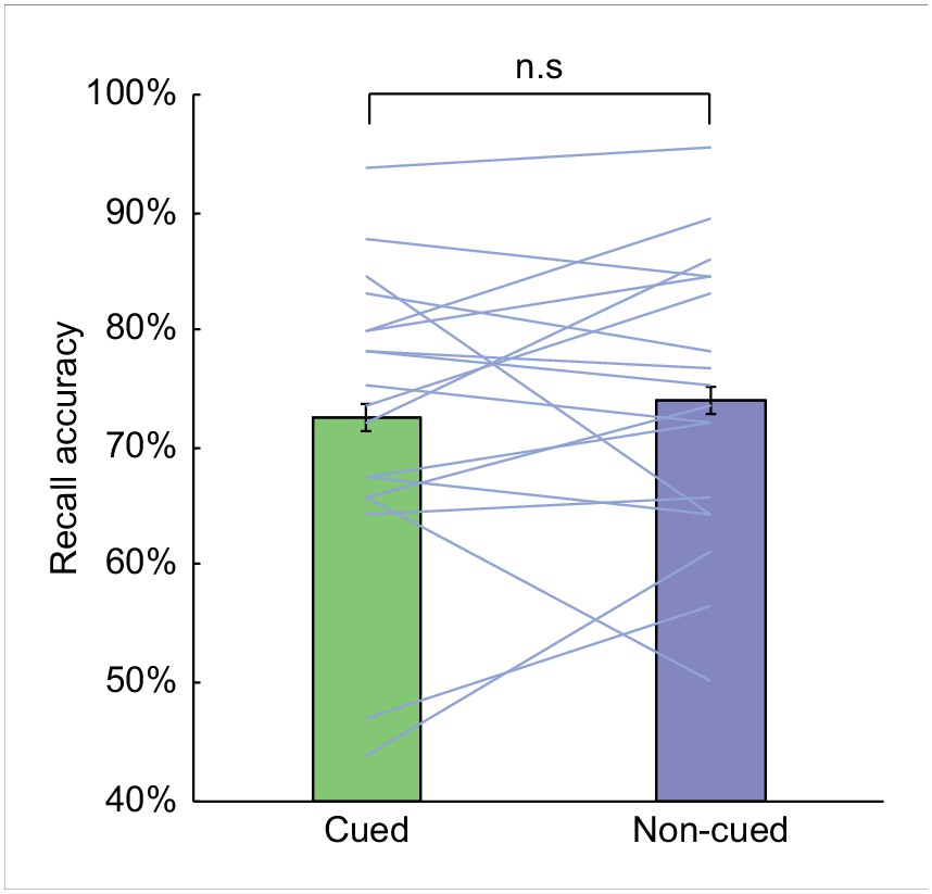
Behavioral results.
(a) Subjects demonstrated robust visuospatial recall (chance = 6.25%; dashed line) at pretest (73.17% correct ±2.71% SEM, t(17) = 24.68, p < 0.001), interference test (41.11% correct ±4.02% SEM, t(17) = 8.66, p < 0.001), posttest (56.70% correct ±2.92% SEM, t(17) = 17.27, p < 0.001), and 1 week follow-up (38.24% correct ±2.92% SEM, t(17) = 10.96, p < 0.001). Performance declined significantly from pretest to posttest (t(17) = 10.06, p < 0.001). (b) During interference testing, subject RTs were significantly slower for objects belonging to categories that were cued in the preceding sleep period, versus non-cued categories. This suggests that memory for object categories that were cued during SWS was more resistant to interference. *p < 0.05, two-tailed t-test. (c) At posttest, recall accuracy (percentage of items recalled at posttest, compared to pretest baseline) was enhanced for cued object locations, compared to non-cued object locations (Z = 1.70, p = 0.04). *p < 0.05, one-tailed Wilcoxon signed-rank test. Error bars in (a) depict mean ±SEM, and error bars in (b–c) depict mean ±within subjects SEM.
-
Figure 2—source data 1
Relates to panel (a).
- https://doi.org/10.7554/eLife.39681.011
-
Figure 2—source data 2
Relates to panel (b).
- https://doi.org/10.7554/eLife.39681.012
-
Figure 2—source data 3
Relates to panel (c).
- https://doi.org/10.7554/eLife.39681.013
Figure 2—figure supplement 1
Visuospatial recall accuracy at pretest.
There was no significant difference in recall accuracy between those object categories that were later cued during sleep, and those that were not (t(17) = 0.62, p = 0.54). n.s., two-tailed t-test. Error bars depict mean ±within subjects SEM.
-
Figure 2—figure supplement 1—source data 1
Relates to entire figure.
- https://doi.org/10.7554/eLife.39681.010
Figure 3 with 1 supplement
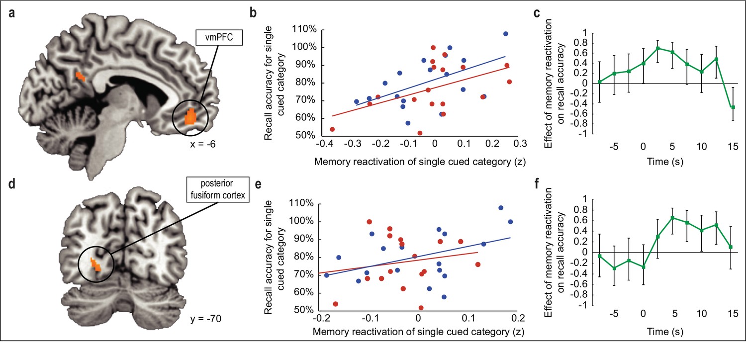
Relationship between odor-evoked memory reactivation in sleep and posttest memory retention.
(a,d) During SWS, the extent to which odors evoked category-specific memory reactivation in vmPFC (a) and posterior fusiform cortex (d) was significantly correlated with behavioral enhancement of cued visuospatial memory retrieval at posttest. fMRI activity is shown at p < 0.001 uncorrected, and images are overlaid on a canonical single-subject T1-weighted MRI scan. (b,e) Correlations between fMRI memory reactivation of an individual object category and recall accuracy for that category at posttest, for the two reactivated object categories taken separately in vmPFC (b) and posterior fusiform cortex (e). Blue and red dots represent individual object categories, where category assignment is arbitrary. (c,f) Illustration of correlation depicted in (a,d) across time points in vmPFC (c) and posterior fusiform cortex (f). Time 0 is aligned to odor onset. Error bars depict 90% confidence intervals.
-
Figure 3—source data 1
Relates to panel (b).
- https://doi.org/10.7554/eLife.39681.019
-
Figure 3—source data 2
Relates to panel (c).
- https://doi.org/10.7554/eLife.39681.020
-
Figure 3—source data 3
Relates to panel (e).
- https://doi.org/10.7554/eLife.39681.021
-
Figure 3—source data 4
Relates to panel (f).
- https://doi.org/10.7554/eLife.39681.022
Figure 3—figure supplement 1
Relationship between within-sleep odor-evoked memory reactivation in hippocampus and posttest memory retention.
(a) The degree to which odors elicited memory reactivation in an anatomical region of interest in hippocampus did not predict enhancement of cued visuospatial recall accuracy at posttest (r(16) = 0.03, p = 0.91). (b–c) Illustration of the correlation between memory reactivation in hippocampus and memory performance for (b) the two reactivated object categories computed separately (blue, r(16) = −0.11, p = 0.66; red, r(16) = 0.14, p = 0.58), and (c) shown across time points. Time 0 is aligned to odor onset. Error bars depict 90% confidence intervals.
-
Figure 3—figure supplement 1—source data 1
Relates to panel (a).
- https://doi.org/10.7554/eLife.39681.016
-
Figure 3—figure supplement 1—source data 2
Relates to panel (b).
- https://doi.org/10.7554/eLife.39681.017
-
Figure 3—figure supplement 1—source data 3
Relates to panel (c).
- https://doi.org/10.7554/eLife.39681.018
Figure 4

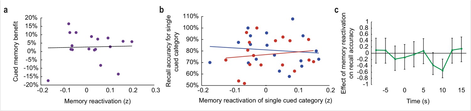
Relationship between odor-evoked memory reactivation and cued memory benefit, restricted to cued categories only.
(a-b) When limiting the analysis of memory reactivation to a comparison of the two cued object categories (without including the two non-cued object category templates from initial learning for comparison), reactivation measures remained significantly correlated with behavioral memory enhancement in a cluster of interest in vmPFC (a; r(16) = 0.61, p = 0.01) and posterior fusiform cortex (b; r(16) = 0.72, p < 0.001).
-
Figure 4—source data 1
Relates to panel (a).
- https://doi.org/10.7554/eLife.39681.024
-
Figure 4—source data 2
Relates to panel (b).
- https://doi.org/10.7554/eLife.39681.025
Figure 5 with 1 supplement
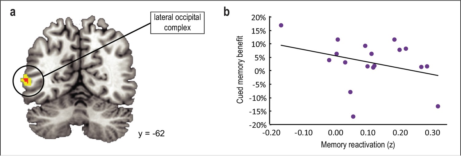
Main effect of odor cueing on category reactivation.
(a) Odor cues evoked reactivation of associated category templates in lateral occipital complex ([−56,–62, 4], t(16) = 3.99, punc = 0.001), although this effect did not survive statistical correction for multiple comparisons. (b) The degree of reactivation in this cluster was not predictive of memory enhancement for cued versus noncued categories upon waking (r(16) = −0.33, p = 0.19). fMRI activity is shown at p < 0.001 uncorrected (red) and p < .005 uncorrected (yellow), and images are overlaid on a canonical single-subject T1-weighted MRI scan.
-
Figure 5—source data 1
Relates to panel (b).
- https://doi.org/10.7554/eLife.39681.028
Figure 5—figure supplement 1
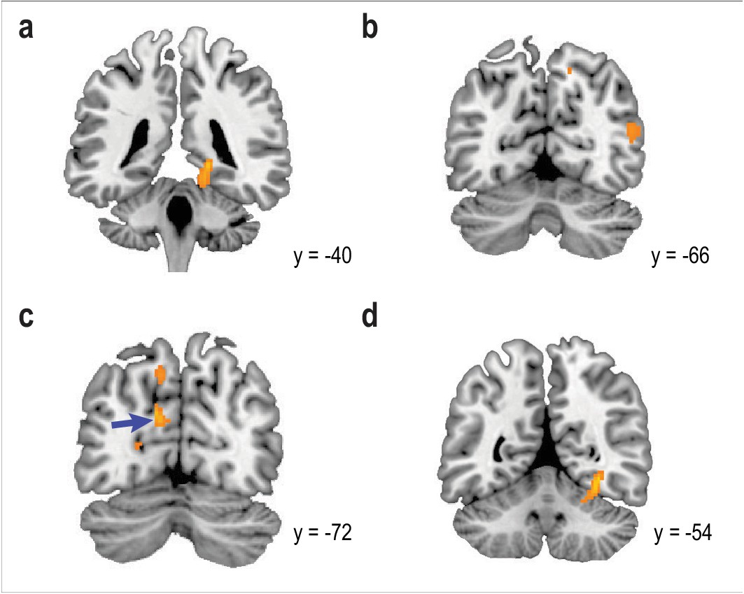
Main effect of odor cueing on reactivation of individual object categories.
Depending on which of the four object categories was cued, odors evoked reactivation of associated category information in discrete brain areas. (a–d) Odor cues induced reactivation of animal information in parahippocampal cortex (a; [18, -40, -4], t(8) = 8.50, punc < 0.001), building information in lateral occipital complex (b; [50, -66, 16], t(8) = 5.76, punc < 0.001), face information in occipital cortex (c, blue arrow; [−12,–72, 24], t(8) = 8.20, punc < 0.001), and tool information in parahippocampal cortex (d; [32, -54, -10], t(8) = 9.38, punc < 0.001). However, these clusters did not survive correction for multiple comparisons. fMRI activity is shown at p < 0.001 uncorrected, and images are overlaid on a canonical single-subject T1-weighted MRI scan.
Tables
Table 1
Time spent in each sleep stage.
Offline sleep scoring revealed that 99.45% of odors were presented during stages 2 and 3 of sleep, and most cues (77.56%) were presented during stage 3 of sleep.
| Sleep stage | Time (min ±SEM) | Percentage (% ±SEM) |
|---|---|---|
| Wake | 9.92 ±2.61 | 13.11 ±3.48 |
| Stage 1 | 8.69 ±1.82 | 11.39 ±2.34 |
| Stage 2 | 26.50 ±3.41 | 34.95 ±4.26 |
| Stage 3 | 30.17 ±4.12 | 40.54 ±5.56 |
| Total sleep time | 65.36 ±2.79 | 86.89 ±3.48 |
| Total time (wake + sleep) | 75.28 ±1.25 | 100 |
-
Table 1—source data 1
Time spent in each sleep stage by subject.
- https://doi.org/10.7554/eLife.39681.030
Additional files
-
Transparent reporting form
- https://doi.org/10.7554/eLife.39681.031
Download links
A two-part list of links to download the article, or parts of the article, in various formats.
Downloads (link to download the article as PDF)
Open citations (links to open the citations from this article in various online reference manager services)
Cite this article (links to download the citations from this article in formats compatible with various reference manager tools)
Odor-evoked category reactivation in human ventromedial prefrontal cortex during sleep promotes memory consolidation
eLife 7:e39681.
https://doi.org/10.7554/eLife.39681


