Light-dependent pathways for dopaminergic amacrine cell development and function
- Northwestern University, United States
- Johns Hopkins University, United States
Figures
Figure 1
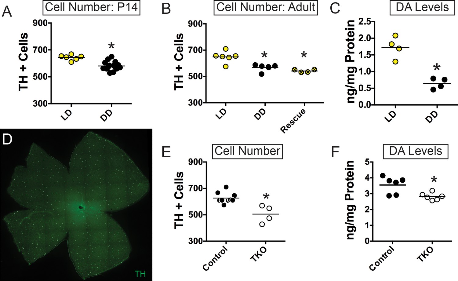
Light exposure during early retinal development is required to set TH-positive cell number and dopamine levels.
(A–B) TH+ cell number in animals bred and reared in either LD (yellow circles) or DD (black circles) from conception at P14 (n = 6 LD, n = 15 DD) (A) or Adult (n = 6 LD, n = 6 DD) (B) stages. Adult TH+ cell number could not be rescued by moving animals from DD to LD at P14 (hatched yellow circles, n = 4) (C) DA levels in adult retinas from animals reared in LD (yellow circles, n = 4) or DD (black circles, n = 4). (D) TH+ cells in whole mount WT adult retina (E–F) TH+ cell number (n = 9 Control, n = 4 TKO) (E) and DA levels (n = 6 Control, n = 6 TKO) (F) in Control (black circles) and TKO (open circles) adult retinas. *p < 0.05. Bars on plots represent mean.
Figure 2 with 1 supplement
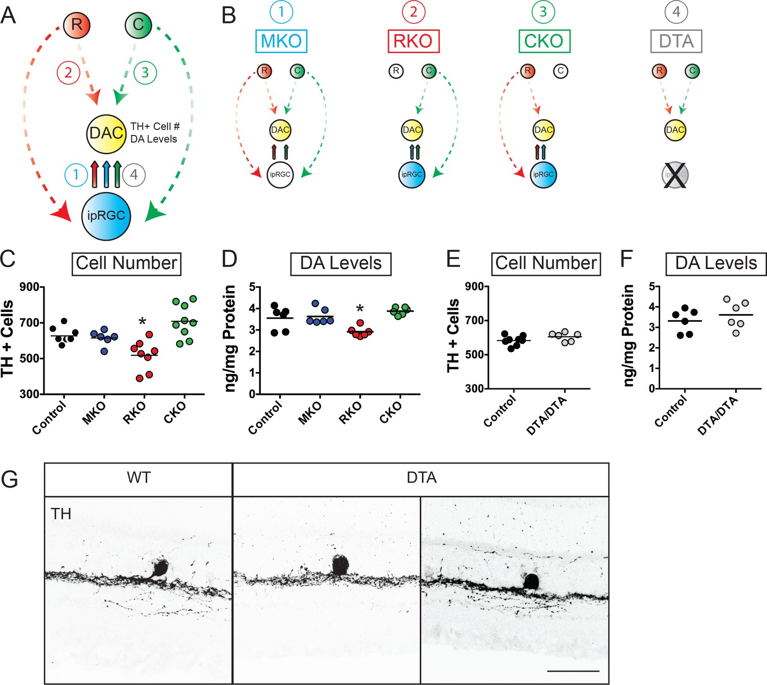
Rod signaling influences TH-positive cell number and dopamine levels through ipRGC-independent pathways.
(A) Diagram depicting potential pathways by which light information could reach DACs (1. Via melanopsin signals relayed by ipRGCs, (2. Via rod signals relayed to DACs, (3. Via cone signals relayed to DACs, or (4. Via rod, cone, and/or melanopsin signals through ipRGC-dependent pathways. Dashed arrows represent indirect influence through multicellular circuits while solid arrows represent direct synaptic connectivity between subtypes. (B) Diagram depicting signaling pathways disrupted in MKO, RKO, CKO, and DTA mouse lines. Dashed arrows represent indirect influence through multicellular circuits while solid arrows represent direct synaptic connectivity between subtypes. (C–D) TH+ cell number and DA levels in Control (black circles, n = 9), MKO (blue circles, n = 6), RKO (red circles, n = 8), and CKO (green circles, n = 9) retinas from adult littermates. (E–F) TH+ cell number (n = 8 Control, n = 6 DTA) and DA levels (n = 6 Control, n = 6 DTA) in Control (black circles) and DTA (gray circles) retinas from adult littermates. (G) TH+ cell anatomy in WT and DTA adult retinal sections. We observed no morphological differences between TH+ cells in these two mouse lines. DA: dopamine, DAC: dopaminergic amacrine cell, MKO: animals lacking melanopsin phototransduction, RKO: animals lacking rod signaling, CKO: animals lacking cone signaling, DTA: animals where ipRGCs are ablated through expression of diphtheria toxin. Scale bar in (G) is 50 μm. *p < 0.05, bars on plots represent mean.
Figure 2—figure supplement 1
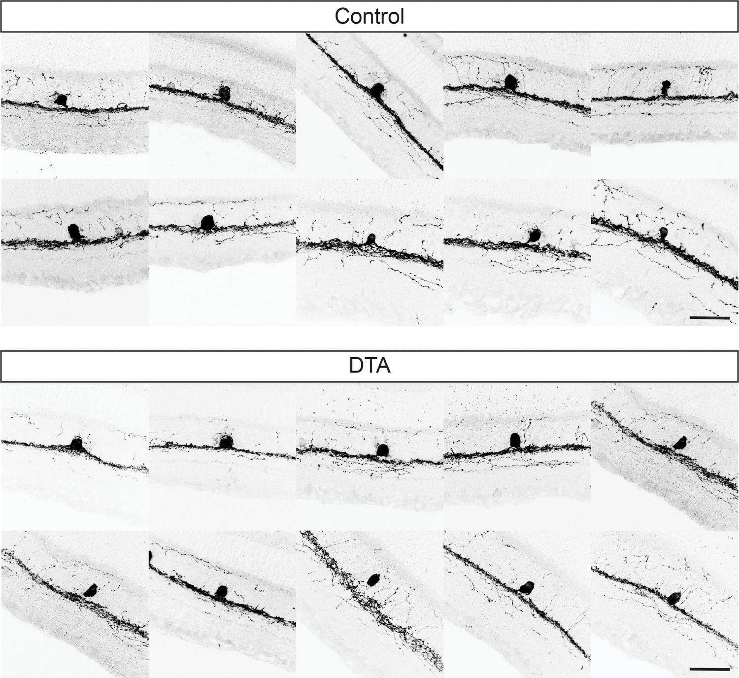
Examples of TH-positive cell stratification in WT and DTA retinas.
TH+ cell stratification in WT (top panels) and DTA (bottom panels) retinas show no differences in morphology. Scale bar is 50 μm.
Figure 3
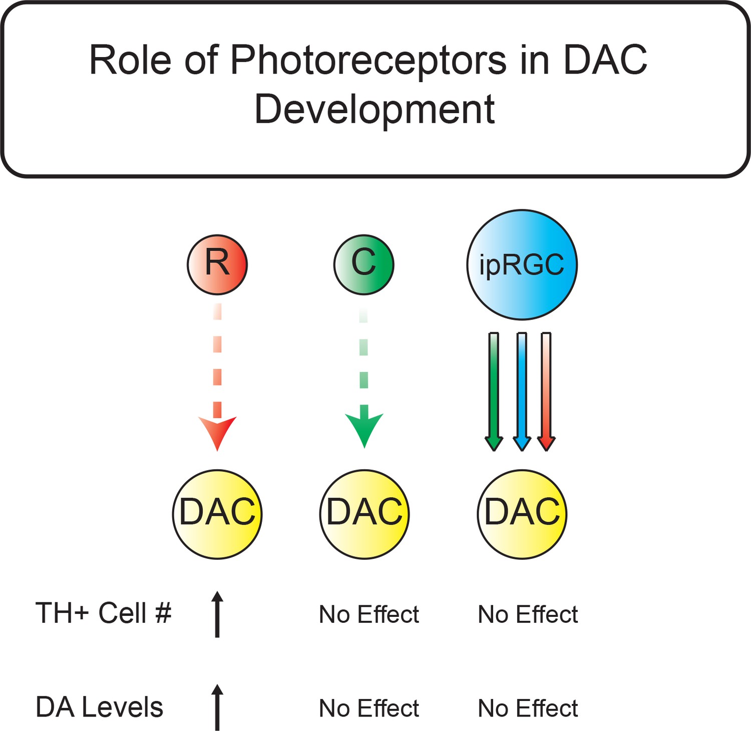
Summary schematic for light-dependent influences on TH-positive cell number and retinal dopamine levels.
Rods signaling serves to increase TH+ cell number and DA levels through ipRGC-independent pathways. Dashed arrows represent indirect influence through multicellular circuits while solid arrows represent direct synaptic connectivity between subtypes. Red represents rod pathway signals, green represents cone pathway signals, and blue represents signals arising from melanopsin phototransduction. Neither ipRGC relay of rod, cone, nor melanopsin signals nor the cone pathway influence TH+ cell number or retinal DA levels. The rod pathway serves to increase the number of TH+ cells and increase retinal DA levels. R: Rod, C: Cone, DAC: dopaminergic amacrine cell, DA: Dopamine.
Tables
Key resources table
| Reagent type (species) or resource | Designation | Source or reference | Identifiers | Additional information |
|---|---|---|---|---|
| Genetic reagent (M. musculus) | Opn4LacZ/LacZ | PMID: 11834834 | RRID: MGI:3797748 | |
| Genetic reagent (M. musculus) | Gnat2cpfl3/cpfl3 | PMID: 17065522 | RRID: MGI:3715214 | |
| Genetic reagent (M. musculus) | Gnat1-/- | PMID: 11095744 | RRID: MGI:3640094 | |
| Genetic reagent (M. musculus) | Opn4DTA/DTA | PMID: 28617242 | n/a | |
| Antibody | Rabbit anti-TH | Millipore | Cat: AB152 | IHC (1:500) |
Additional files
-
Transparent reporting form
- https://doi.org/10.7554/eLife.39866.006
Download links
A two-part list of links to download the article, or parts of the article, in various formats.
Downloads (link to download the article as PDF)
Open citations (links to open the citations from this article in various online reference manager services)
Cite this article (links to download the citations from this article in formats compatible with various reference manager tools)
Light-dependent pathways for dopaminergic amacrine cell development and function
eLife 7:e39866.
https://doi.org/10.7554/eLife.39866


