Balance between BDNF and Semaphorins gates the innervation of the mammary gland
- Weizmann Institute of Science, Israel
Figures
Figure 1 with 2 supplements

TrkB displays sexually dimorphic expression in the mammary gland while members of the Semaphorin family display a non-dimorphic expression.
(A, B) Mammary gland sections of late E13 female and male embryos were stained with TrkBECD antibody and counterstained with DAPI for visualization of the mammary gland structure. Arrows indicate TrkB-expressing sensory axons innervating the mammary gland; arrowheads point to the male-specific expression of TrkB in the mesenchymal cells surrounding the gland. (C–H) In-situ hybridization of mammary gland sections of late E13 female (C, E, G) and male (D, F, H) embryos using specific digoxigenin (DIG)-labeled RNA probes for Sema3d, Sema3f and Sema6f, as indicated. The mammary epithelium is marked by a circle according to the DAPI staining. Scale bar: 50 µm. (I, J) Whole mount X-gal staining of late E12 Sema6A Het female and male mutant embryos. Glands 2–4 are marked by arrows, glands 1 and 5 are hidden by the limbs.
Figure 1—figure supplement 1

In-Situ hybridization screen for Semaphorins expression in the mammary gland.
In-situ hybridization of mammary gland sections of late E13 female embryos using specific digoxigenin (DIG)-labeled RNA probes against Semaphorins of Class 3, 4, 5 and 6. Mammary epithelium is marked by a circle according to the DAPI staining. Scale bar: 50 µm. Semaphorins expression in male embryos is identical (data not shown).
Figure 1—figure supplement 2
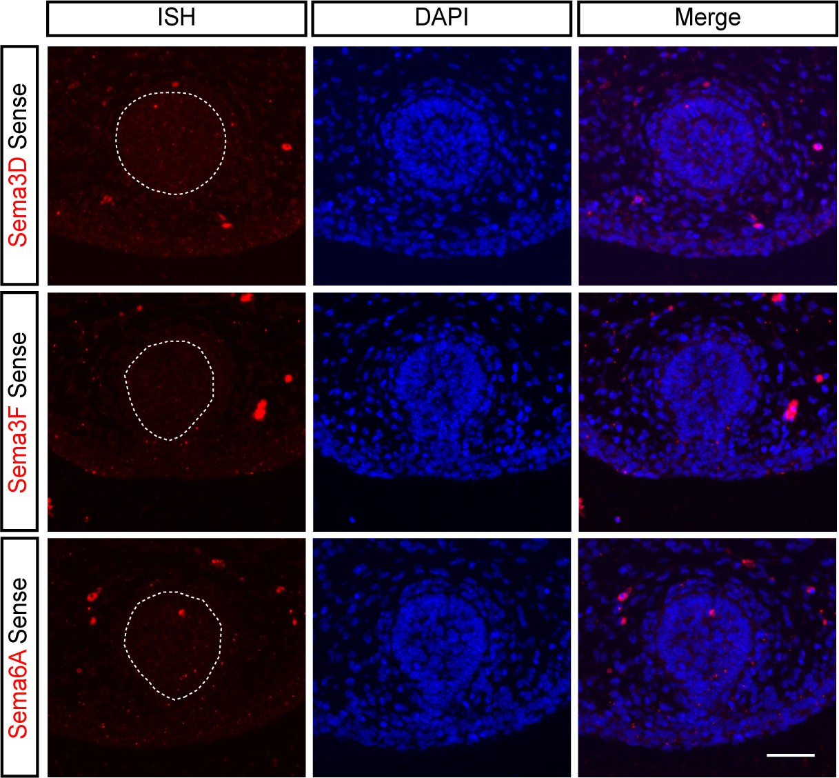
Sense controls of Sema3d, Sema3f and Sema6a.
In-situ hybridization of mammary gland sections of late E13 female embryos using specific digoxigenin (DIG)-labeled Sense RNA probes against Semaphorins 3D, 3F and 6A. Mammary epithelium is marked by a circle according to the DAPI staining. Scale bar: 50 µm. Male embryos gave the same signal (data not shown).
Figure 2 with 1 supplement
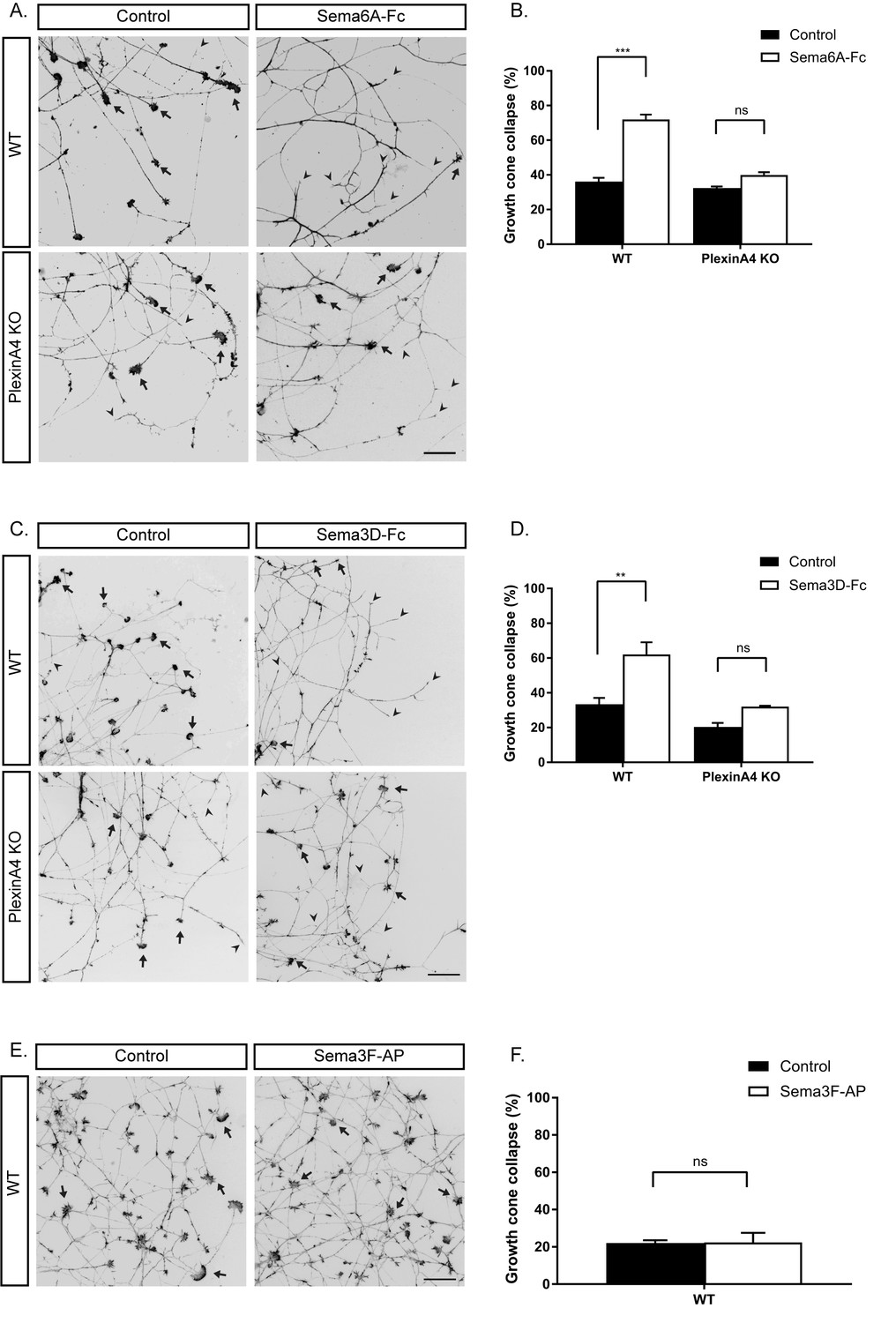
In vitro growth cone collapse of BDNF-responsive neurons by Sema6A and Sema3D is PlexinA4 dependent.
(A, C, E) DRG explants from early E13 WT or PlexinA4 KO embryos were grown for 48 hr, treated with 1 ng/µl soluble Sema6A-Fc (A), 0.02 ng/µl soluble Sema3D-Fc (C), 1 nM of Sema3F-AP (E), or control media for 30 min, fixed, and stained with Phalloidin-Rhodamine to visualize growth cones. Black arrowheads indicate collapsed growth cones and arrows indicate intact growth cones. Scale bar: 50 µm. (B, D, F) Quantification of collapsed growth cones as percentage of total, means ±SEM of more than 1000 growth cones obtained from three independent experiments. (B) WT: control vs Sema6A-Fc, p<0.0001; PlexinA4 KO: control vs Sema6A-Fc, p=0.110307. (D) WT: control vs Sema3D-Fc, p=0.0053; PlexinA4 KO: control vs Sema3D-Fc, p=0.2666. (F) Control vs Sema3F-AP, p=0.954. ***p<0.001, **p<0.01, ns - non-significant, two-way ANOVA with post hoc analysis.
-
Figure 2—source data 1
Percentage of collapsed growth cones in WT and PlexinA4 KO.
- https://doi.org/10.7554/eLife.41162.008
Figure 2—figure supplement 1

Sema6A binds solely to PlexinA4 in BDNF-responsive sensory neurons.
DRG explants from early E13 embryos or PlexinA4 KO embryos grown for 48 hr were incubated with 1 ng/µl soluble Sema6A-Fc for 90 min containing 1:1000 goat anti-human IgG (Fc specific), Alkaline Phosphatase (AP)-conjugated antibody. Binding was detected by alkaline phosphatase reaction.
Figure 3 with 1 supplement
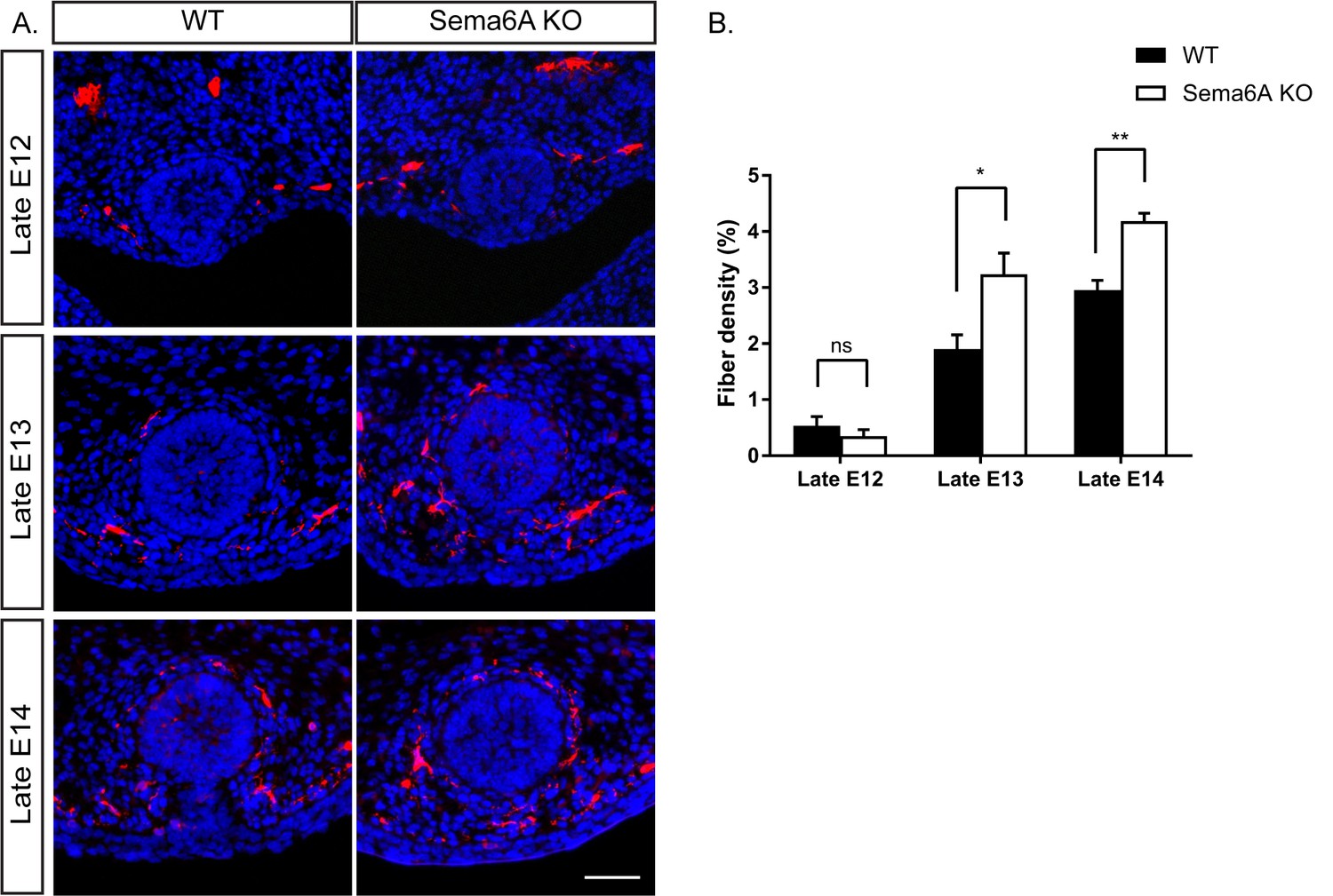
Ablation of Sema6A causes hyperinnervation of the mammary gland by sensory axons in female embryos.
(A) Mammary gland sections of WT and Sema6A KO female embryos at different embryonic stages were stained with Tuj1 (red) and DAPI (blue) for visualization of sensory neuron innervations. Scale bar: 50 µm. (B) Quantification of mammary gland innervation, means ±SEM, n = 5 embryos for each bar. Late E12: p=0.3872; Late E13: p=0.019; Late E14: p=0.0026. *p<0.05, **p<0.01, ns - non-significant, two-way ANOVA followed by separate t-tests per stage.
-
Figure 3—source data 1
Percentage of mammary gland innervation density in WT and Sema6A KO female embryos.
- https://doi.org/10.7554/eLife.41162.012
Figure 3—figure supplement 1
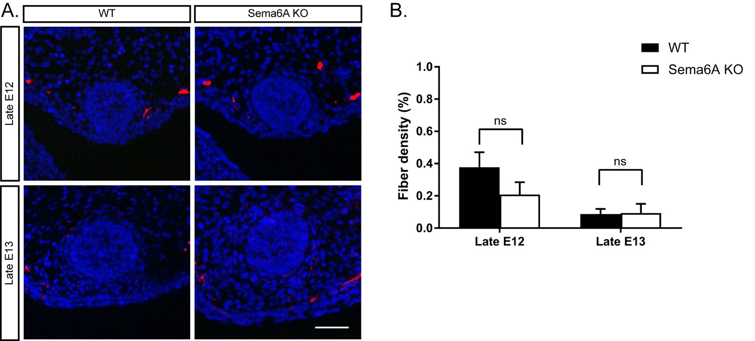
Ablation of Sema6A has no effect on mammary gland innervation in male embryos.
(A) Mammary gland sections of WT and Sema6A KO male embryos at different embryonic stages were stained with Tuj1 (red) and DAPI (blue) for visualization of sensory neuron innervations. Scale bar: 50 µm. (B) Quantification of mammary gland innervation, means ± SEM, n = 5 embryos for each bar. Late E12 p=0.1837, Late E13 p=0.9392. ns – non-significant, two-way ANOVA followed by separate t-tests per stage.
-
Figure 3—figure supplement 1—source data 1
Percentage of mammary gland innervation density in WT and Sema6A KO male embryos.
- https://doi.org/10.7554/eLife.41162.011
Figure 4 with 1 supplement
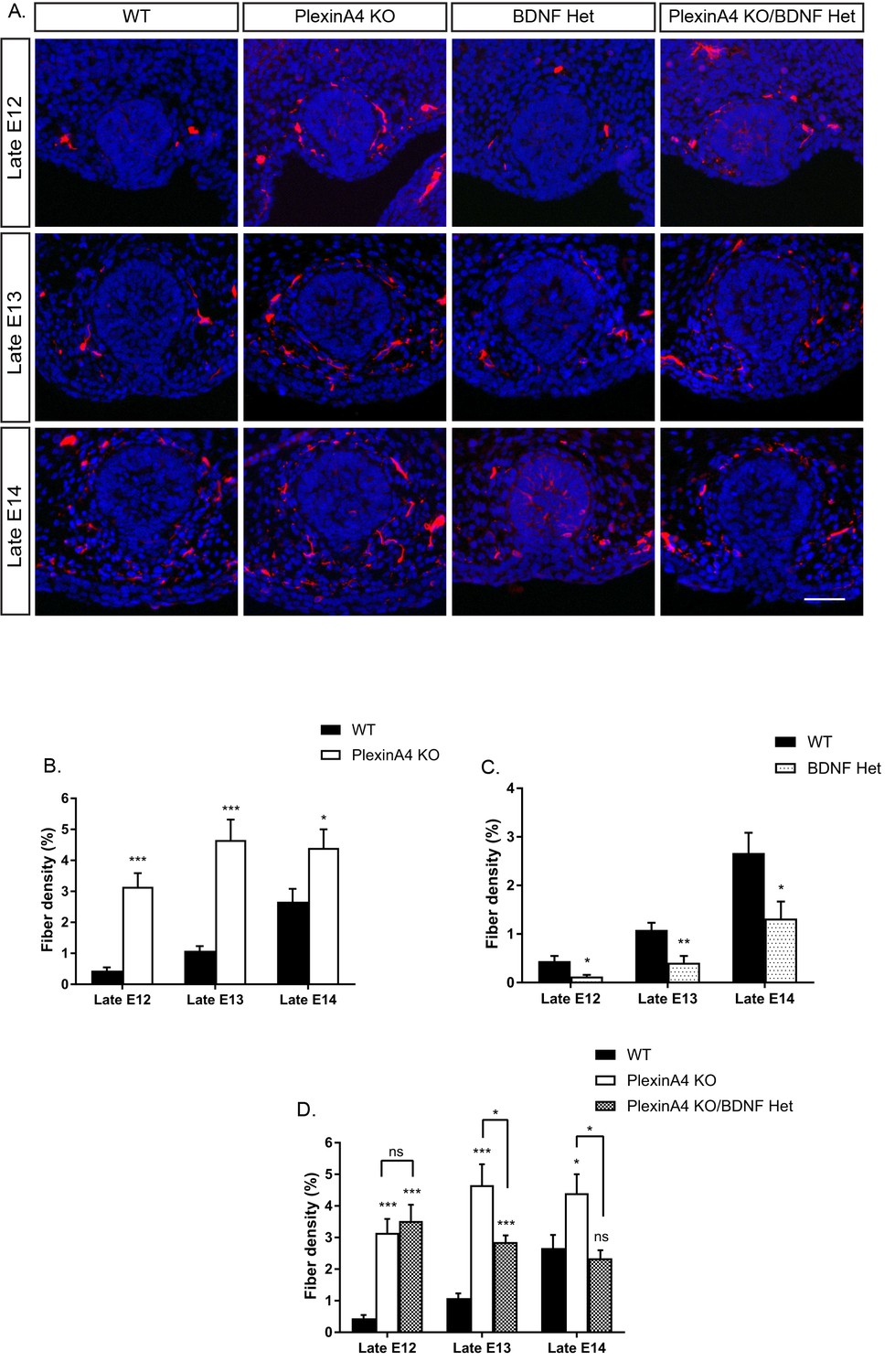
Ablation of PlexinA4 in females causes axonal hyperinnervation of the mammary gland which is balanced by genetic reduction in BDNF.
(A) Mammary gland sections from WT, PlexinA4 KO, BDNF Het, and PlexinA4 KO/BDNF Het female embryos at different embryonic stages were stained with Tuj1 (red) and DAPI (blue) to visualize sensory neuron innervations. Scale bar: 50 µm. (B–D) Quantification of mammary gland innervation, means ± SEM. Late E12: n = 9, 7, 8, 11; Late E13: n = 9, 10, 6, 8; Late E14: n = 5, 8, 7, 6 for WT, PlexinA4 KO, BDNF Het and PlexinA4 KO/BDNF Het, respectively. (B) WT vs PlexinA4 KO: Late E12 p<0.0001, Late E13 p<0.0001, Late E14 p=0.0484. (C) WT vs BDNF Het: Late E12 p=0.0178, Late E13 p=0.0084, Late E14 p=0.0359. (D) WT vs PlexinA4 KO/BDNF Het: Late E12 p<0.0001, Late E13 p=0.0003, Late E14 p=0.8856. PlexinA4 KO vs PlexinA4 KO/BDNF Het: Late E12 p=0.9940, Late E13 p=0.0480, Late E14 p=0.0149. *p<0.05, **p<0.01, ***p<0.001, ns – non-significant, two-way ANOVA followed by separate t-tests per stage. P values compared to WT are marked on top of each bar.
-
Figure 4—source data 1
Percentage of mammary gland innervation density in WT, PlexinA4 KO, BDNF Het and PlexinA4 KO/BDNF Het female embryos.
- https://doi.org/10.7554/eLife.41162.015
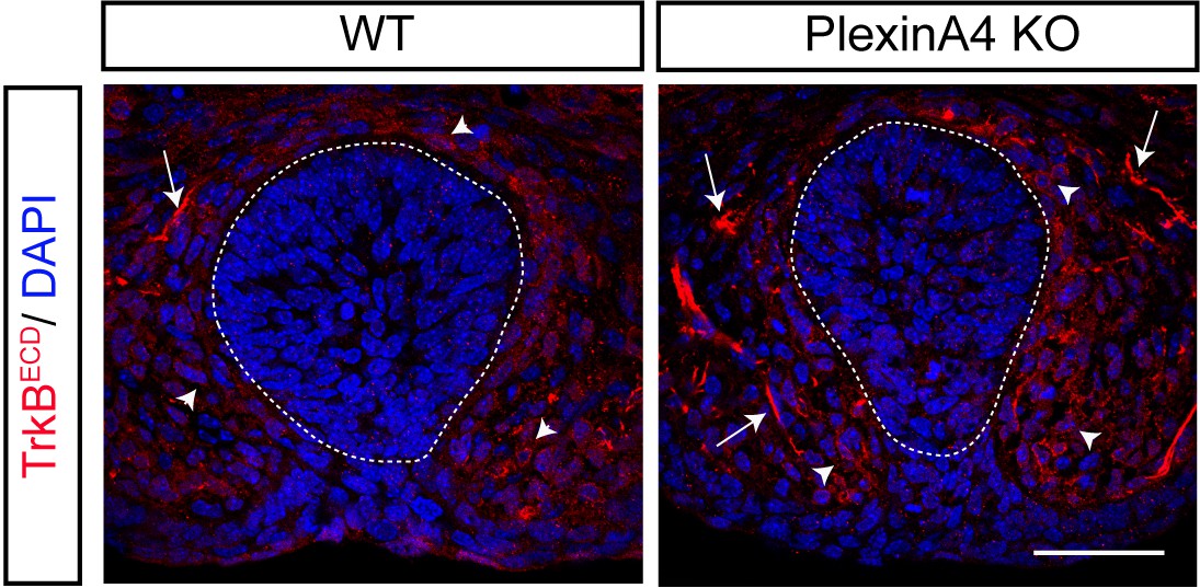
Figure 4—figure supplement 1

Excess innervations in female PlexinA4 KO are TrkB positive.
Mammary gland sections of late E13 WT and PlexinA4 KO female embryos were stained with TrkBECD antibody (red), Tuj1 (green) and counterstained with DAPI for visualization of the mammary gland structure. Scale bar: 50 µm.
Figure 5 with 2 supplements
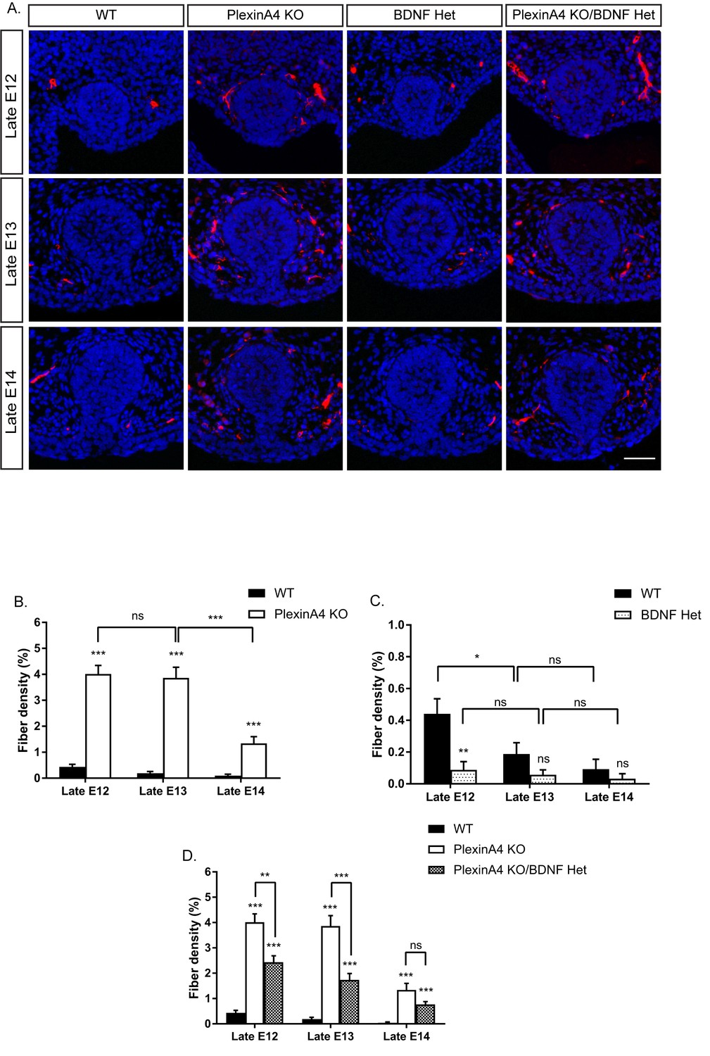
Ablation of PlexinA4 delays axonal pruning in males independent of the initial levels of the mammary gland innervation.
(A) Mammary gland sections of WT, PlexinA4 KO, BDNF Het, and PlexinA4 KO/BDNF Het male embryos at different embryonic stages were stained with Tuj1 (red) and DAPI (blue) to visualize sensory neuron innervations. Scale bar: 50 µm. (B–D) Quantification of mammary gland innervation, means ±SEM. Late E12: n = 11, 9, 9, 10; Late E13: n = 10, 7, 7, 8; Late E14: n = 6, 11, 6, 6 for WT, PlexinA4 KO, BDNF Het and PlexinA4 KO/BDNF Het, respectively. (B) WT vs PlexinA4 KO: Late E12 p<0.0001, Late E13 p<0.0001, Late E14 p<0.0001. PlexinA4 KO: Late E12 vs Late E13 p=0.7803, Late E13 vs Late E14 p<0.0001. (C) WT vs BDNF Het: Late E12 p=0.0013, Late E13 p=0.1156, Late E14 p=0.3739. WT: Late E12 vs late E13 p=0.0473, Late E13 vs late E14 p=0.3282. BDNF Het: Late E12 vs Late E13 p=0.6117, Late E13 vs Late E14 p=0.5833 (D) WT vs PlexinA4 KO/BDNF Het: Late E12 p<0.0001, Late E13 p<0.0001, Late E14 p=0.00011. PlexinA4 KO vs PlexinA4 KO/BDNF Het: Late E12 p=0.0010, Late E13 p=0.0003, Late E14 p=0.1049. *p<0.05, **p<0.01, ***p<0.001, ns – non-significant, two-way ANOVA followed by separate t-tests per stage. P values compared to WT are marked on top of each bar.
-
Figure 5—source data 1
Percentage of mammary gland innervation density in WT, PlexinA4 KO, BDNF Het and PlexinA4 KO/BDNF Het male embryos.
- https://doi.org/10.7554/eLife.41162.019
Figure 5—figure supplement 1

Excess innervations in male PlexinA4 KO are TrkB positive.
Mammary gland sections of late E13 WT and PlexinA4 KO male embryos were stained with TrkBECD antibody (red), Tuj1 (green) and counterstained with DAPI for visualization of the mammary gland structure. Scale bar: 50 µm.
Figure 5—figure supplement 2
PlexinA4 does not control the expression of TrkB.T1.
Mammary gland sections of late E13 WT and PlexinA4 KO male embryos were stained with TrkBECD antibody, and counterstained with DAPI for visualization of the mammary gland structure. Arrows indicate TrkB-expressing sensory axons innervating the mammary gland; arrow heads point to membrane expression of TrkBECD in mesenchymal cells surrounding the gland, which is not changed in the PlexinA4 KO. Scale bar: 50 µm.
Figure 6
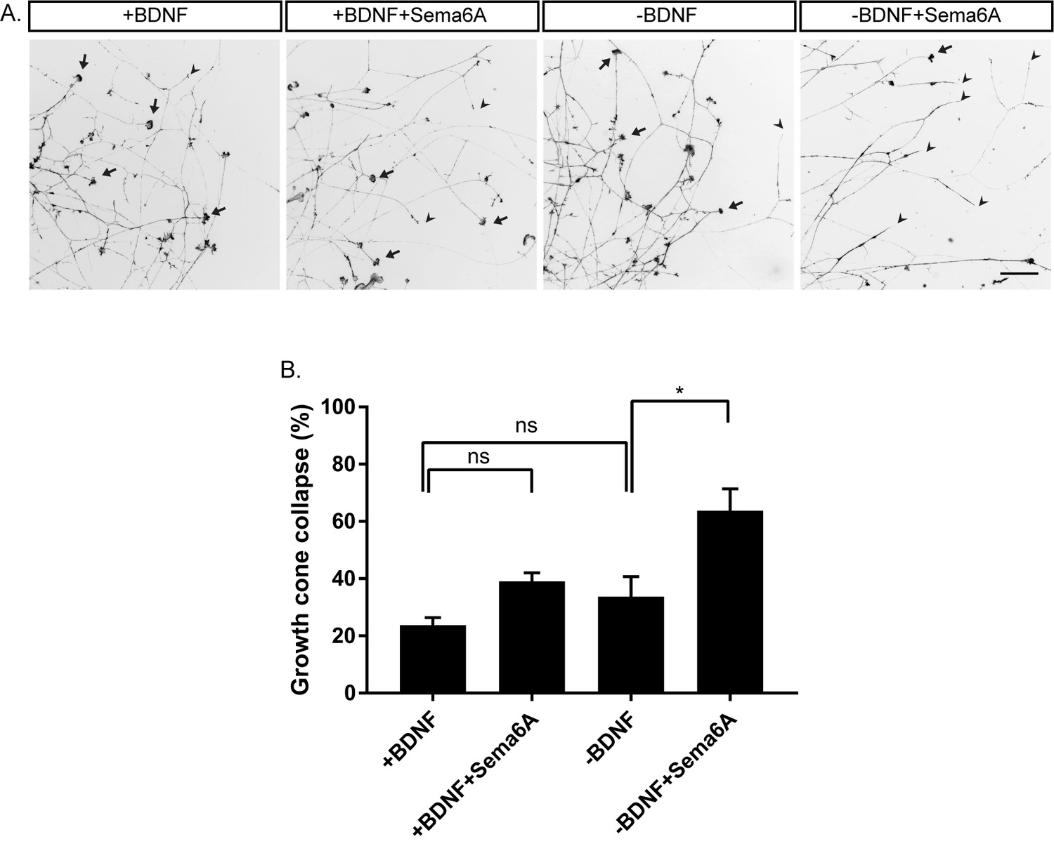
Hypersensitivity to PlexinA4 signaling induced by reduction in BDNF.
(A) Explants of early E13 embryonic DRGs were cultured in media containing 50 ng/ml BDNF for 48 hr, then the media was replaced to 50 ng/ml BDNF (+BDNF), or media without BDNF (-BDNF) both with low concentration of Sema6A (0.025 ng/µl) for 30 min. Growth cones were visualized with Phalloidin- Rhodamine staining. Black arrowheads refer to collapsed growth cones and arrows indicate non-collapsed. Scale bar: 50 µm. (B) Quantification of percent of collapsed growth cones, means ± SEM of more than 1000 growth cones obtained from three independent experiments. +BDNF vs +BDNF+Sema6A p=0.2872, +BDNF vs -BDNF p=0.6095, -BDNF vs -BDNF+Sema6A p=0.0223. *p<0.05, ns – non-significant, one-way ANOVA with post hoc analysis.
-
Figure 6—source data 1
Percentage of collapsed growth cones.
- https://doi.org/10.7554/eLife.41162.021
Figure 7
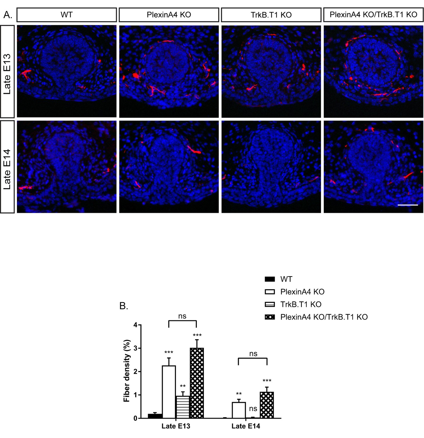
Co-ablation of TrkB.T1 and PlexinA4 has no additive effect on axonal pruning.
(A) Mammary gland sections of WT, PlexinA4 KO, TrkB.T1 KO, and PlexinA4 KO/TrkB.T1 KO male embryos at different embryonic stages were stained with Tuj1 (red) and DAPI (blue) to visualize sensory neuron innervations. Scale bar: 50 µm. (B) Quantification of mammary gland innervation, means ±SEM. Late E13: n = 5, 8, 6, 7, Late E14: n = 5, 5, 9, 6 for WT, PlexinA4 KO, TrkB.T1 KO and PlexinA4 KO/TrkB.T1 KO, respectively. WT vs PlexinA4 KO: Late E13 p<0.0001, Late E14 p=0.0015. WT vs TrkB.T1 KO: Late E13 p=0.0020, Late E14 p=0.0867. WT vs PlexinA4 KO/TrkB.T1 KO: Late E13 p<0.0001, Late E14 p=0.0006. PlexinA4 KO vs PlexinA4 KO/TrkB.T1 KO: Late E13 p=0.1114, Late E14 p=0.1037. **p<0.01, ***p<0.001, ns – non-significant, two-way ANOVA followed by separate t-tests per stage. P values compared to WT are marked on top of each bar.
-
Figure 7—source data 1
Percentage of mammary gland innervation density in WT, PlexinA4 KO, TrkB.T1 KO and PlexinA4 KO/TrkB.T1 KO male embryos.
- https://doi.org/10.7554/eLife.41162.023
Figure 8
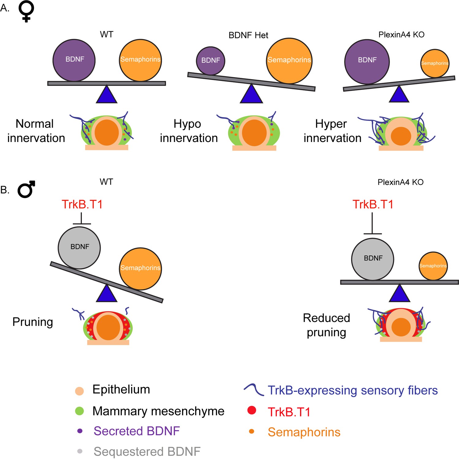
The relative balance between BDNF and Semaphorins regulates the level of mammary gland innervation in females and may promote axonal pruning in males.
(A) In females: a balance between BDNF and Semaphorins specifies normal innervation of the mammary gland. Decreased levels of BDNF cause hypoinnervation and decreased Semaphorins cause hyperinnervation. (B) In males: Sequestering of BDNF by TrkB.T1 results in pruning, while decreased Semaphorin signaling results in reduced pruning.
Tables
Key resources table
| Reagent type (species) or resource | Designation | Source or reference | Identifiers | Additional information |
|---|---|---|---|---|
| Genetic reagent (M. Musculus) | Sema6a-/- | PMID: 11242070 | Dr. Marc Tessier-Lavigne (Stanford) | |
| Genetic reagent (M. Musculus) | Plxna4-/- | PMID: 15721238 | Dr. Marc Tessier-Lavigne (Stanford) | |
| Genetic reagent (M. Musculus) | Ntrk2.T1-/- | PMID: 16815329 | Dr. Lino Tessarollo (NIH) | |
| Genetic reagent (M. Musculus) | Bdnf-/- | Jackson Laboratory | stock #: 002266 RRID: MGI:2175720 | PMID: 8139657 |
| Cell line (Homo sapiens) | HEK293 | ATCC | ATCC CRL-1573 RRID:CVCL_0045 | |
| Cell line (Cercopithecus aethiops) | COS1 | ATCC | ATCC CRL-1650 RRID: CVCL_0223 | |
| Antibody | Mouse monoclonal anti-tubulin β-III | R and D | Cat. #: MAB1195 RRID:AB_357520 | IHC (1:500) |
| Antibody | Rabbit polyclonal anti-tubulin β-III | Biolegend | Cat. # 802001 RRID:AB_2564645 | IHC-Fr (1:1000) |
| Antibody | Polyclonal Goat anti-Mouse Trkb | R and D | Cat. # AF1494 RRID:AB_2155264 | IHC-Fr (1:100) |
| Peptide, recombinant protein | Human Sema6A-FC | PMID: 20606624 | ||
| Peptide, recombinant protein | Mouse Sema3D-FC | This paper | ||
| Peptide, recombinant protein | Mouse AP-Sema3F | PMID: 9331348 | ||
| Chemical compound, drug | Phalloidin-Rhodamine | Sigma | p1951 | 1:250 |
| Chemical compound, drug | Ara-C | Sigma | C1768 | 30 uM |
Additional files
-
Transparent reporting form
- https://doi.org/10.7554/eLife.41162.025
Download links
A two-part list of links to download the article, or parts of the article, in various formats.
Downloads (link to download the article as PDF)
Open citations (links to open the citations from this article in various online reference manager services)
Cite this article (links to download the citations from this article in formats compatible with various reference manager tools)
Balance between BDNF and Semaphorins gates the innervation of the mammary gland
eLife 8:e41162.
https://doi.org/10.7554/eLife.41162


