Increased anxiety and decreased sociability induced by paternal deprivation involve the PVN-PrL OTergic pathway
- Shaanxi Normal University, China
- Emory University, United States
- University of Tsukuba, Japan
- University of Connecticut Health Center, United States
Figures
Figure 1
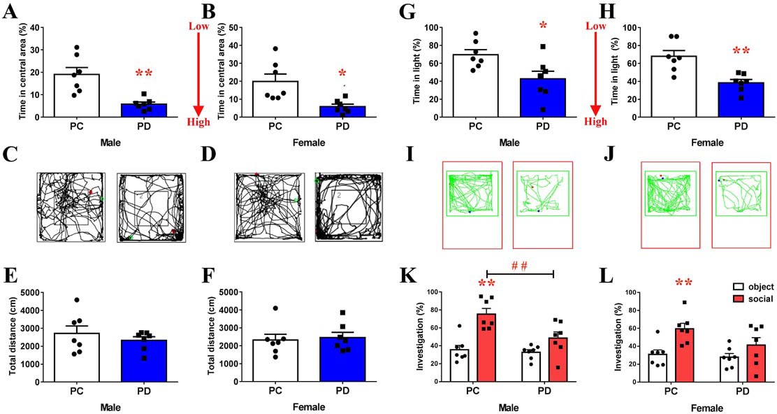
Effect of paternal deprivation on anxiety-like behavior and social preference in adult mandarin voles (n = 7).
(A, B) Percentage of time in the central area, (C, D) representative path and (E, F) total distance of mandarin voles in the open field test. *p<0.05; **p<0.01. Independent sample t-tests. (G, H) Percentage of time in the light area and (I, J) animal traces of mandarin voles in the light and dark box. *p<0.05; **p<0.01. Independent sample t-tests. Effect of PD on social preference in (K) males and (L) females. Error bars indicate SEM. **p<0.025 vs. object stimulus. ##p<0.025 vs. PC. Two-way ANOVA (factors: treatment × stimulus). PC, biparental care; PD, paternal deprivation.
-
Figure 1—source data 1
Statistical results of the percentage of time spent in the central area and total distance in the open field, percentage of time in the light area in the light-dark box, and the percentage of time in investigating the social stimulus or object stimulus in the social preference test.
- https://doi.org/10.7554/eLife.44026.004
Figure 2 with 1 supplement
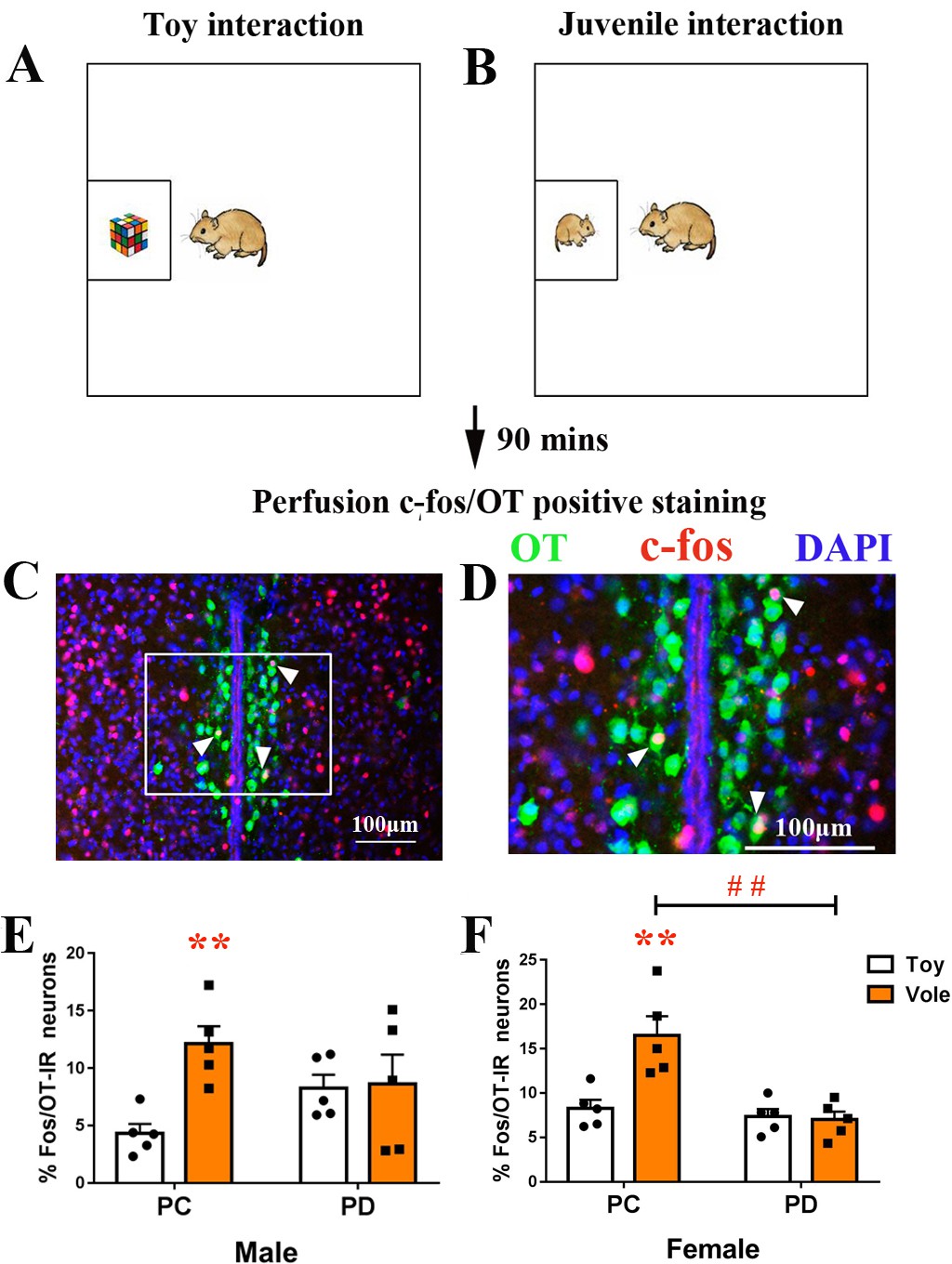
Effect of paternal deprivation on the percentage of c-fos/OT double-labeled neurons in the PVN in adult mandarin voles (n = 5).
Voles were subjected to interaction with either (A) a juvenile voles or (B) a magic cube. (C, D) Double-immunohistochemical staining of c-fos (red) and OT (blue). Effect of PD on the percentage of neurons double-labeled for OT and c-fos in (E) males and (F) females. Error bars indicate SEM. **p<0.01 vs. object stimulus. ##p<0.01 vs. PC. Two-way ANOVA (factors: treatment × stimulus). PC, biparental care; PD, paternal deprivation.
-
Figure 2—source data 1
Numbers of c-fos/OT double-labeled positive cells in the PVN.
- https://doi.org/10.7554/eLife.44026.007
Figure 2—figure supplement 1
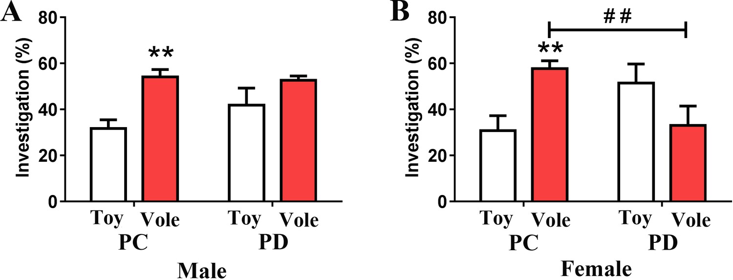
Paternal deprivation diminishes social approach in (A) male and (B) female mandarin voles (n = 5).
Error bars indicate SEM. **p<0.025 vs. Toy. ## p<0.025 vs. PC. PC, biparental care; PD, paternal deprivation.
Figure 3
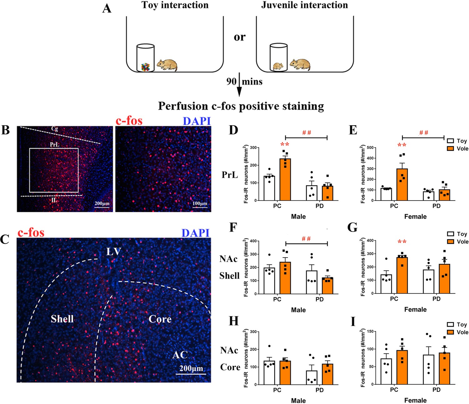
Effect of paternal deprivation on c-fos expression in the PrL and the NAc of adult mandarin voles (n = 5).
(A) Voles were subjected to interaction with either a juvenile vole or a magic cube. (B) Immunohistochemical staining of c-fos (red) and 4',6-diamidine-2'-phenylindole dihydrochloride (DAPI) (blue). The effect of PD on c-fos expression in the PrL of (D) males and (E) females. (C) Images show c-fos immunoreactivity in the NAc shell and the NAc core. Effect of PD on c-fos expression in (F) males and (G) females of the NAc shell. Effect of PD on c-fos expression in the NAc core of (H) males and (I) females. Error bars indicate SEM. **p<0.01 vs. object stimulus. ##p<0.01 vs. PC. Two-way ANOVA (factors: treatment × stimulus). PC, biparental care; PD, paternal deprivation.
-
Figure 3—source data 1
Numbers of c-fos-positive cells in the PrL, NAc shell and NAc core.
- https://doi.org/10.7554/eLife.44026.009
Figure 4

Effect of paternal deprivation on PVN OT-IR neurons.
(A) PC males, (B) PD males, (C) PC females and (D) PD females. 3V, 3rd ventricle. (E) Schematic drawing illustrating tissue in the PVN. (F) Quantification of OT-IR neurons in the PVN. Error bars indicate SEM. n = 4. **p<0.01 vs. PC. ##p<0.01 vs. male. Two-way ANOVA (factors: treatment × sex). PC, biparental care; PD, paternal deprivation; PVN, paraventricular nucleus.
-
Figure 4—source data 1
Numbers of OT-positive cells in the PVN.
- https://doi.org/10.7554/eLife.44026.011
Figure 5
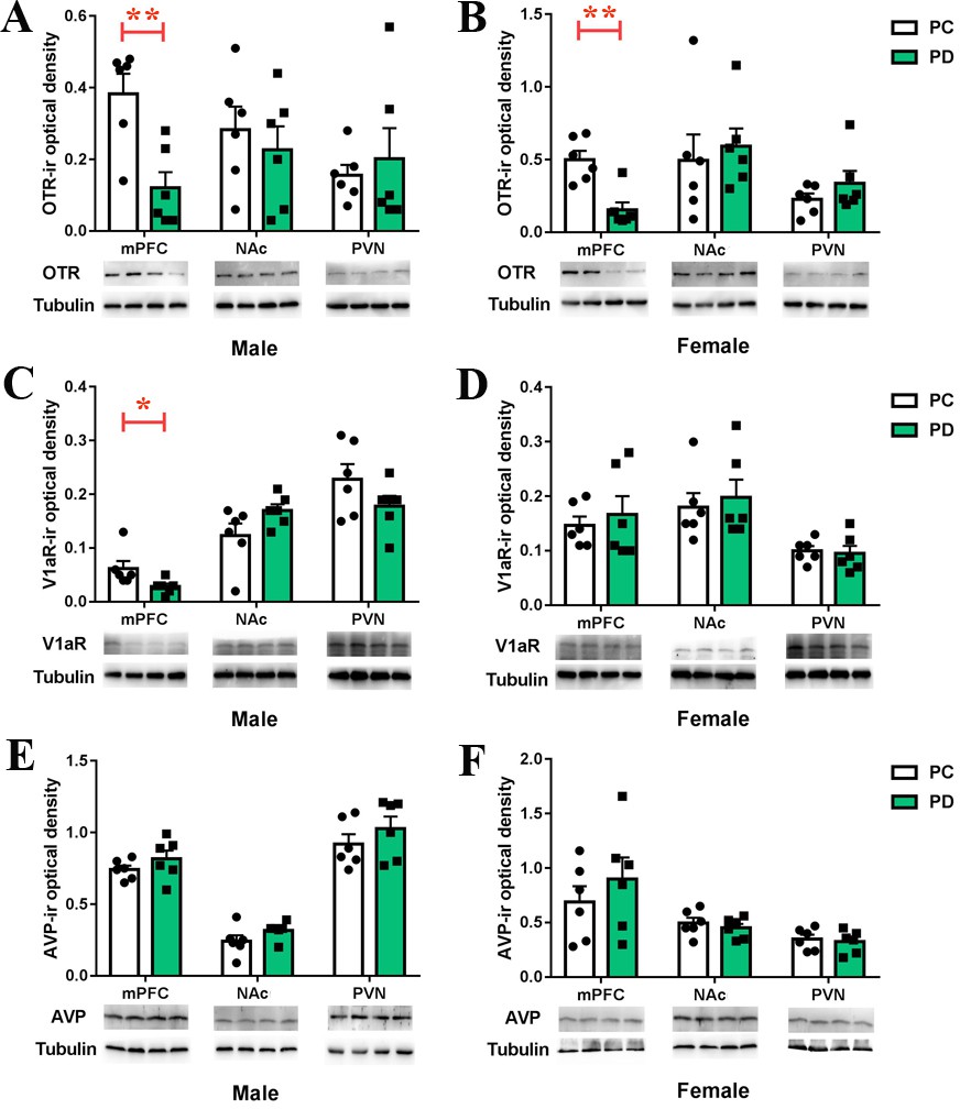
Effects of paternal deprivation on mesocorticolimbic.
(A–B) OTR, (C–D) V1aR and (E–F) AVP immunoreactivity in male (n = 6) and female (n = 6) mandarin voles. Error bars indicate SEM. *p<0.05; **p<0.01. Independent sample t-tests. AVP, arginine vasopressin; mPFC, medial prefrontal cortex; NAc, nucleus accumbens; OTR,oxytocin receptor; PC, biparental care; PD, paternal deprivation; PVN, paraventricular nucleus; V1aR, vasopressin 1a receptor.
-
Figure 5—source data 1
Levels of OTR, V1aR or AVP in the mPFC, NAc and PVN measured by Western Blot assay.
- https://doi.org/10.7554/eLife.44026.013
Figure 6

Effects of PrL OT administration on anxiety-like behavior and social preference in paternal deprivation mandarin voles.
(A) Experimental schematics. (B) Histological representations of the microinjection site and (C, D) schematic diagrams showing the location of injector tips in the PrL. ×: missed. OT in the PrL is anxiolytic in both of sexes. (E, H) Percentage of time in the central area and (F, I) total distance in the open field test. One-way ANOVA. *p<0.05. OT in the PrL promotes a social preference in (G) males and (J) females. #p<0.0083 vs object stimulus. Two-way ANOVA (factors: treatment × sex). (Male: CSF – n = 6; 1 ng OT – n = 6; 10 ng OT – n = 6; 10 ng OT/10 ng OTA – n = 6; 10 ng OT/100 ng OTA – n = 5; 10 ng OT/10 ng V1aRA – n = 6. Female: CSF – n = 6; 1 ng OT – n = 6; 10 ng OT – n = 6; 1 ng OT/10 ng OTA – n = 5; 1 ng OT/100 ng OTA – n = 5; 10 ng OT/10 ng V1aRA – n = 6).
-
Figure 6—source data 1
The percentage of time spent in the central area, the total distance in the open field, and the percentage of time spent investigating the social stimulus and the object stimulus after administration of CSF, OT, OT/OTA, or OT/V1aRA to the PrL.
- https://doi.org/10.7554/eLife.44026.015
Figure 7 with 4 supplements
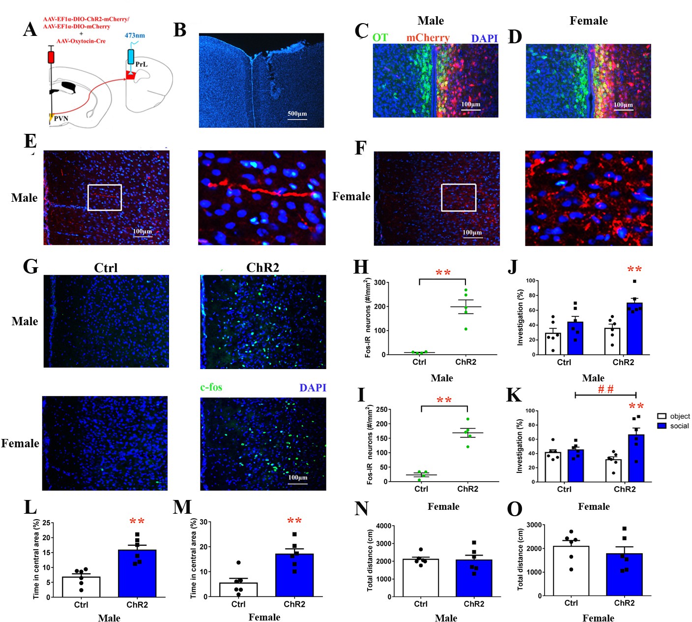
Optogenetic stimulation of OT terminals rescued changes in emotion and social preference behavior that were induced by paternal deprivation.
(A) Schematic drawing of the locations of rAAV-Ef1α-DIO-ChR2-mCherry plus rAAV-Oxytocin-Cre or rAAV-Ef1α-DIO-mCherry plus rAAV-Oxytocin-Cre injection into the right PVN and optic fiber implants. (B) Immunohistological image showing the targetting of fiber implants in the right PrL. Colocalization of ChR2-mCherry (red), OT neurons (green) and DAPI (blue) in the PVN of (C) males and (D) females. Confocal images of axonal mCherry signal in the PrL of (E) males and (F) females. (G) Images show expression of c-fos in the PrL after photostimulation. Quantification of c-fos in the PrL of (H) males (Ctrl: n = 4; ChR2: n = 5) and (I) females (Ctrl: n = 4; ChR2: n = 5) after photostimulation. Optogenetic activation of oxytocinergic fibers in the PrL increases social preference of (J) males (n = 6) and (K) females (n = 6). **p<0.01 vs. object stimulus. ## p<0.01 vs. Ctrl. Two-way ANOVA (factors: photostimulation treatment × stimulus type). Activation of PVN-PrL oxytocinergic projection significantly increased the percentage of time in the central area for both (L) males (n = 6) and (M) females (n = 6), but did not influence total distance traveled for (N) males (n = 6) or (O) females (n = 6). *p<0.05; **p<0.01. Independent sample t-tests. Error bars indicate SEM.
-
Figure 7—source data 1
Statistical result of levels of c-fos in the PrL after optogenetic activation of PrL-projecting PVN OTergic neurons during the open field, the social preference test.
- https://doi.org/10.7554/eLife.44026.021
Figure 7—figure supplement 1
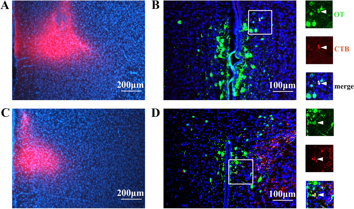
Histology and immunostaining.
The histology of CTB injecting into the right PrL of (A) male and (C) female voles. Immunostaining showing colocalization of OT neurons (green) and CTB (red) in the PVN of (B) male and (D) female voles.
Figure 7—figure supplement 2
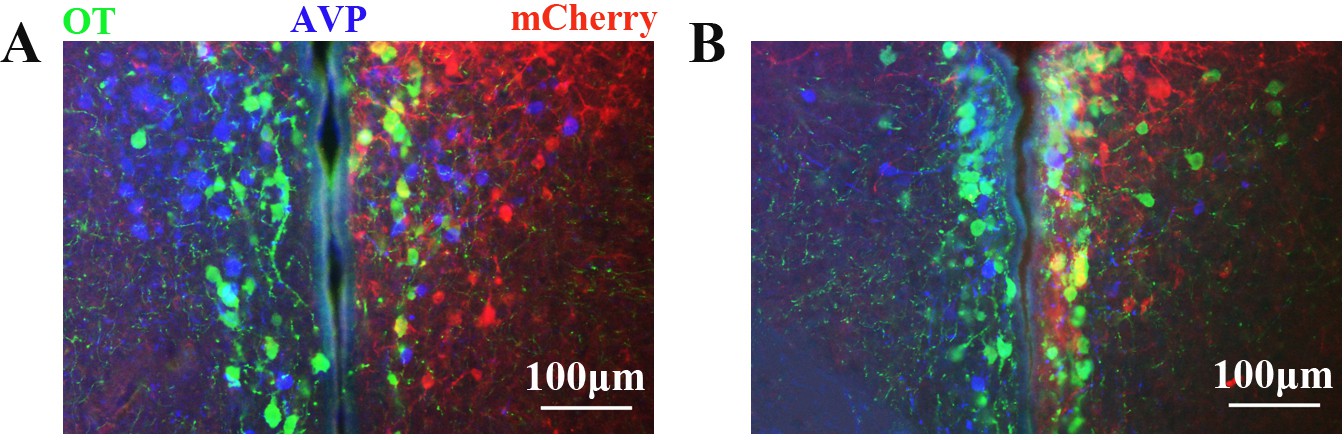
Immunostaining showing colocalization of mCherry (red) with OT (green) but not AVP (blue) in PVN neurons of (A) male and (B) female voles.
https://doi.org/10.7554/eLife.44026.018
Figure 7—figure supplement 3
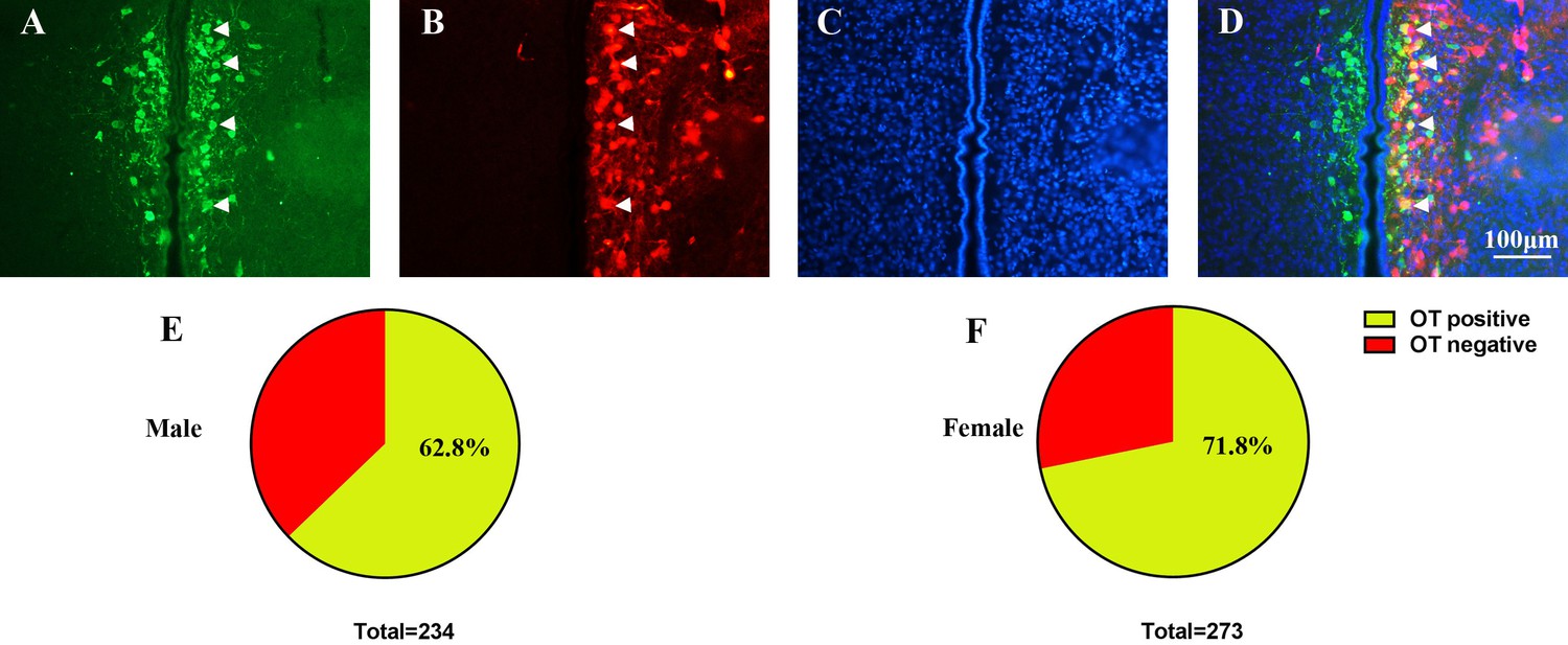
Representative immunostaining allowing the vizualization of (A) OT expression (green), (B) mCherry (red) and (C) DAPI (blue) in PVN neurons of PD voles.
(D) Colocalization image including bands (A), (B) and (C). Arrowheads indicate the OT, mCherry and DAPI. Quantification of OT–meCherry colocalization in (E) males (147/234 = 62.8%, from two voles) and (F) females (203/283 = 71.7%, from two voles).
Figure 7—figure supplement 4
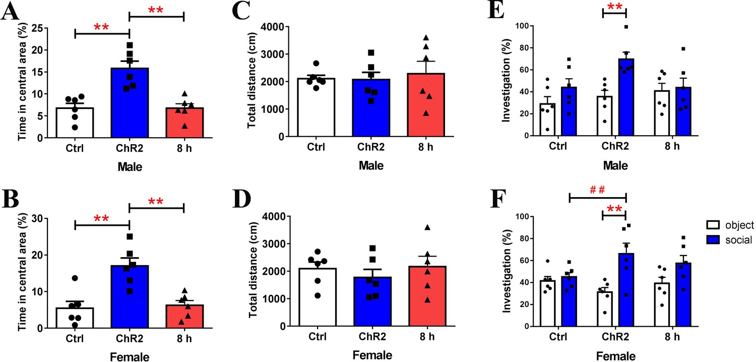
After activation of PVN-to-PrL OT terminals does not elicit long-lasting effects on anxiety-like behavior and social preference in PD voles (n = 6).
The behavioral changes disappeared 8 hr after blue light photostimulation. Quantification of the percentage of time spent in the central area ((A): male; (B): female) and total distance traveled ((C): male; (D): female). **p<0.01. One-way ANOVA. Quantification of social preference ((E): male; F): female)). **p<0.01 vs. object stimulus. ## p<0.01 vs. Ctrl. Two-way ANOVA. Error bars indicate SEM.
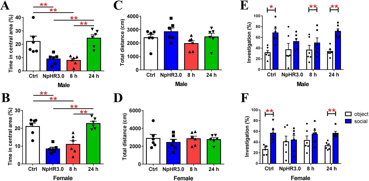
Figure 8 with 2 supplements
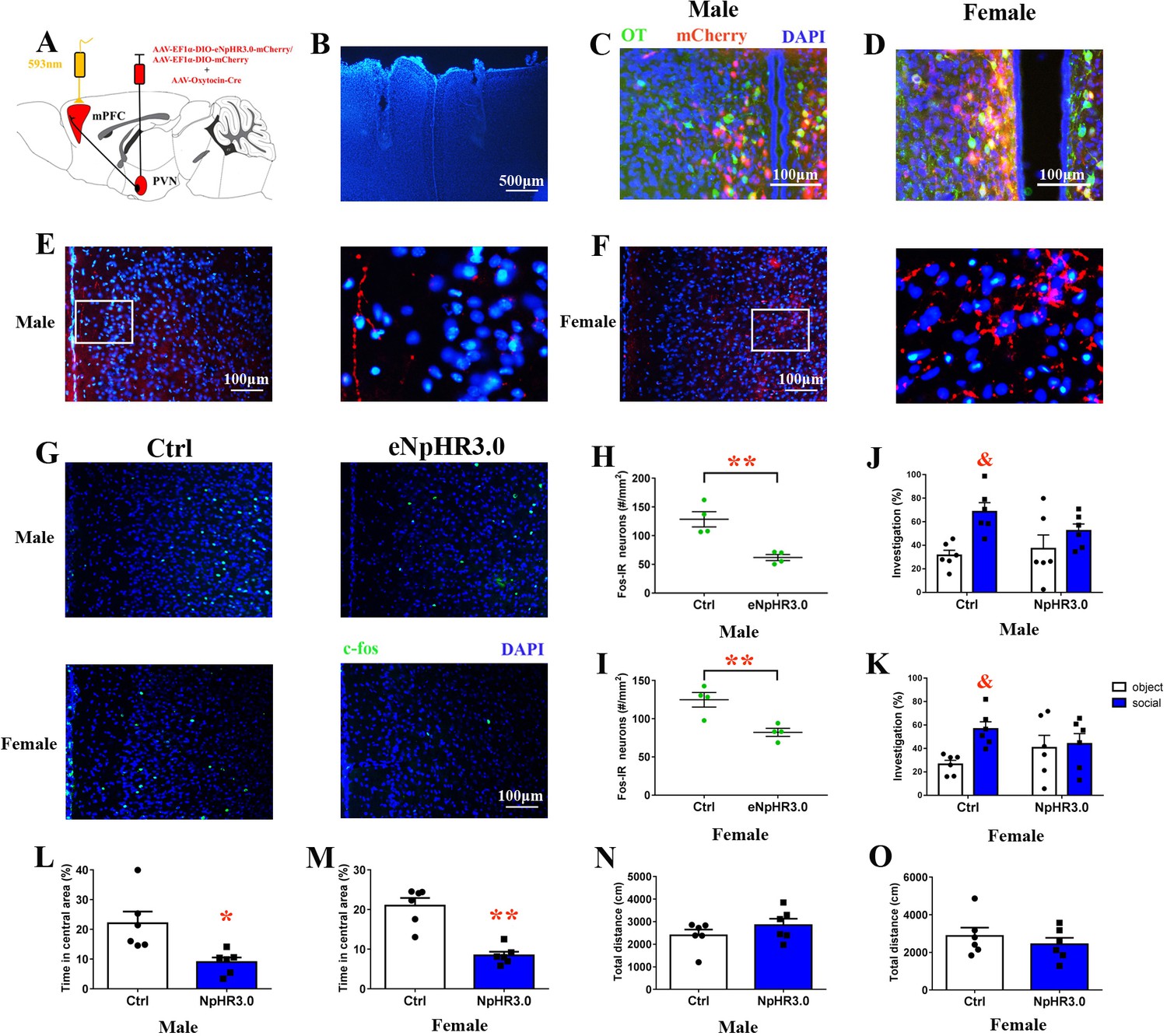
Optogenetic inhibition of OT terminals elicits anxiety-like behavior and attenuated social preference in naive voles.
(A) Schematic drawing of the locations of rAAV-Ef1α-DIO-eNpHR3.0-mCherry plus rAAV-Oxytocin-Cre or rAAV-Ef1α-DIO-mCherry plus rAAV-Oxytocin-Cre injection bilaterally into the PVN and optic fiber implants. (B) Immunohistological image showing target of fiber implants in the bilateral PrL. Colocalization of eNpHR3.0-mCherry (red), OT neurons (green) and DAPI (blue) in the PVN of (C) males and (D) females. Confocal images of axonal mCherry signal in the PrL of (E) males and (F) females. (G) Images showing the expression of c-fos in the PrL after yellow light photostimulation. Quantification of c-fos in the PrL of (H) males (n = 4) and (I) females (n = 4) after photostimulation. **p<0.01. Independent sample t-tests. Optogenetic inhibition of oxytocinergic fibers in the PrL decreases the social preference of (J) males (n = 6) and (K) females (n = 6); p<0.025. Two-way ANOVA (factors: optogenetic inhibition treatment × stimulus type). Inhibition of PVN-PrL oxytocinergic projection significantly decreases the percentage of time spent in the central area for both (L) males (n = 6) and (M) females (n = 6), but did not influence total distance traveled by (N) males (n = 6) or (O) females (n = 6). **p<0.01. Independent sample t-tests. Error bars indicate SEM.
-
Figure 8—source data 1
Results of statistical analysis of the levels of c-fos in the PrL after optogenetic inhibition of PrL-projecting PVN OTergic neurons during the open field and the social preference test.
- https://doi.org/10.7554/eLife.44026.025
Figure 8—figure supplement 1
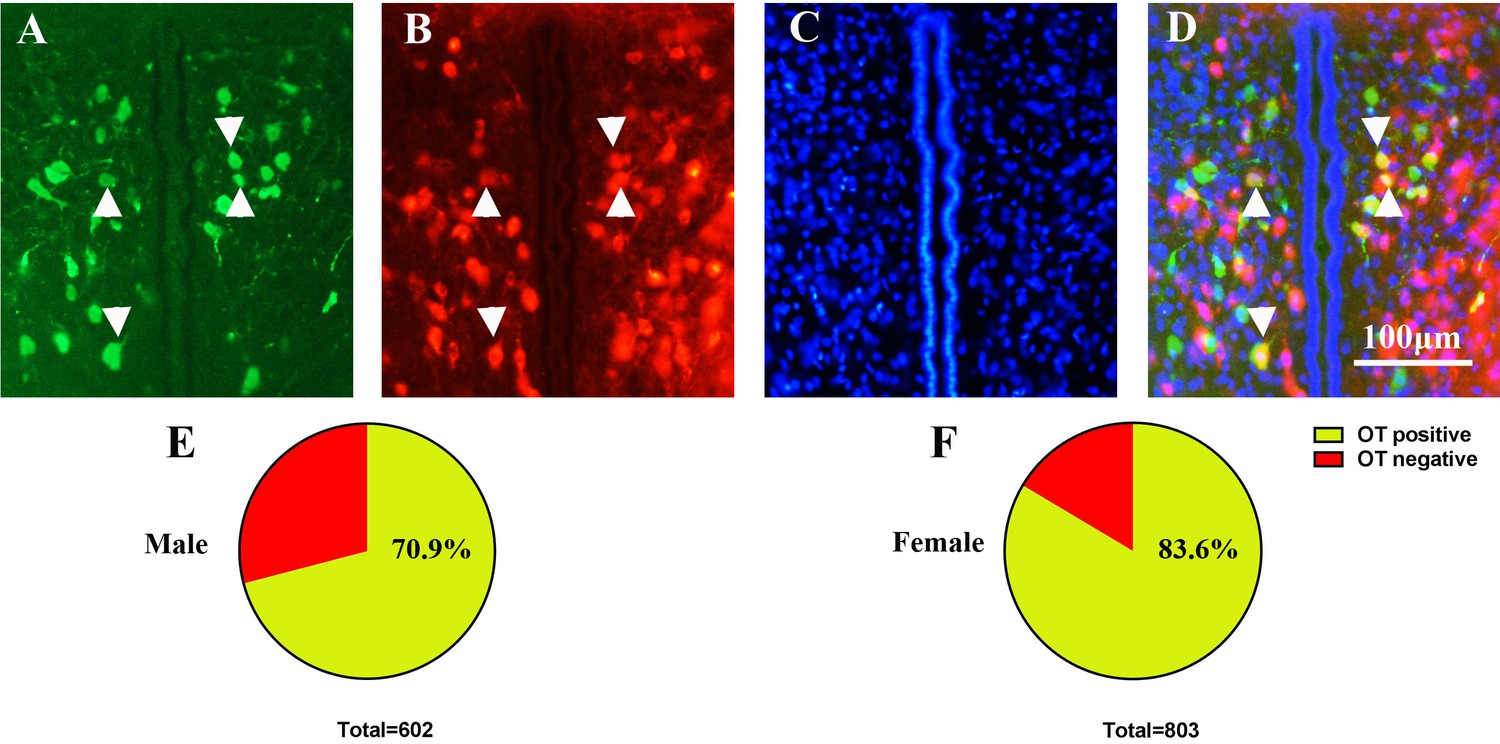
Representative immunostaining of (A) OT expression (green), (B) mCherry (red) and (C) DAPI (blue) in PVN neurons of naïve voles.
(D) Colocalization image for bands (A), (B) and (C). Arrowheads indicate the OT, mCherry and DAPI. Quantification of OT/meCherry colocalization in (E) males (427/602 = 70.9%, from two voles) and (F) females (672/803 = 83.6%, from two voles).
Figure 8—figure supplement 2
After inhibition of PVN-to-PrL OT terminals does not elicit long-lasting effects on anxiety-like behavior and social preference in naïve voles (n = 6).
The behavioral changes (except social preference of male: within 8 hr) occur within 8–24 hr following yellow light photostimulation. Quantification of the percentage of time in the central area ((A): male; (B): female) and total distance traveled ((C): male; (D): female). **p<0.01. One-way ANOVA. Quantification of social preference ((E): male; (F): female). *p<0.05; **p<0.01 vs. object stimulus. Two-way ANOVA. Error bars indicate SEM.
Author response image 1
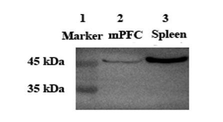
Lane 2 and Lane 3: Anti-Oxytocin Receptor antibody [EPR12789] (ab181077, Abcam) at 1/2000 dilution.
Lane 1: Protein marker. Lane 2: Voles mPFC tissue lysate. Lane 3: Mouse spleen tissue lysate.
Tables
Key resources table
| Reagent type (species) or resource | Designation | Source or reference | Identifiers | Additional information |
|---|---|---|---|---|
| Antibody | anti-oxytocin mouse monoclonal antibody | Millipore | Cat#MAB5296 RRID:AB_2157626 | concentration: 1:7500 |
| Antibody | anti-vasopressin rabbit polyclonal antibody | Millipore | Cat#AB1565 RRID:AB_90782 | concentration:1:4000 |
| Antibody | anti-c-fos rabbit polyclonal antibody | Abcam | Cat#ab190289 RRID:AB_2737414 | concentration:1:1500 |
| Antibody | anti-oxytocin receptor rabbit monoclonal antibody | Abcam | Cat#ab181077 | concentration:1:2000 |
| Antibody | anti-vasopressin receptor 1A (V1aR) goat polyclonal antibody | GeneTex | Cat#GTX89114 RRID:AB_10724608 | concentration:1:7000 |
| Antibody | anti-β-tubulin mouse monoclonal antibody | ComWin Biotechnology | Cat#CW0098M | concentration:1:5000 |
| Antibody | anti-rabbit goat conjugated with TRITC | Jackson ImmunoResearch | Cat#111-025-003 RRID:AB_2337926 | concentration:1:200 |
| Antibody | anti-rabbit goat conjugated with DyLight 405 | Jackson Immunoresearch | Cat#111-475-003 RRID:AB_2338035 | concentration:1:200 |
| Antibody | anti-rabbit goat conjugated with DyLight 488 | Boster | Cat#BA1127 | concentration:1:200 |
| Antibody | anti-mouse goat antibody conjugated with DyLight 488 | Boster | Cat#BA1126 | concentration:1:200 |
| Antibody | anti-rabbit goat conjugated with horseradish peroxidase | ZhongShan Goldenbridge | ZB-2301 RRID:AB_2747412 | concentration:1:10,000 |
| Antibody | anti-mouse goat conjugated with horseradish peroxidase | ZhongShan Goldenbridge | ZB-2305 RRID:AB_2747415 | concentration:1:10,000 |
| Antibody | anti-goat rabbit conjugated with horseradish peroxidase | ZhongShan Goldenbridge | ZB-2306 | concentration:1:10,000 |
| Reagent | normal goat serum | Boster | Cat#AR0009 | |
| Virus | AAV-Ef1α-DIO-ChR2-mCherry | BrainVTA | Cat#PT-0002 | |
| Virus | AAV-Ef1α-DIO-eNpHR3.0-mCherry | BrainVTA | Cat#PT-0007 | |
| Virus | AAV-Ef1α-DIO-mCherry | BrainVTA | Cat#PT-0013 | |
| Virus | AAV-Oxytocin-Cre | BrainVTA | Cat#PT-0263 | |
| Chemical compound, drug | radioimmunoprecipitation assay buffer (RIPA) | Solarbio | Cat#R0010 | |
| Chemical compound, drug | enhanced chemiluminescence (ECL) reagent | Millipore | Cat#WBKLS0500 | |
| Chemical compound, drug | antifade solution | Boster | Cat#AR1109 | |
| Chemical compound, drug | 4',6-diamidine-2'-phenylindole dihydrochloride (DAPI) | Boster | Cat#AR1176 | |
| Chemical compound, drug | Cholera Toxin Subunit B (CTB)−594 | Thermo Fisher Scientific | Cat#C34777 | |
| Software, algorithm | OBSERVER v5.0 | Noldus | https://www.noldus.com/knowledge-base/observer-50 | |
| Software, algorithm | SPSS | IBM | RRID:SCR_002865 | |
| Software, algorithm | ImageJ | NIH | RRID:SCR_003070 | |
| Software, algorithm | GraphPad Prism 5 | GraphPad | RRID:SCR_002798 | |
| Software, algorithm | SuperMaze | Shanghai XinRuan | XR-XJ117 | |
| Software, algorithm | SocialScan | Clever Sys | http://cleversysinc.com/CleverSysInc/home/software/socialscan/ | |
| Other | PVDF membranes | Millipore | C3117 |
Additional files
-
Transparent reporting form
- https://doi.org/10.7554/eLife.44026.026
Download links
A two-part list of links to download the article, or parts of the article, in various formats.
Downloads (link to download the article as PDF)
Open citations (links to open the citations from this article in various online reference manager services)
Cite this article (links to download the citations from this article in formats compatible with various reference manager tools)
Increased anxiety and decreased sociability induced by paternal deprivation involve the PVN-PrL OTergic pathway
eLife 8:e44026.
https://doi.org/10.7554/eLife.44026


