Sexual transmission of murine papillomavirus (MmuPV1) in Mus musculus
- University of Wisconsin-Madison School of Medicine and Public Health, United States
Figures
Figure 1
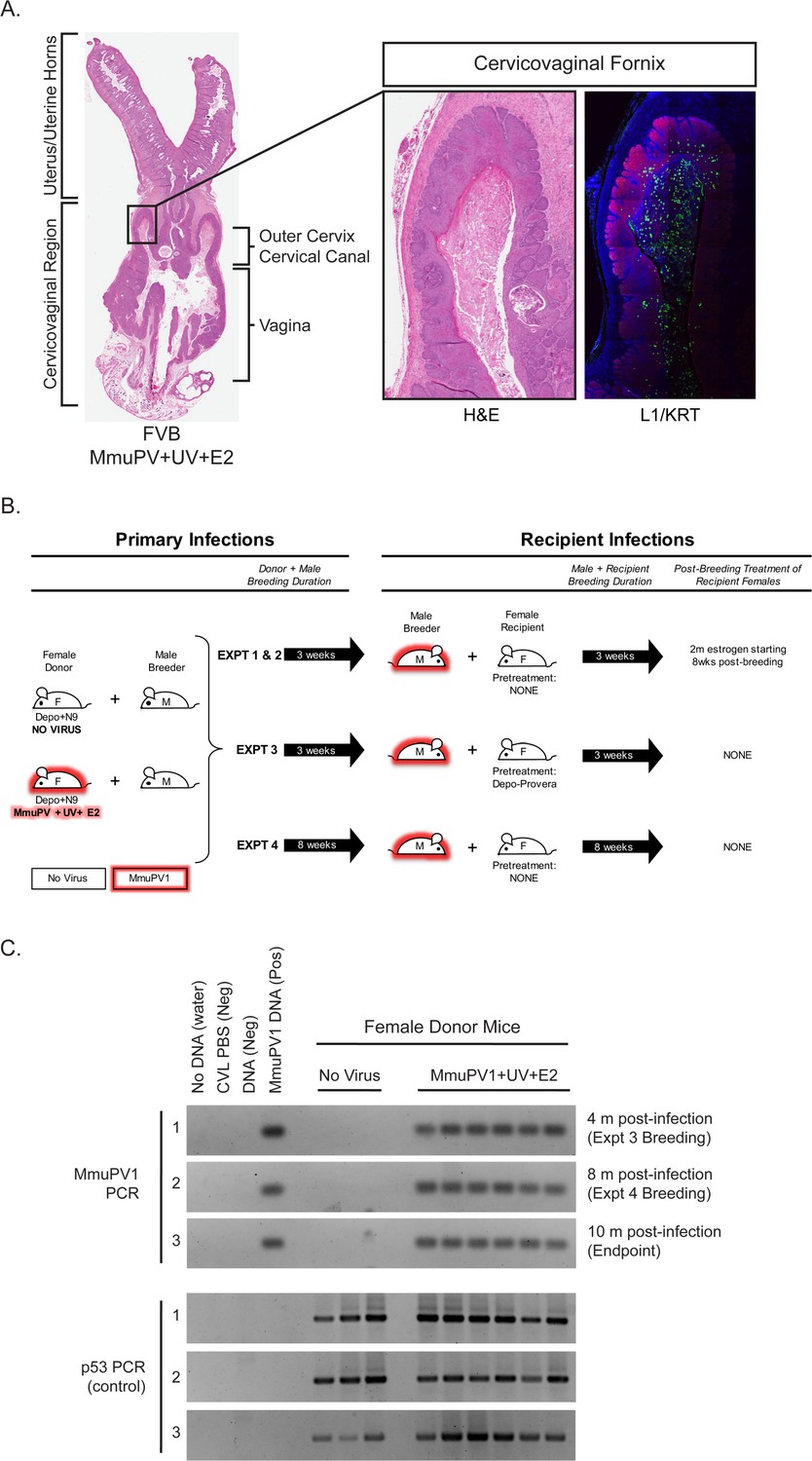
Rationale and experimental design for MmuPV1 sexual transmission studies.
(A) A full-slide scan of a representative H and E-stained female reproductive tract from a Donor infected for 4 months with MmuPV1+UV+E2 with anatomical regions labeled. On the right, higher magnification images of the cervicovaginal fornix (inset) stained with H and E (left) or immunofluorescence for keratin (KRT; red) and MmuPV1 L1 capsid protein (L1; green). (B) Schematic of MmuPV1 sexual transmission experimental design. Mice infected or potentially infected are indicated in red. (C) DNA was isolated from cervicovaginal lavage samples from a group of representative MmuPV1+UV+E2-infected females that were used as Donors in Experiments 3 and 4. Lavages were conducted at the onset of Experiment 3 (4 months post-infection), the onset of Experiment 4 (8 months post-infection) and Experiment four endpoint (10 months post-infection. DNA was analyzed by PCR for the MmuPV1 E2 gene (top) or for the p53 gene (bottom) to verify DNA presence/quality.
Figure 2
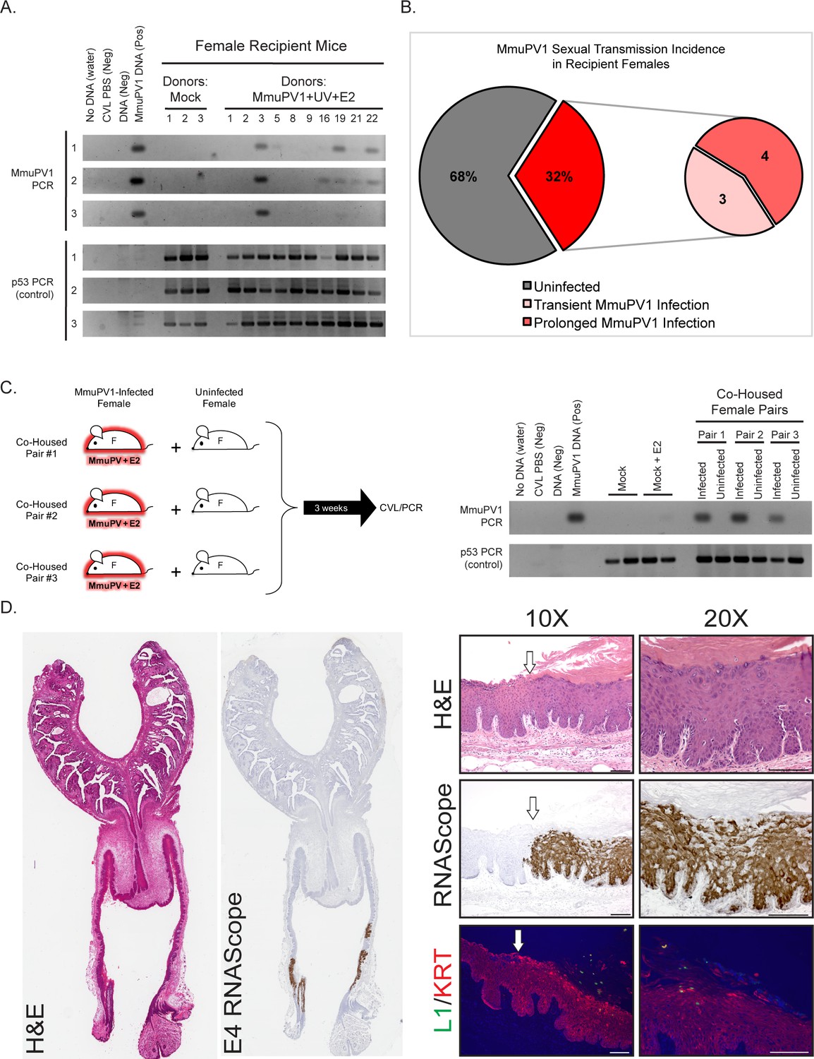
Evidence for sexual transmission: Assessment of MmuPV1 infection status in female Recipient reproductive organs.
(A) DNA was isolated from cervicovaginal lavage (CVL) samples collected from a group of representative Recipient female mice at three different occasions. The numerical mouse identifiers correspond to mice listed in Table 1. The three different CVL time points are as follows (time is listed in weeks following introduction to male Breeder): Mock Recipient mice #1–3 and Recipients #1–3 from Experiment 1 (CVL1: 6 weeks, CVL2: 13 weeks, CVL3: 17 weeks), Recipient mice #5, #8, and #9 from Experiment 2 (CVL1: 8 weeks, CVL2: 11 weeks, CVL3: 13 weeks), Recipient mouse #16 from Experiment 3* (CVL1: 3 weeks, CVL2: 4.5 weeks, CVL3: 9 weeks), and Recipient mice #19, #21, and #22 from Experiment 4 (CVL1: 4.5 weeks, CVL2: 7 weeks, CVL3: 9 weeks). DNA was analyzed by PCR for the MmuPV1 E2 gene (top) or for the p53 gene (bottom) to verify DNA presence/quality. (B) Incidence of MmuPV1 infection via sexual transmission in Recipient females as determined by CVL for MmuPV1 E2 gene. (C) Schematic of co-habitation study in which each co-housed pair consisted of an experimentally MmuPV1-infected female mouse and an uninfected female mouse. After 3 weeks of co-habitation, DNA isolated from cervicovaginal lavages was analyzed by PCR for the MmuPV1 E2 gene (top) or for the p53 gene (bottom) to verify DNA presence/quality. (D) Full-slide scans of the female reproductive tract harvested from Recipient Mouse #3 with a prolonged MmuPV1 infection as a result of sexual contact. Tissue is stained with H and E (left) or for the MmuPV1 E4 viral transcript using RNAscope (right). Higher magnification images of the infected regions of epithelia are shown stained with H and E (top), RNAscope for the MmuPV1 E4 transcript (middle), and the MmuPV1 L1 protein (green) and keratin 14 (red) by immunofluorescence (bottom). White arrow indicates junction between uninfected and MmuPV1-infected epithelia. All scale bars = 100 µM.
Figure 3
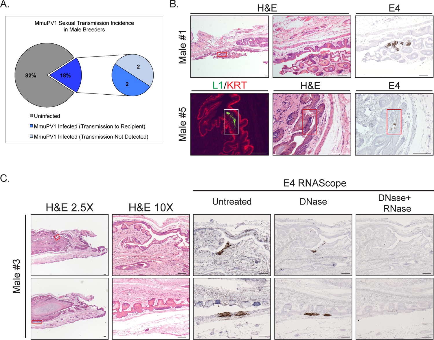
Male Breeders harbor infections in their reproductive organs.
(A) Incidence of MmuPV1 infection via sexual transmission in male Breeders as determined by staining tissue for MmuPV1 E4 transcript using RNAscope. (B) Various regions of the penis in MmuPV1-positive male Breeders stained with H and E or RNAscope for MmuPV1 E4 viral transcripts. Lower magnification images on the left for Male #1 include inset boxes (red) indicating the region staining positive for E4, which is shown on the far right. Higher magnification H and E-stained image of the region is shown in the center. Top: Male #1 (did not transmit to Recipient) with MmuPV1-positive region in the glans penile epithelium (red inset). Bottom: Male #5 (transmitted to Recipient) with MmuPV1-positive region in the glans penile epithelium stained for L1 (green) and K14 (KRT; red) (white inset; left), H and E (red inset; middle), and E4 by RNAscope (red inset; right). (C) Tissue from Male #3 (transmitted to Recipient #3 shown in Figure 2C) is stained with H and E (left two columns) or for the MmuPV1 E4 viral transcript using RNAscope (right) three panels). Lower magnification images on the left include inset boxes (red) indicating the region staining positive for E4. Higher magnification images of the infected regions of epithelia are shown stained with H and E and RNAscope for the MmuPV1 E4 transcript (bottom). In the RNAscope analysis, slides were left untreated, treated with DNase to remove any signal from viral DNA, and with DNase+RNase to verify signal is specific for viral RNA transcripts. Top: Male #3 (transmitted to Recipient) with MmuPV1-positive region in the mump ridge groove of the glans penis (red inset). Bottom: Male #3 (transmitted to Recipient) with MmuPV1-positive region in the prepuce (foreskin)/inner preputial space (red inset). All scale bars = 100 µM.
Tables
Table 1
Overview of MmuPV1 sexual transmission experiments and results
https://doi.org/10.7554/eLife.50056.004| Expt. | Experimental Conditions | Treatment of DONOR Female | DONOR pregnancy | Infection status of MALE BREEDER | Infection status of RECIPIENT female | RECIPIENT pregnancy (# if > 1) | # Positive CVL (total # of CVLs) in RECIPIENT Female |
|---|---|---|---|---|---|---|---|
| 1 | 3 weeks breeding; Recipients untreated prior to breeding, placed on E2 8 weeks after introduction of male. | No Virus #1 | Yes | Negative | Negative | Yes | 0 (4) |
| No Virus #2 | No | Negative | Negative | Yes | 0 (4) | ||
| No Virus #3 | Yes | Negative | Negative | Yes | 0 (4) | ||
| 3 | 3 weeks breeding; Recipients untreated prior to breeding. | No Virus #4 | Yes | Negative | Negative | Yes | 0 (3) |
| No Virus #5 | Yes | Negative | Negative | No | 0 (3) | ||
| No Virus #6 | Yes | Negative | Negative | No | 0 (3) | ||
| 4 | Prolonged Donor and Recipient breeding with male (8 weeks). Recipients untreated prior to breeding. | No Virus #7 | Yes | Negative | Negative | Yes (2) | 0 (4) |
| No Virus #8 | Yes | Negative | Negative | Yes (2) | 0 (4) | ||
| No Virus #9 | Yes | Negative | Negative | Yes (2) | 0 (4) | ||
| 1 | 3 weeks breeding, Recipients untreated prior to breeding, placed on E2 8 weeks after introduction of male. | MmuPV1+UV+E2 #1 | No | Positive | Negative | Yes | 0 (4) |
| MmuPV1+UV+E2 #2 | Yes | Negative | Negative | Yes | 0 (4) | ||
| MmuPV1+UV+E2 #3 | Yes | Positive | Positive (Prolonged) | Yes | 3 (4) | ||
| 2 | 3 weeks breeding, Recipients untreated prior to breeding, placed on E2 8 weeks after introduction of male. | MmuPV1+UV+E2 #4 | No | Negative | Negative | Yes | 0 (5) |
| MmuPV1+UV+E2 #5 | No | Positive | Positive (Transient) | Yes | 1 (5) | ||
| MmuPV1+UV+E2 #6 | No | Negative | Negative | Yes | 0 (5) | ||
| MmuPV1+UV+E2 #7 | No | Negative | Negative | Yes | 0 (5) | ||
| MmuPV1+UV+E2 #8 | No | Negative | Negative | Yes | 0 (5) | ||
| MmuPV1+UV+E2 #9 | Yes | Negative | Positive (Transient) | Yes | 1 (5) | ||
| 3 | 3 weeks breeding; Recipients untreated prior to breeding, not treated with E2. | MmuPV1+UV+E2 #10 | No | Positive | Negative | Yes | 0 (3) |
| MmuPV1+UV+E2 #11 | No | Negative | Negative | Yes (2) | 0 (3) | ||
| MmuPV1+UV+E2 #12 | Yes | Negative | Negative | Yes | 0 (3) | ||
| 3* | 3 weeks breeding; female Recipients treated with Depo-Provera 5d prior to breeding, not treated with E2. | MmuPV1+UV+E2 #13 | No | Negative | Negative | No | 0 (3) |
| MmuPV1+UV+E2 #14 | No | Negative | Negative | No | 0 (3) | ||
| MmuPV1+UV+E2 #15 | Yes | Negative | Negative | No | 0 (3) | ||
| MmuPV1+UV+E2 #16 | Yes | Negative | Positive (Prolonged) | No | 2 (3) | ||
| 4 | Prolonged Donor and Recipient breeding with male (8 weeks). Recipients untreated prior to breeding, not treated with E2. | MmuPV1+UV+E2 #17 | No | Negative | Negative | Yes (2) | 0 (4) |
| MmuPV1+UV+E2 #18 | No | Negative | Negative | Yes (2) | 0 (4) | ||
| MmuPV1+UV+E2 #19 | Yes | Negative | Positive (Prolonged) | Yes (3) | 3 (4) | ||
| MmuPV1+UV+E2 #20 | No | Negative | Negative | Yes | 0 (4) | ||
| MmuPV1+UV+E2 #21 | No | Negative | Positive (Transient) | Yes (3) | 1 (4) | ||
| MmuPV1+UV+E2 #22 | No | Negative | Positive (Prolonged) | Yes (2) | 2 (4) |
Key resources table
| Reagent type (species) or resource | Designation | Source or reference | Identifiers | Additional information |
|---|---|---|---|---|
| Strain, strain background | FVB/N | Taconic Biosciences | RRID:IMSR_TAC:fvb | Males (n = 31) Females (n = 31) |
| Strain, strain background | MmuPV1 | Joh et al., 2012 Uberoi et al., 2016 | GenBank: GU808564.1 | In-lab stock ‘AU 11/13’, pAU.4 |
| Antibody | Anti-MusPV1 L1 (rabbit polyclonal immune serum) | Chris Buck, NCI/NIH | IF (1:5000) | |
| Antibody | Anti-K14 (rabbit polyclonal) | BioLegend | Cat#905301; RRID:AB_2565048 | IF (1:1000) |
| Sequence-based reagent | MmuPV1_E2_1 | Hu et al., 2015 Cladel et al., 2017a Spurgeon et al., 2019 | PCR primers | GCCCGAAGACAACACCGCCACG |
| Sequenced-based reagent | MmuPV1_E2_2 | Hu et al., 2015 Cladel et al., 2017a Spurgeon et al., 2019 | PCR primers | CCTCCGCCTCGTCCCCAAATGG |
| Sequenced-based reagent | p53-1 | Spurgeon et al., 2019 | PCR primers | TATACTCAGAGCCGGCCT |
| Sequenced-based reagent | p53-2 | Spurgeon et al., 2019 | PCR primers | ACAGCGTGGTGGTACCTTAT |
| Sequenced-based reagent | p53-3 | Spurgeon et al., 2019 | PCR primers | TCCTCGTGCTTTACGGTATC |
| Sequenced-based reagent | MusPV-E4 | Xue et al., 2017 | RNAscope probe Cat#473281 | |
| Commercial assay or kit | RNAscope 2.5 HD Detection Kit Brown | ACDBio | Cat# 322300 | |
| Chemical compound, drug | Tyramide signal amplification (TSA)-related reagents | Online protocol: https://doi.org/10.17504/protocols.io.i8cchsw | ||
| Chemical compound, drug | medroxyprogesterone acetate | Amphastar Pharmaceuticals | Depo-Provera | 3 mg/animal, subcutaneous injection |
| Chemical compound, drug | Nonoxynol-9 (4%) | Options Conceptrol | Cat#247149 | 50 µl/mouse, intravaginal |
| Chemical compound, drug | Carboxyl methylcellulose (4%) | Sigma Aldrich | Cat# C4888 | 25 µl/mouse, intravaginal |
| Chemical compound, drug | 17β-estradiol pellet, 0.05 mg/60 days | Innovative Research of America | Cat#SE-121 | 0.05 mg/60 days, subcutaneous pellet |
| Other | Hematoxylin QS | Vector | Cat#H-3404 | Counterstain |
| Other | Shandon Instant Hematoxylin | Thermo Fisher | Cat#6765015 | H and E stain |
| Other | Eosin | Sigma Aldrich | Cat#E4382 | H and E stain |
| Other | DNase I | Thermo Fisher Scientific | Cat#EN0521 | 20 units/sample |
| Other | RNase A | Qiagen | Cat#1006657 | 500 µg/sample |
| Other | RNase T1 | Fermentas | Cat#EN0542 | 2000 units/sample |
Additional files
-
Transparent reporting form
- https://doi.org/10.7554/eLife.50056.007
Download links
A two-part list of links to download the article, or parts of the article, in various formats.
Downloads (link to download the article as PDF)
Open citations (links to open the citations from this article in various online reference manager services)
Cite this article (links to download the citations from this article in formats compatible with various reference manager tools)
Sexual transmission of murine papillomavirus (MmuPV1) in Mus musculus
eLife 8:e50056.
https://doi.org/10.7554/eLife.50056


