New light shed on the early evolution of limb-bone growth plate and bone marrow
- Department of Organismal Biology, Evolution and Development, Uppsala University, Sweden
- European Synchrotron Radiation Facility, France
- Flinders University, College of Science and Engineering, Australia
- Comenius University in Bratislava, Faculty of Natural Sciences, Department of Ecology, Slovakia
Figures
Figure 1
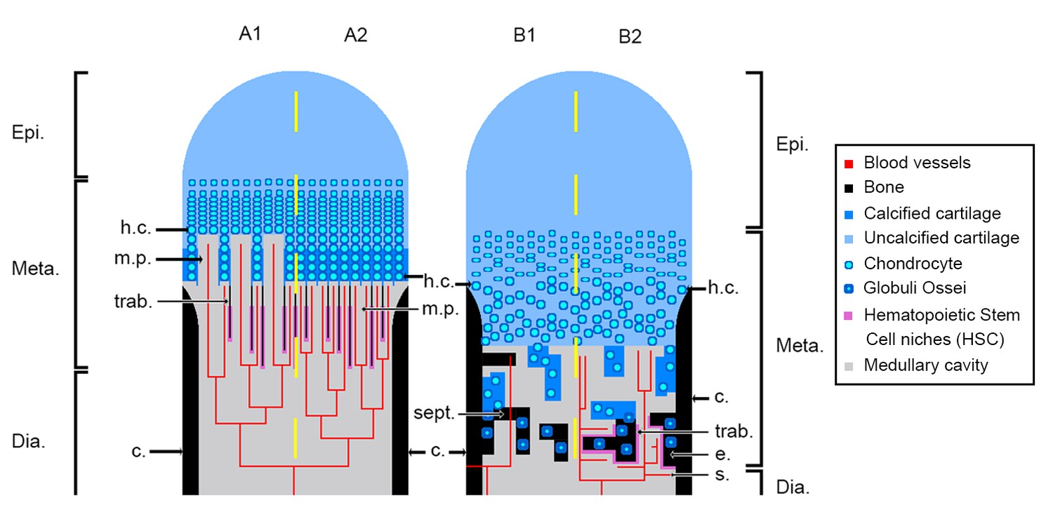
Schematic drawing of the long-bone epiphyses of extant amniotes (A) and amphibians (B).
Four conditions are figured here. They are separated by yellow dashed lines: A1, condition in crocodiles (interpreted from Haines, 1938); A2, condition in mammals at an early developmental stage before the appearance of the secondary ossification centre (Anderson and Shapiro, 2010; Tanaka, 1976); B1, condition in Triturus (Cynops) pyrrhogaster (Quilhac et al., 2014; Tanaka, 1976); B2, condition in Rana catesbeiana (Francillon, 1981; Tanaka, 1976). Abbreviations: c., cortex; Dia., diaphysis; e., endosteal bone; Epi., epiphysis; h.c., hypertrophied chondrocytes; Meta., metaphysis; m.p., marrow process; s., sinusoids; sept., septum; trab., trabeculae.
Figure 2

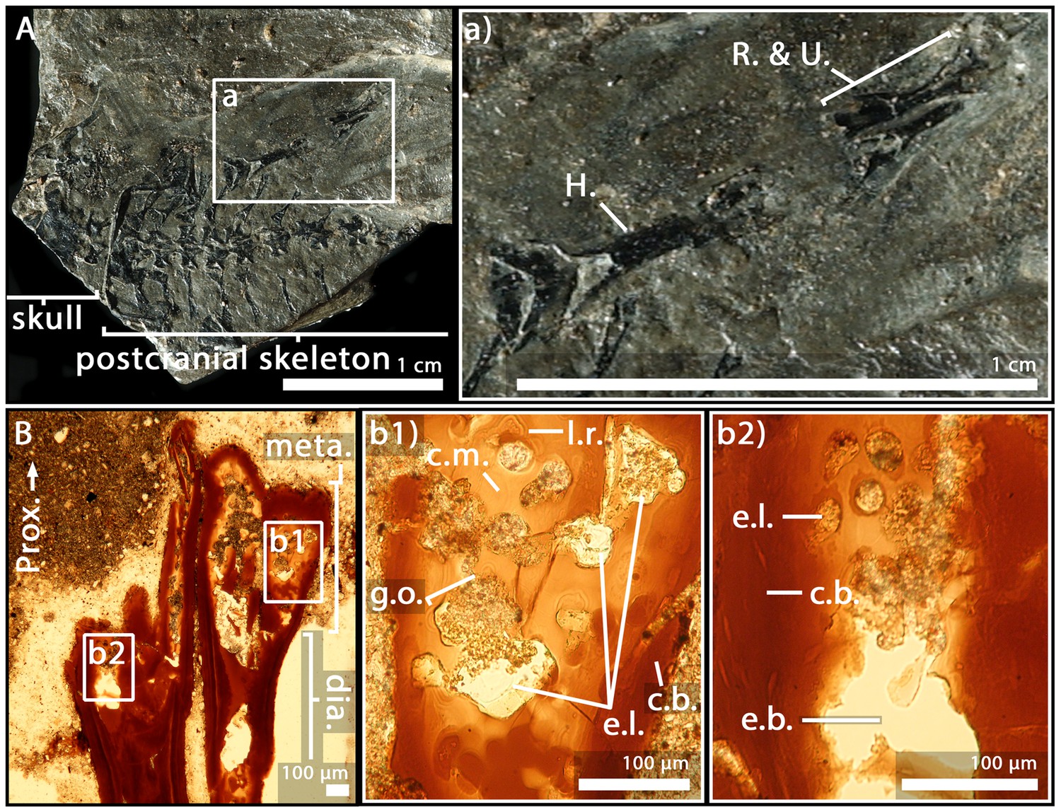
Juvenile specimen of Apateon caducus, GPIM-N 1297.
(A) Skeleton. (a) Right limb. (B) Epiphyseal and metaphyseal histology of the proximal end of the humerus. (C) Epiphyseal and metaphyseal histology of the proximal end of the radius (c2-3) and ulna (c1). Abbreviations: c.b., cortical bone; c.c., cluster of chondrocytes; c.f., calcification front; c.m., cartilage matrix; dia., diaphysis; e.b., erosion bay; e.l., erosion lacunae; g.o., globuli ossei; H., humerus; l.r., Liesegang’s rings; meta., metaphysis; m.f., mineralisation front; o.n., ossification notch; Prox., proximal end; R. and U., radius and ulna; t., trabeculae.
Figure 3
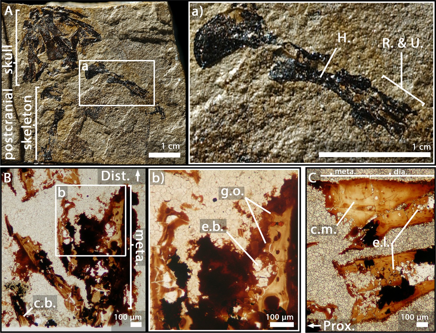
Adult specimen of Apateon caducus, GPIM-N 1572.
(A) Skeleton. (a) Right limb. (B) Epiphyseal and metaphyseal histology of the proximal end of the radius (b1) and ulna (b2). Abbreviations: c.b., cortical bone; c.m., cartilage matrix; dia., diaphysis; e.b., erosion bay; e.l., erosion lacunae; g.o., globuli ossei; H., humerus; l.r., Liesegang’s rings; meta., metaphysis; Prox., proximal end; R. and U., radius and ulna.
Figure 4

Adult specimen of Apateon pedestris, SMNS 54981.
(A) Skeleton. (a) Right limb. (B) Epiphyseal and metaphyseal histology of the distal end of the humerus. (C) Epiphyseal and metaphyseal histology of the proximal end of the radius (c2) and ulna (c1). Abbreviations: c.b., cortical bone; c.c.t., calcified-cartilage trabecula; Dist., distal end; e.b., erosion bay; g.o., globuli ossei; H., humerus; l.r., Liesegang’s rings; meta., metaphysis; m.f., mineralisation front; m.t., mineralised trabecula; Prox., proximal end; R. and U., radius and ulna.
Figure 5
Adult specimen of Apateon pedestris, SMNS 54988.
(A) Skeleton. (a) Right limb. (B) Epiphyseal and metaphyseal histology of the distal end of the humerus. (C) Epiphyseal and metaphyseal histology of the proximal end of the radius and ulna. Abbreviations: c.b., cortical bone; c.m., cartilage matrix; dia., diaphysis; Dist., distal end; e.b., erosion bay; e.l., erosion lacunae; g.o., globuli ossei; H., humerus; meta., metaphysis; Prox., proximal end; R. and U., radius and ulna.
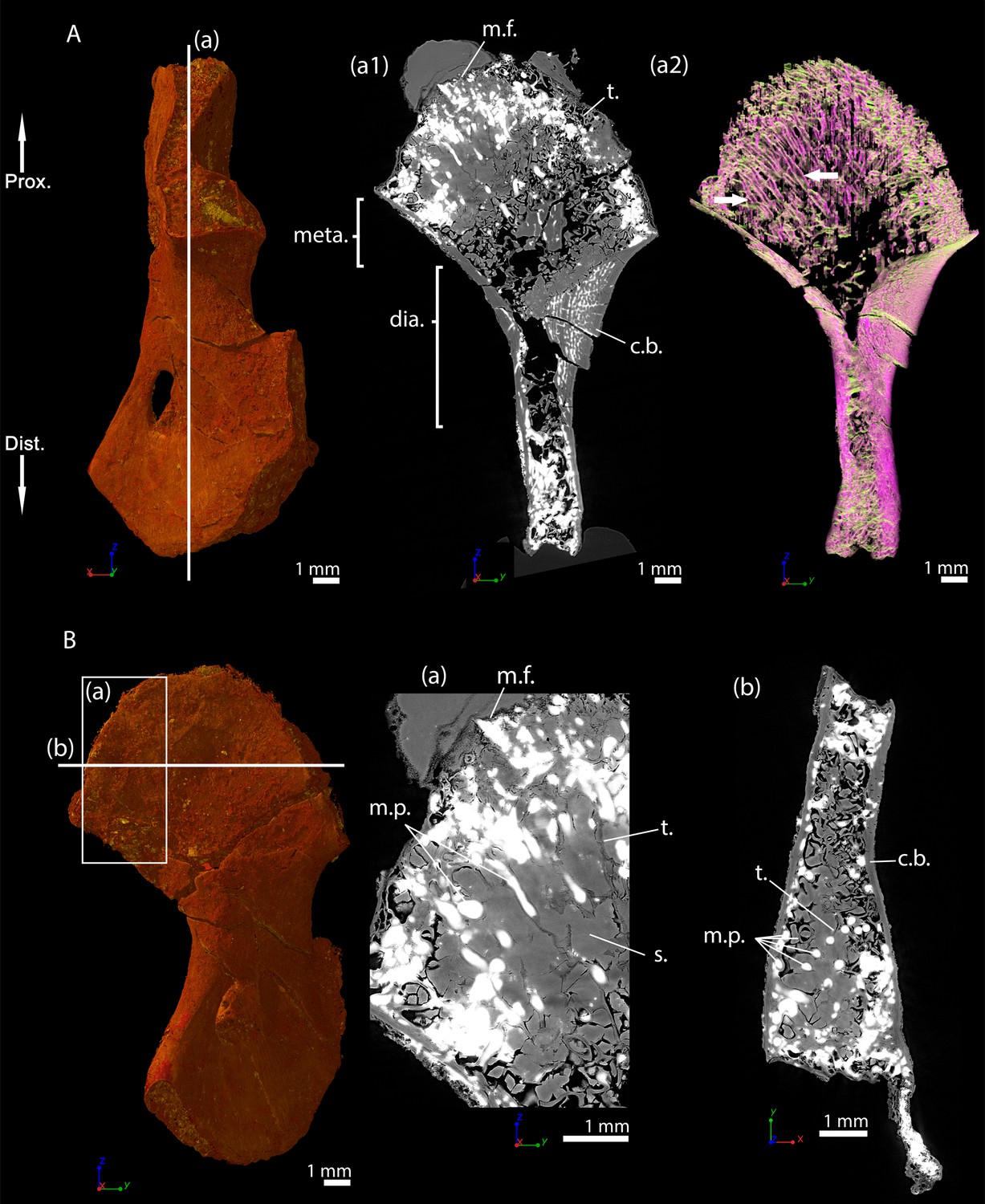
Figure 6
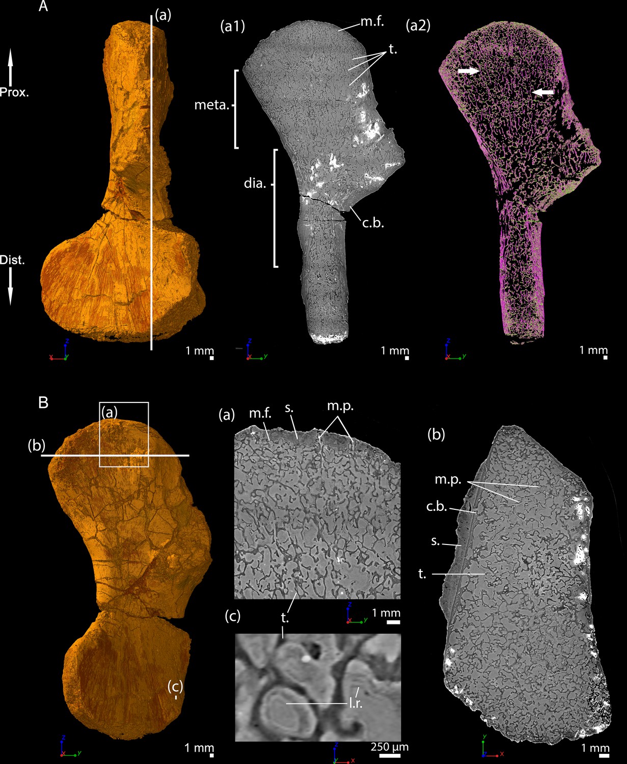
Left humerus of a (sub-)adult specimen of Metoposaurus sp., MUZ PGI OS-220/171 imaged using PPC-SRµCT.
(A) Frontal view. (a1) Longitudinal virtual thin section (40 µm thick) and (a2) longitudinal virtual thin section of the segmented model of the bone (50 µm thick). The longitudinally-oriented trabeculae are highlighted in purple (white arrows), while the transversally-oriented trabeculae appear in green. (B) Ventral view. (a) Longitudinal virtual thin section of the proximal metaphysis (40 µm thick), (b) transverse virtual thin section made in the metaphysis and (c) longitudinal thin section made in the distal metaphysis. Abbreviations: c.b., cortical bone; dia., diaphysis; Dist., distal end; l.r., Liesegang’s rings; meta., metaphysis; m.f., mineralisation front; m.p., marrow process; Prox., proximal end; s., sediment; t., trabeculae.
Figure 7
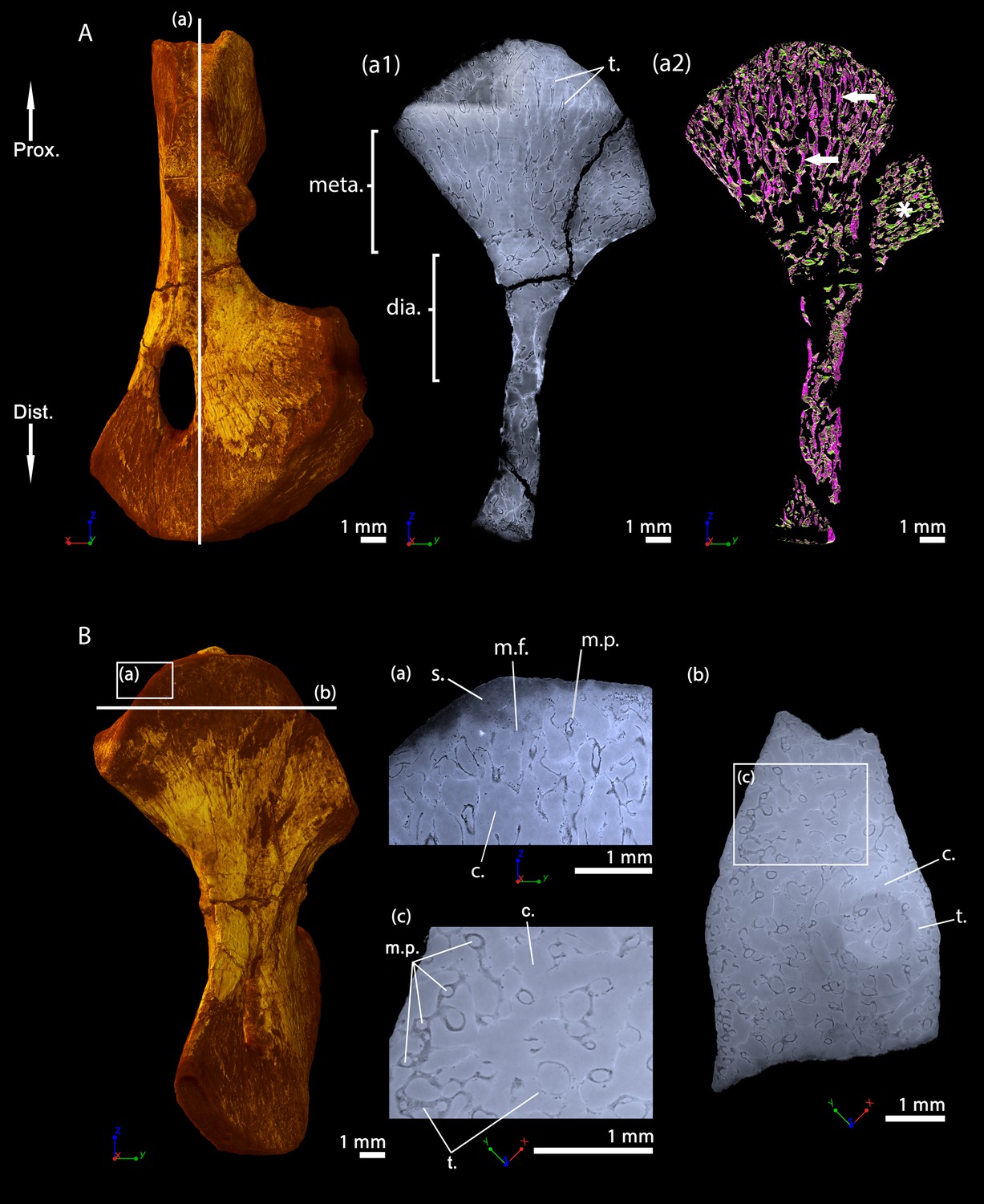
Left humerus of a juvenile specimen of Seymouria sanjuanensis, MNG 7747 imaged using PPC-SRµCT.
(A) Frontal view. (a1) Longitudinal virtual thin section (40 µm thick), the darker part is an artefact in the original data due to electron reinjection in the synchrotron storage ring (refilling) during the scan and (a2) longitudinal virtual thin section of the segmented model of the bone (250 µm thick). The longitudinally-oriented trabeculae (pointed by horizontal arrows) are highlighted in purple, while the transversally-oriented trabeculae appear in green. Note that, due to the shape of the metaphysis, the trabeculae exhibit an overall fan-like configuration which progressively tilts to 90 degrees at the location of the deltopectoral crest (Asterisk). For that reason, the longitudinal trabeculae appear green and the transverse trabeculae appear purple at this location. (B) Ventral view. (a) Longitudinal virtual thin section in the proximal metaphysis (40 µm thick), (b) transverse virtual thin section in the metaphysis, the large ring artefact results from the synchrotron electron refilling visible in a1, (c) detail of (b) showing marrow processes and cavities in transverse section. Abbreviations: c., cavity; dia., diaphysis; Dist., distal end; meta., metaphysis; m.f., mineralisation front; m.p., marrow process; Prox., proximal end; s., sediment; t., trabeculae.
Figure 8 with 1 supplement

Right humerus of an adult Seymouria sanjuanensis, CM 28597 imaged using PPC-SRµCT.
(A) Frontal view. (a1) Longitudinal virtual thin section (40 µm thick) and (a2) longitudinal section of the segmented model of the bone (450 µm thick). The longitudinally-oriented trabeculae (pointed by horizontal arrows) are highlighted in purple, while the transversally-oriented trabeculae appear in green. Note that, due to the shape of the metaphysis, the trabeculae exhibit an overall fan-like configuration which progressively tilts to 90 degrees at the location of the deltopectoral crest (Asterisk). For that reason, the longitudinal trabeculae appear green and the transverse trabeculae appear purple at this location. (B) Dorsal view. (a) Longitudinal virtual thin section of the proximal metaphysis (40 µm thick), (b) transverse virtual thin section in the metaphysis (40 µm thick), (c) detail of (b) showing marrow processes and cavities in transverse section. Abbreviations: c., cavity; dia., diaphysis; Dist., distal end; meta., metaphysis; m.f., mineralisation front; m.p., marrow process; Prox., proximal end; s., sediment; t., trabeculae.
Figure 8—figure supplement 1
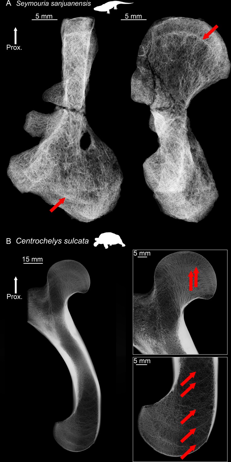
Humeral microanatomical architecture of the stem amniote Seymouria sanjuanensis (CM 28597) and the tortoise Centrochelys sulcata.
(A) Radiography of the humerus of S. sanjuanensis in anterior view (on the left) and ventral view (on the right). The red arrows show the presence of a resting surface both in the proximal and distal metaphyses. (B) Virtual 5 mm-thick section made in the humerus of C. sulcata. Both metaphyses are zoomed in (as 2-mm-thick sections) to show the presence of multiple resting surfaces (red arrows). Abbreviations: Prox., proximal end.
Figure 9
Right humerus of a subadult Discosauriscus austriacus, SNM Z 15568 imaged using PPC-SRµCT.
Due to processing to convert the scan data into a stack of images, the images have been flipped, thereby resulting in a flipped 3D model. (A) Frontal view. (a1) Longitudinal virtual thin section (40 µm thick) and (a2) longitudinal section of the segmented model of the bone (160 µm thick). The longitudinally-oriented trabeculae are highlighted in purple, while the transversally-oriented trabeculae appear in green. (B) Ventral view. (a) Longitudinal virtual thin section of the proximal metaphysis (40 µm thick) and (b) transverse virtual thin section in the proximal metaphysis (40 µm thick). Abbreviations: c.b., cortical bone; dia., diaphysis; Dist., distal end; meta., metaphysis; m.f., mineralisation front; m.p., marrow process; Prox., proximal end; s., sediment; t., trabeculae.
Figure 10 with 1 supplement
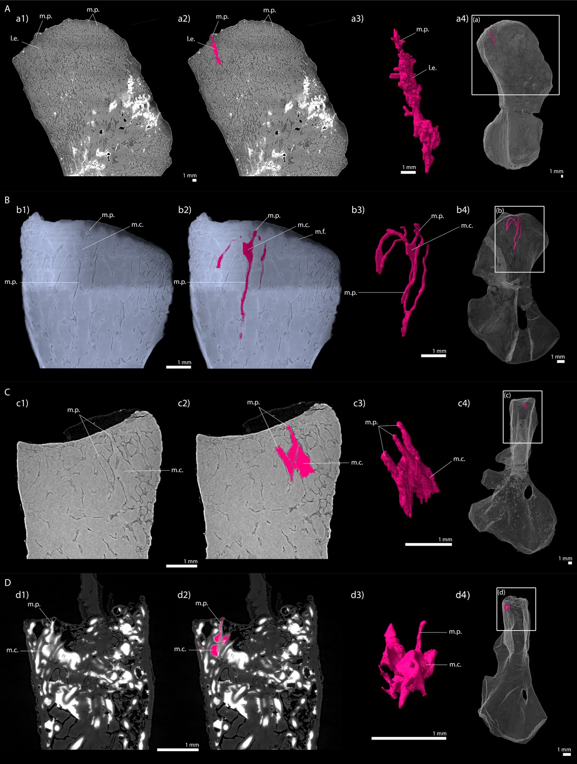
Longitudinal virtual sections and three-dimensional (3D) segmentation from PPC-SRµCT of marrow processes and marrow cavities in the humeral proximal ends of: A, Metoposaurus sp.
(MUZ PGI OS-220/171); B, Seymouria sanjuanensis (MNG 7747); C, S. sanjuanensis (CM 28597); D, Discosauriscus austriacus (SNM Z 15568). (a1, b1, c1, d1) Longitudinal virtual thin section (60 µm thick); (a2, b2, c2, d2) marrow processes and cavities segmented; (a3, b3, c3, d3) 3D models of the segmentations. Note that the marrow cavities have not been completely segmented in 3D to allow the full visualisation of the marrow processes; (a4, b4, c4, d4) respective locations of a3, b3, c3, d3 in the humeri. Abbreviations: l.e., region of local erosion; m.c., marrow cavity; m.f., mineralisation front; m.p., marrow process.
Figure 10—figure supplement 1
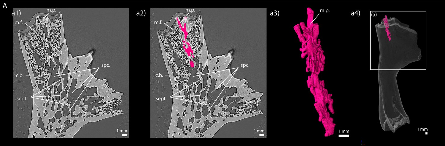
Longitudinal virtual sections and three-dimensional (3D) segmentation from PPC-SRµCT of marrow processes in the humeral proximal end of (A) Andrias sp.
(MNHN-ZA-AC-2005–72, Museum national d’Histoire naturelle, Paris, France). (a1) Longitudinal virtual thin section (60 µm thick), (a2) marrow processes segmented, (a3) 3D model of the segmentation and (a4) location of a3 in the humerus. The humerus of Andrias has been imaged using the protocol published by Sanchez et al., 2014. Abbreviations: c.b., cortical bone; m.f., mineralisation front; m.p., marrow process; sept., septa; spc., spaces.
Figure 11
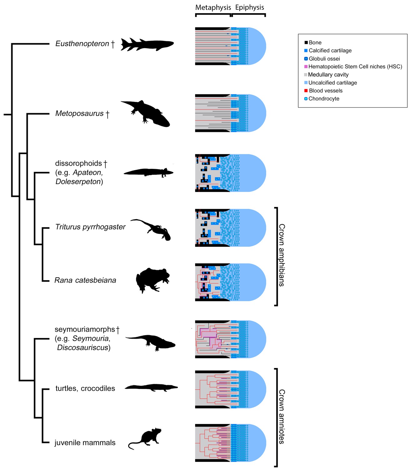
Evolution of growth-plate patterns and metaphyseal organisations of the long bones of the studied taxa in a phylogenetic context (e.g., Ruta and Coates, 2007; Schoch, 2019).
Hypothesis on haematopoietic activity is herein contextualised. Black silhouettes represent the taxa studied. Crosses (†) have been attributed to fossil taxa.
Figure 12
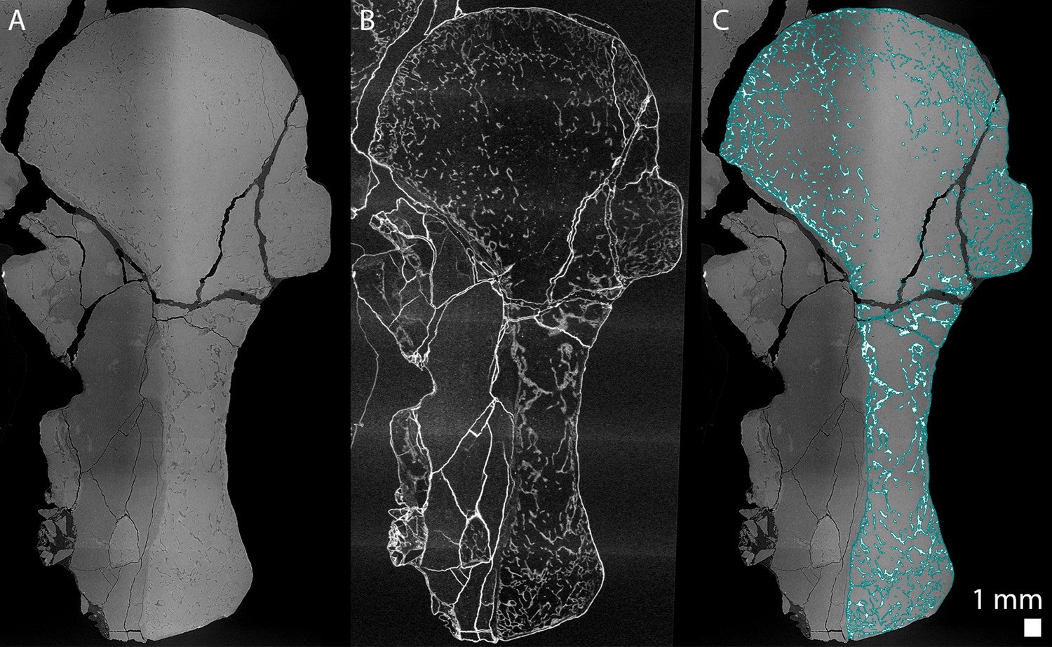
Longitudinal virtual thin sections of the right humerus of an adult Seymouria sanjuanensis (CM 28597).
(A) Tomogram showing low frequency artefacts resulting in an image divided into a darker and brighter part, (B) image processed with a filter for tomographic texture enhancement to remove low frequency artefacts (Cau et al., 2017) and (C) overlap of A and the bone segmentation with the aid of B used to produce Figure 8Aa2. The segmentation is highlighted by the blue line in C.
Tables
Table 1
Table summarising the material used.
Skull length measurements and ontogenetic stages determined by Berman et al., 1987b; Sanchez et al., 2008; Sanchez et al., 2010a and Klembara et al., 2006.
| Species | Collection number | Skull length (cm) | Ontogenetic stage | Bone |
|---|---|---|---|---|
| Apateon caducus | GPIM-N 1297 | 1.52 | Juvenile | Humerus |
| Radius | ||||
| Ulna | ||||
| GPIM-N 1572 | Estimated to 1.60 | Adult | Radius | |
| Ulna | ||||
| Apateon pedestris | SMNS 54981 | 0.86 | Adult | Humerus |
| Radius | ||||
| Ulna | ||||
| SMNS 54988 | 1.06 | Adult | Humerus | |
| Radius | ||||
| Ulna | ||||
| Seymouria sanjuanensis | MNG 7747 | 5.6 | Juvenile | Humerus |
| CM 28597 | 8.8 | Adult | Humerus | |
| Discosauriscus austriacus | SNM Z 15568 | 6.2 | Subadult | Humerus |
| Metoposaurus sp. | MUZ PGI OS-220/171 | - | Subadult or adult | Humerus |
Table 2
Microanatomical measurements made on the samples using VGStudio MAX (version 3.2, Volume Graphics Inc, Germany).
The protocol details are provided by Estefa et al., 2020.
| Species | Thickness of the trabeculae (µm) | Diameter of the marrow processes (µm) | |
|---|---|---|---|
| Diaphysis | Metaphysis | Metaphysis | |
| Metoposaurus sp. (Subadult or Adult, MUZ PGI OS-220/171) | 131 | 117 | 248 |
| Seymouria sanjuanensis (Juvenile, MNG 7747) | 94 | 25 | 100 |
| S. sanjuanensis (Adult, CM 28597) | 79 | 30 | 100 |
| Discosauriscus austriacus (Subadult, SNM Z 15568) | 80 | 54 | 111 |
Additional files
Download links
A two-part list of links to download the article, or parts of the article, in various formats.
Downloads (link to download the article as PDF)
Open citations (links to open the citations from this article in various online reference manager services)
Cite this article (links to download the citations from this article in formats compatible with various reference manager tools)
New light shed on the early evolution of limb-bone growth plate and bone marrow
eLife 10:e51581.
https://doi.org/10.7554/eLife.51581


