USP16 counteracts mono-ubiquitination of RPS27a and promotes maturation of the 40S ribosomal subunit
- Institute of Biochemistry, ETH Zurich, Switzerland
- Molecular Life Sciences Ph.D. Program, Switzerland
- Institute of Molecular Systems Biology, ETH Zurich, Switzerland
Figures
Figure 1
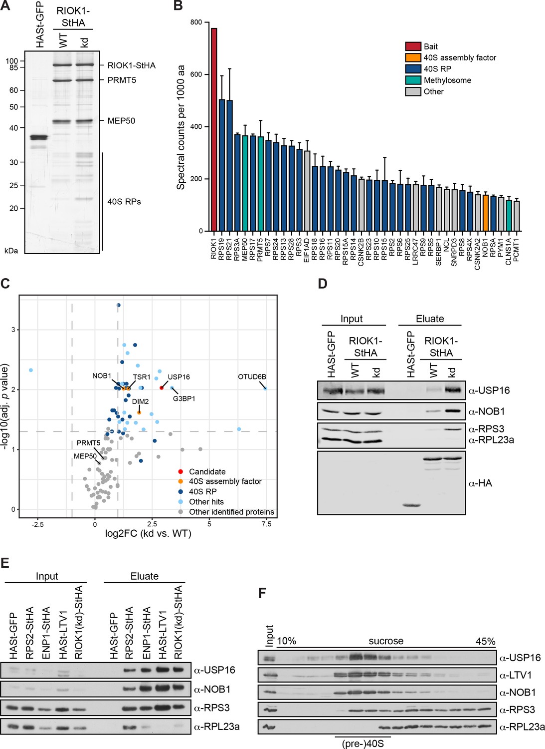
USP16 is a pre-40S associated factor.
(A) StrepTactin affinity purification of HASt-GFP, wild-type (WT) and kinase-dead (kd) RIOK1-StHA from HEK293 cell lysates. Eluates were analyzed by SDS-PAGE and silver staining or mass spectrometry. (B) Proteomic analysis of three independent RIOK1(kd)-StHA StrepTactin affinity purifications. The plot shows spectral counts (mean ± SD) of the 40 top identified hits after filtering against the HASt-GFP control and after normalization to spectral counts of the bait protein as well as to the size of the protein in amino acids (aa). Data before and after normalization are shown in Supplementary file 2. (C) Proteomic analysis of three independent experiments as in (A). The plot shows the log2 fold change (log2FC) of the average number of spectral counts of proteins identified in the RIOK1(kd)-StHA versus the RIOK1(WT)-StHA pull down against the negative log10 of the adjusted p value. Proteins with a fold change <2 and an adjusted p value > 0.05, demarcated by dashed lines, were considered nonsignificant. (D) StrepTactin pull-downs of HASt-GFP, RIOK1(WT)- and RIOK1(kd)-StHA from HEK293 cell lysates. Eluates were analyzed by immunoblotting using the indicated antibodies. Load corresponds to 0.05% of the input and 20% of the eluates. (E) StrepTactin pull-downs of HASt-GFP, RPS2-StHA, ENP1-StHA, HASt-LTV1, and RIOK1(kd)-StHA from HEK293 cell lysates. Eluates were analyzed by immunoblotting using the indicated antibodies. Load corresponds to 0.05% of the input and 20% of the eluates. (F) HEK293 cell extract was separated on a linear 10–45% sucrose gradient by centrifugation. Input and gradient fractions were analyzed by immunoblotting using the indicated antibodies.
Figure 2
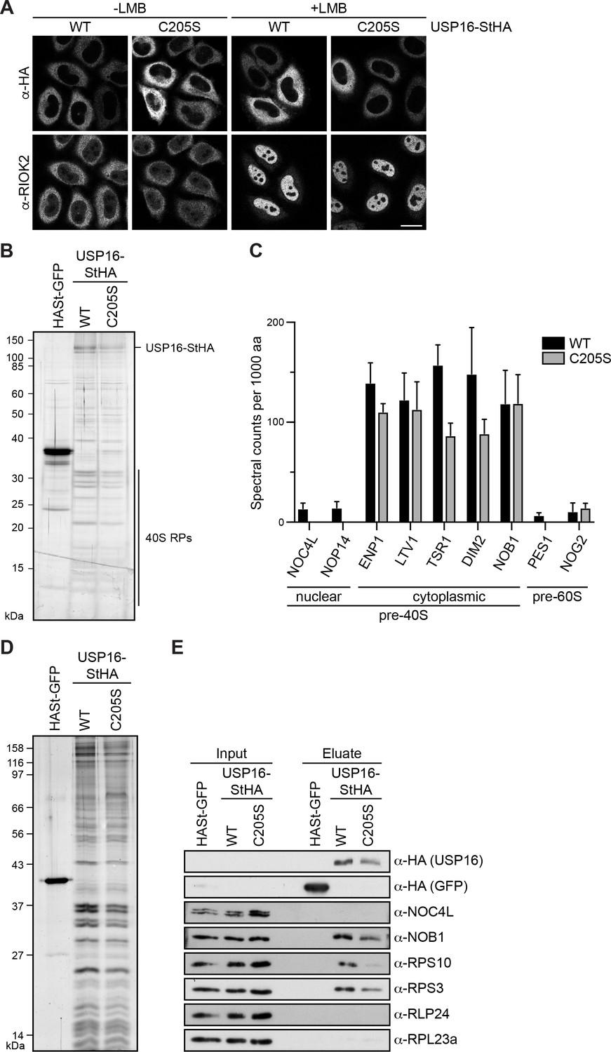
USP16 is associated with cytoplasmic pre-40S subunits.
(A) USP16(WT)- and USP16(C205S)-StHA constructs were transiently transfected into HeLa cells and their localization was analyzed by immunostaining using an HA antibody 24 hr after transfection. CRM1-mediated nuclear export was inhibited by treatment with leptomycin B (LMB; 20 nM, 120 min) as indicated. Immunostaining of the 40S trans-acting factor RIOK2 served as positive control for the LMB treatment. Scale bar, 20 µm. (B) One-step StrepTactin affinity purification of HASt-GFP, wild-type (WT) and catalytically inactive (C205S) USP16-StHA from HEK293 cell lysates. Eluates were analyzed by SDS-PAGE and silver staining or mass spectrometry. (C) Proteomic analysis of three independent experiments as in (B). The plot shows spectral counts (mean ± SD) of identified candidate proteins normalized to spectral counts of the bait protein as well as to the size of the protein in amino acids (aa). Candidate proteins are categorized into 40S and 60S trans-acting factors. (D) Tandem affinity purification (TAP) from HEK293 cells expressing HASt-GFP, USP16(WT)- or USP16(C205S)-StHA. Eluates were analyzed by SDS-PAGE and silver staining. (E) Immunoblot analysis of (D) using the indicated antibodies against the bait (α-HA), 40S (NOB1, NOC4L) and 60S (RLP24) trans-acting factors, and RPs. Load corresponds to 0.05% of the input and to 20% of the eluates.
Figure 3
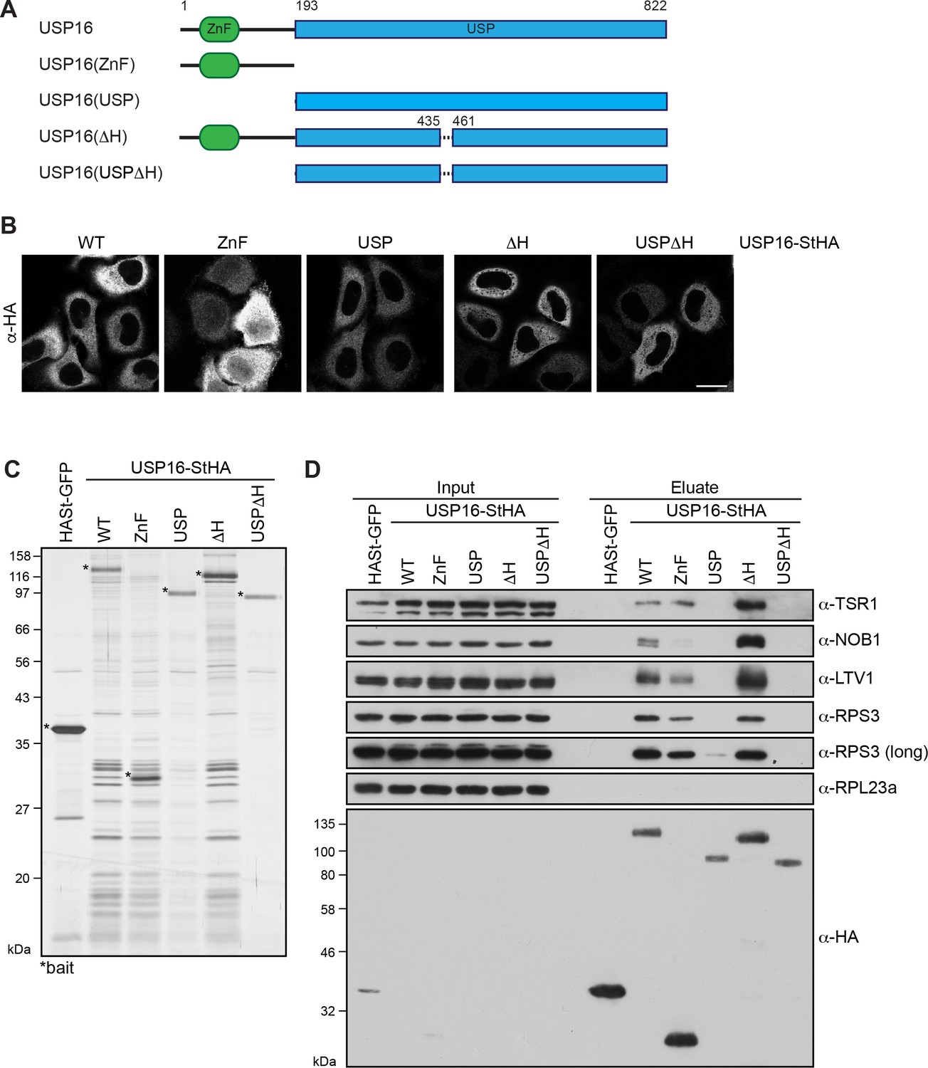
Pre-40S association of USP16 depends on its ZnF-UBP domain and a USP16-specific insertion in the USP domain.
(A) Scheme of protein domains present in human USP16 and generated deletion constructs. USP16 contains an N-terminal zinc-finger ubiquitin binding domain (ZnF-UBP) domain and a C-terminal ubiquitin-specific protease (USP) domain. The amino acid numbers indicate the positions of the truncations, dashed lines represent deletions. (B) C-terminally StHA-tagged USP16 constructs, depicted in (A), were transiently transfected into HeLa cells and their localization was analyzed by immunostaining using an HA antibody 24 hr after transfection. Scale bar, 20 µm. (C) TAP from HEK293 cell lines expressing HASt-GFP or the USP16 constructs depicted in (A). Eluates were analyzed by SDS-PAGE and silver staining. Bait proteins are marked with an asterisk (*). (D) Immunoblot analysis of experiment in (C) using the indicated antibodies against 40S trans-acting factors, ribosomal proteins or the bait (α-HA). Two different exposures are shown for the RPS3 immunoblot. Load corresponds to 0.05% of the input and to 20% of the eluates.
Figure 4 with 1 supplement
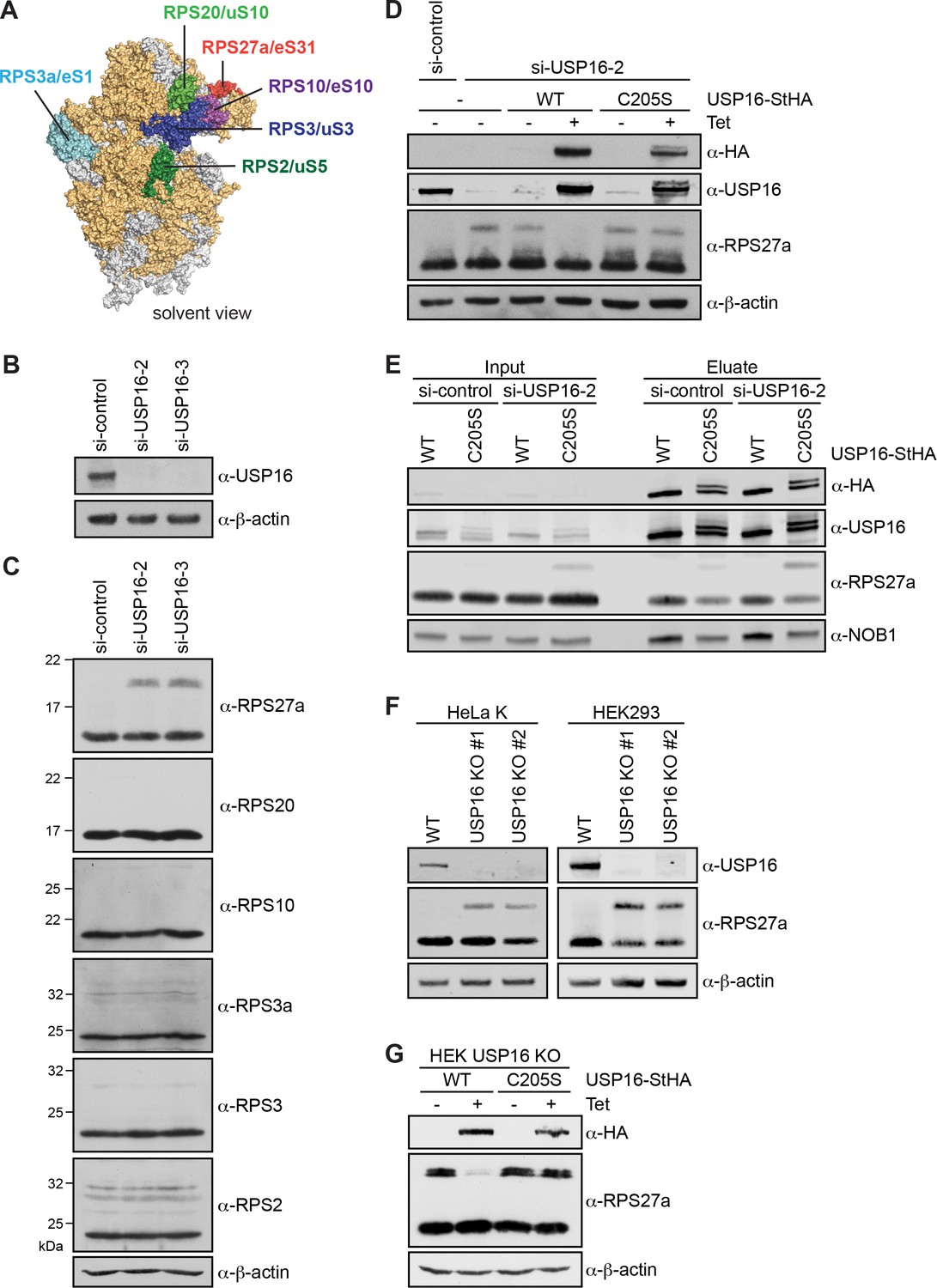
Depletion of USP16 leads to accumulation of modified RPS27a.
(A) Structural model of the human 40S subunit shown from the solvent side (adapted from Khatter et al., 2015, PDB ID: 4UG0). Ribosomal proteins analyzed for mono-ubiquitination are highlighted, others RPs are shown in orange and 18S rRNA is shown in gray. (B) HeLa cells were treated with either control siRNA (si-control) or two different siRNAs against USP16 for 72 hr. To control for downregulation of USP16, cell extracts were analyzed by immunoblotting using a USP16 antibody. (C) Immunoblot analysis of cell extracts from (A) using the indicated antibodies against RPs of the 40S subunit. (D) Parental HEK293 cells or HEK293 cells expressing USP16(WT)- or USP16(C205S)-StHA were treated with a control siRNA or an siRNA targeting the 3’UTR of the USP16 mRNA for 72 hr. Cells expressing USP16(WT)- or USP16(C205S)-StHA were either left uninduced (-Tet) or induced (+Tet) with tetracycline (0.5 µg/ml) for the last 48 hr of the siRNA treatment. Cell extracts were analyzed by immunoblotting using the indicated antibodies. (E) StrepTactin pull-down from lysates of HEK293 cells that had been transfected with either a control or a USP16 siRNA 72 hr before harvest and induced to express either USP16(WT)- or USP16(C205S)-StHA (0.5 µg/ml tetracycline) for the last 48 hr of the siRNA treatment. Inputs and eluates were analyzed by immunoblotting using the indicated antibodies. Note that the USP16(C205S)-StHA protein is potentially modified as it runs as two bands in SDS-PAGE. Load corresponds to 0.05% of the input and to 20% of the eluates. (F) USP16 knockout (KO) cell lines were generated using the CRISPR/Cas9 system in HeLa and HEK293 backgrounds using two different guide RNAs targeting exon 3 (KO #1) or exon 5 (KO #2) of USP16, respectively. Cell extracts were analyzed by immunoblotting using the indicated antibodies. (G) HEK293 USP16 KO cells expressing USP16(WT)- or USP16(C205S) were either left uninduced (-Tet) or induced (+Tet) with tetracycline (0.5 µg/ml) for 24 hr. Cell extracts were analyzed by immunoblotting using the indicated antibodies. Note that monoubiquitinated RPS27a sometimes runs as a double band in SDS-PAGE.
Figure 4—figure supplement 1
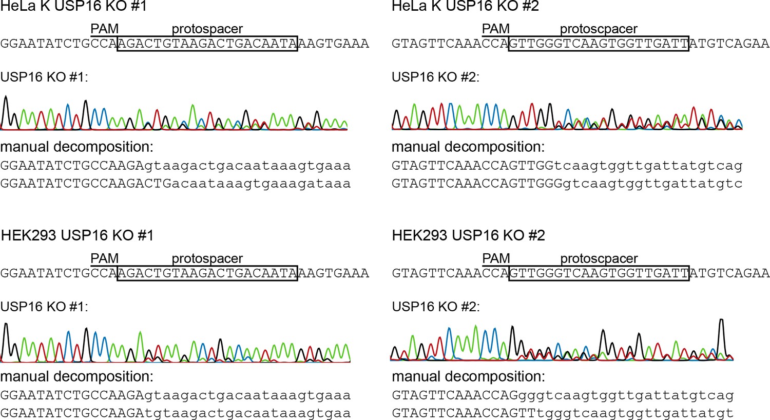
Validation of USP16 knockout cell lines generated by CRISPR/Cas9.
Genomic sequences of the USP16-/- isogenic HeLa and HEK293 KO clones generated in this study using two different guide RNAs targeting different exons (exon three for KO #1 and exon five for KO #2). Sanger sequencing chromatograms show indel mutations in the USP16 gene targeted by CRISPR. The top sequence indicates the WT sequence of the target region and the location of the protospacer and the protospacer adjacent motif (PAM). The overlapping peaks in the chromatogram of the sequenced clone indicate the presence of two different indel mutations in the two copies of the USP16 gene. Manual decomposition of the chromatogram shows the two resulting sequences in the target region and confirms frame-shift indels in both alleles.
Figure 5 with 1 supplement
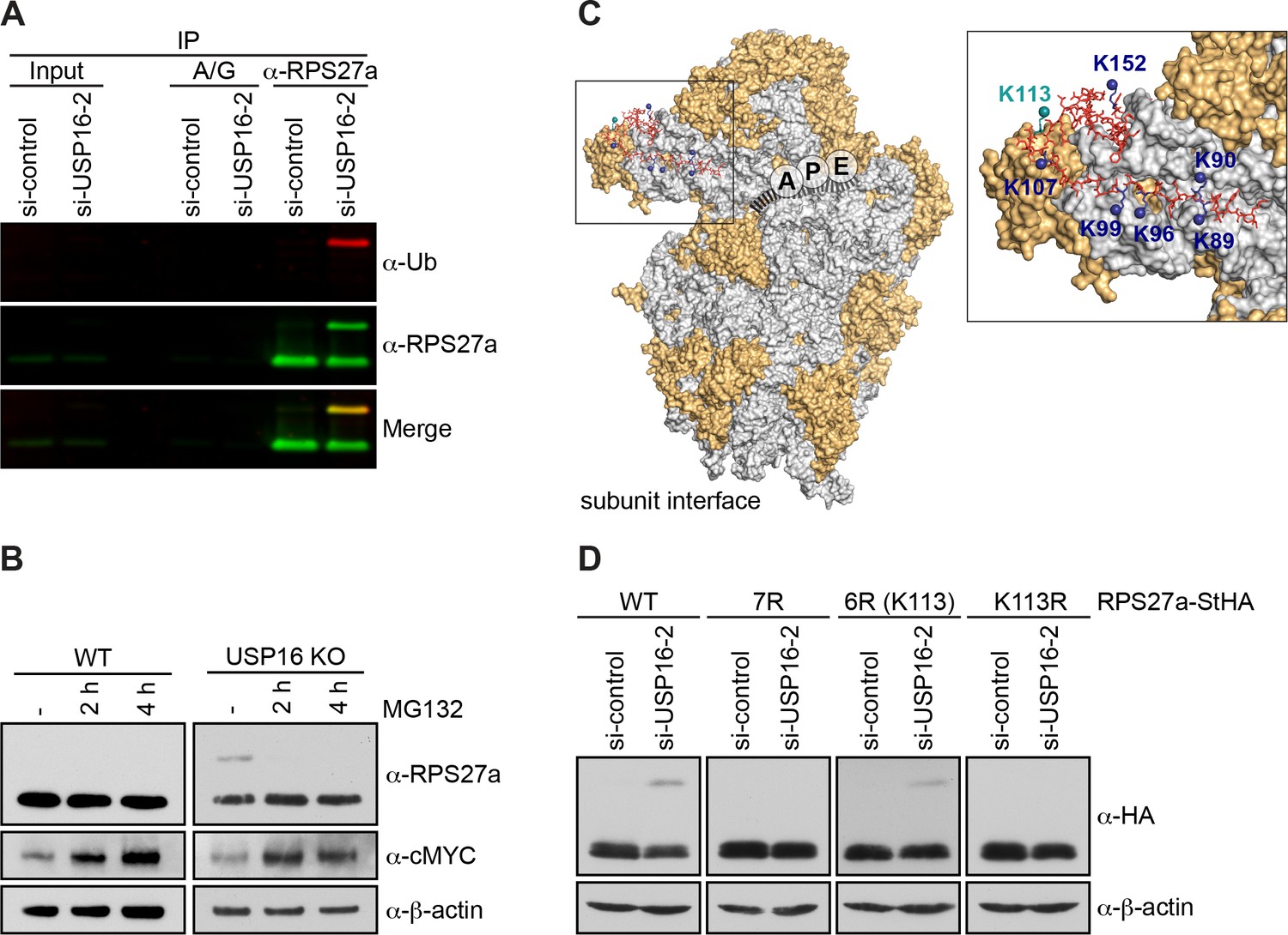
RPS27a is trans-ubiquitinated on lysine 113.
(A) Denaturing immunoprecipitation (IP) of RPS27a from lysates of HeLa cells treated with control or USP16 siRNA for 72 hr. Protein A/G beads without antibody were used as negative control. Eluates were analyzed by immunoblotting using RPS27a and ubiquitin (Ub) antibodies and simultaneous detection of fluorescently labeled rabbit (for USP16) or mouse (for Ub) secondary antibodies using an Odyssey (LI-COR) imaging system. Load corresponds to 0.1% of the input and to 20% of the eluates. (B) HeLa WT and USP16 KO cells were treated with MG132 (20 µM) for the indicated times and cell extracts were analyzed by immunoblotting using the indicated antibodies. (C) Structure of the human 40S subunit shown from the subunit interface side (adapted from Khatter et al., 2015, PDB ID: 4UG0), highlighting the mRNA path (dashed line), the positions of the ribosomal A, P, and E sites, and RPS27a at the 40S beak. The surfaces of RPs and rRNA are depicted in orange and light gray, respectively. RPS27a is depicted as red sticks. Mutated lysine residues are indicated in blue and teal (K113). In the inset, the positions of the mutated lysine residues are highlighted. (D) HeLa FlpIn cell lines expressing C-terminally StHA-tagged RPS27a WT and mutants (7R: K89/90/96/99/107/113/152R; 6R(K113): K89/90/96/99/107/152R; K113R) were treated with control or USP16 siRNA for 72 hr. Extracts were analyzed by immunoblotting using the indicated antibodies.
Figure 5—figure supplement 1
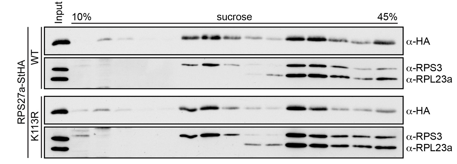
RPS27a(K113R)-StHA is incorporated into ribosomes.
Extracts from HeLa FlpIn cell lines expressing RPS27a-StHA or RPS27a(K113R)-StHA were separated on a linear 10–45% sucrose gradient by centrifugation and the fractions were analyzed by immunoblotting using the indicated antibodies.
Figure 6

Deletion of USP16 leads to late cytoplasmic ribosome biogenesis defects.
(A) Northern blot analysis of total RNA extracted from HeLa WT and USP16 KO cells. A radioactively labeled 5’ ITS1 probe was used to detect the indicated pre-rRNA precursors. Mature 18S and 28S rRNA were visualized by GelRed staining of the gel. (B) Quantification of 18S-E/18S and 18S-E/28S (pre-)rRNA levels (mean ± SD) of three independent experiments as in (A). **p≤0.01 (unpaired t-test). (C) HeLa WT and USP16 KO cells were transfected with USP16(WT)- or USP16(C205S)-StHA. After 24 hr, cells were analyzed by immunostaining using an antibody against the transfected construct (α-HA) and the indicated antibodies recognizing the 40S trans-acting factors ENP1, DIM2, NOB1, and RIOK1. For analyses of NOB1 and RIOK1, cells were treated with leptomycin B (LMB; 20 nM, 90 min) to inhibit CRM1-dependent nuclear export. Arrows mark cells expressing the USP16 constructs and showing phenotypic rescue in the USP16 KO cell line. Scale bar, 20 µM. (D) Biological replicates of experiments in (C) were analyzed for the efficiency of rescue by transfection of USP16(WT)- or USP16(C205S)-StHA into USP16 KO cells regarding the localization of DIM2, NOB1 and RIOK1. Example pictures of cells showing either the USP16 KO or the rescue phenotype are shown along with the quantification of the rescue efficiency (DIM2: N = 3, n ≥ 42; NOB1: N = 4, n ≥ 67; RIOK1: N = 3, n ≥ 61). Scale bar, 10 µM.
Figure 7 with 2 supplements
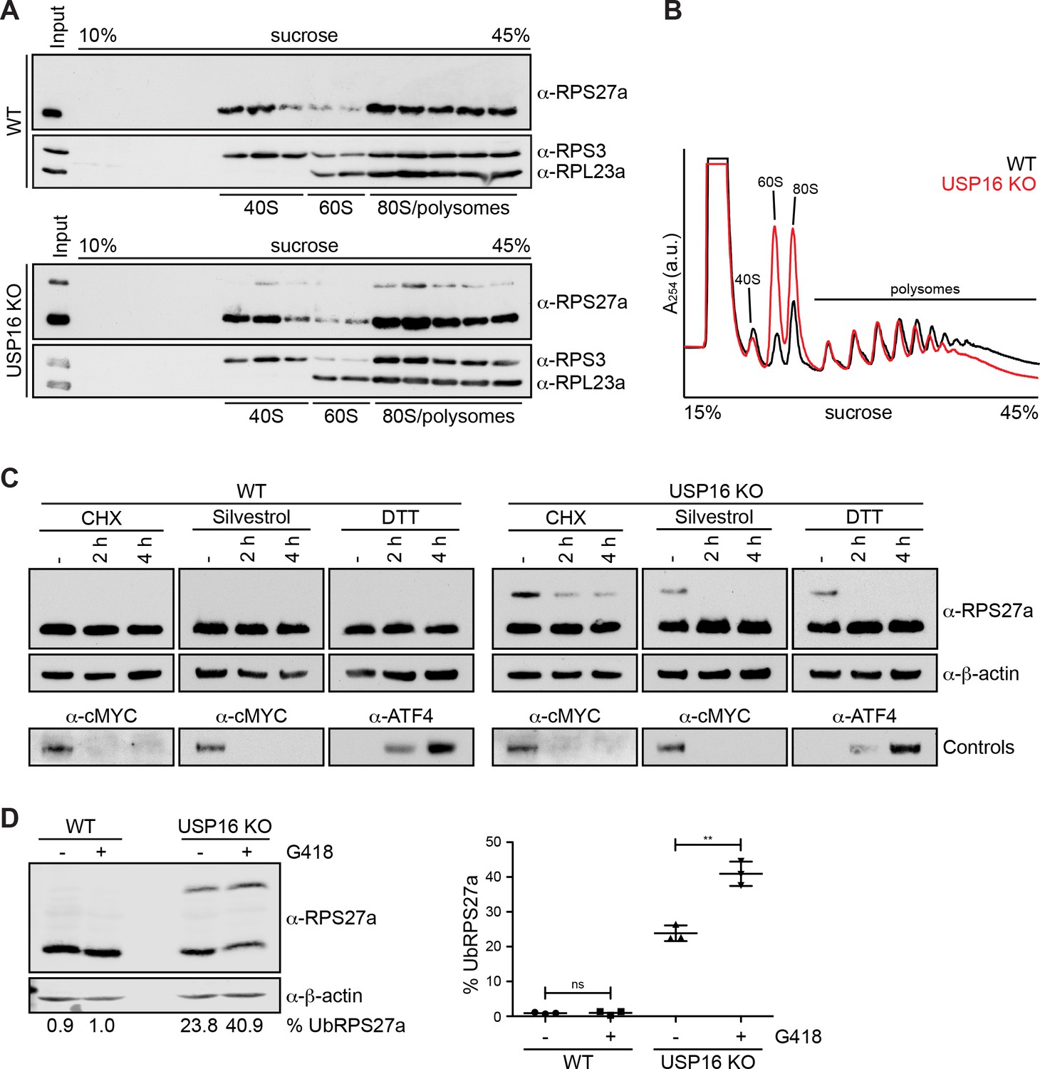
RPS27a ubiquitination is altered by interference with mRNA translation.
(A) Extracts from HEK293 WT and USP16 KO cells were separated on a linear 10–45% sucrose gradient by centrifugation. Input and gradient fractions were analyzed by immunoblotting using the indicated antibodies. (B) Extracts from HeLa WT (black line) and USP16 KO (red line) cells were separated on a linear 10–45% sucrose gradient by centrifugation and analyzed by polysome profiling. (C) HeLa WT and USP16 KO cells were treated with cycloheximide (CHX; 100 µg/ml), silvestrol (1 µM) or DTT (5 mM) for the specified times and cell extracts were analyzed by immunoblotting using the indicated antibodies. Efficiency of the treatments was assessed by immunoblotting showing rapid loss of short-lived cMYC after CHX and silvestrol treatments, and accumulation of the UPR-induced transcription factor ATF4 after DTT treatment. (D) HeLa WT and USP16 KO cells were treated with G418 (400 µg/ml) for 24 hr and cell extracts were analyzed by immunoblotting using the indicated antibodies (left panel). The signal of three independent experiments was quantified and the percentage of ubiquitinated RPS27a (% UbRPS27a) of total RPS27a (modified plus unmodified RPS27a) was determined using an Odyssey (LI-COR) imaging system (mean ± SD; right panel). Values below the left panel indicate the mean of three independent experiments. **p≤0.01 (unpaired t-test).
Figure 7—figure supplement 1
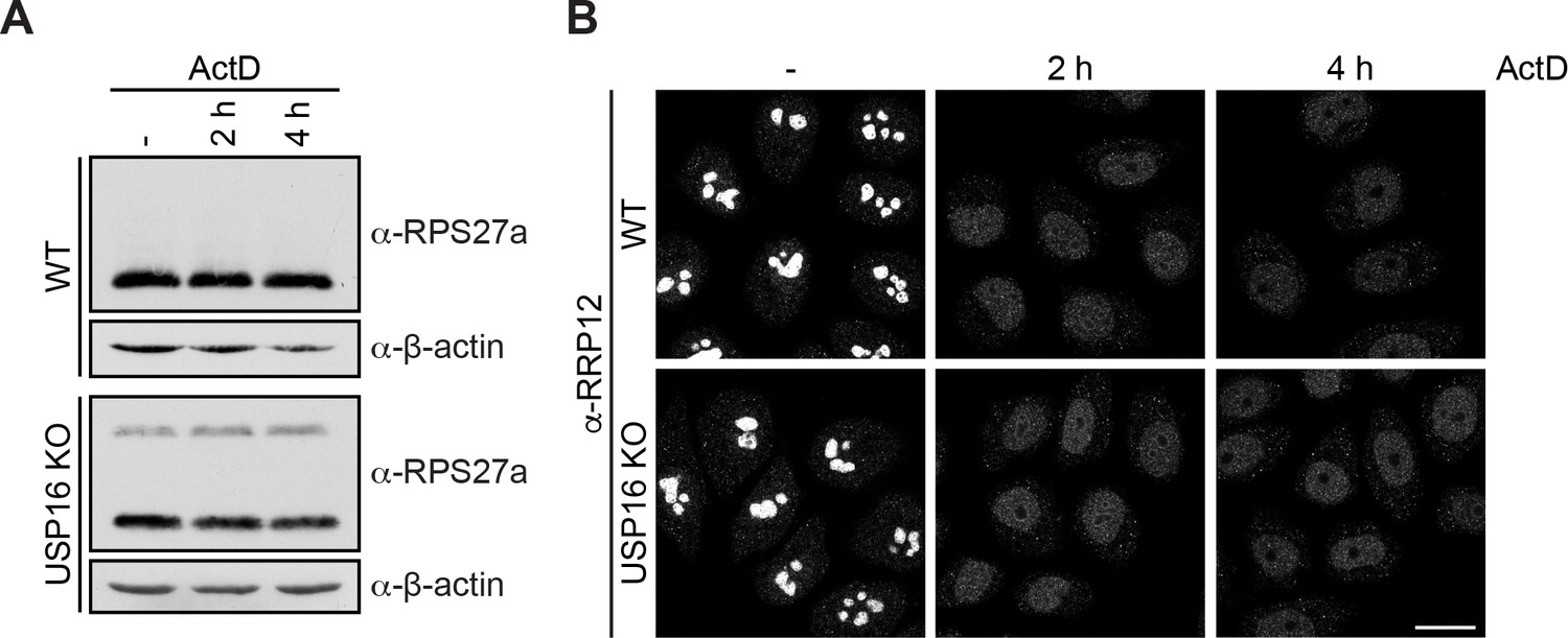
Actinomycin D treatment does not affect RPS27a ubiquitination.
(A) HeLa WT and USP16 KO cells were treated with actinomycin D (ActD; 10 nM) for the specified times and cell extracts were analyzed by immunoblotting using the indicated antibodies. (B) Immunofluorescence analysis of the 40S trans-acting factor RRP12 of the experiment in (A) to control for efficiency of the ActD treatment. Scale bar, 20 µM.
Figure 7—figure supplement 2
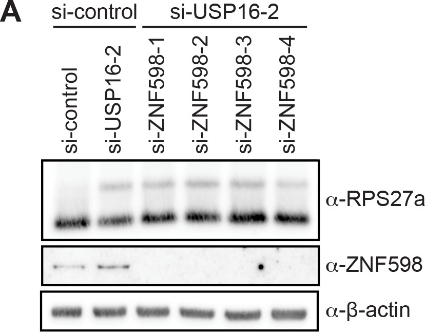
Depletion of the E3 ligase ZNF598 does not affect RPS27a ubiquitination.
HeLa cells were treated with control siRNA, a combination of control and USP16-2 siRNA, or a combination of USP16-2 and four different ZNF598 siRNAs for 72 hr. Cell extracts were analyzed by immunoblotting using the indicated antibodies to check for RPS27a ubiquitination and ZNF598 depletion.
Additional files
-
Supplementary file 1
Key Resources Table.
- https://cdn.elifesciences.org/articles/54435/elife-54435-supp1-v2.docx
-
Supplementary file 2
Proteomic analysis of the RIOK1 wild-type and kinase-dead interactome S2-1 Spectral counts of proteins identified on HASt-GFP, RIOK1(WT)- and RIOK1(kd)-StHA in three independent biological replicates.
S2-2 Spectral counts of proteins identified on RIOK1(WT)- and RIOK1(kd)-StHA after normalization to protein length (spectral counts per 1000 amino acids) and to spectral counts of the RIOK1(WT)-StHA bait in replicate II after filtering against the HASt-GFP control. S2-3 Comparison of the normalized spectral counts of proteins identified on RIOK1(WT)- and RIOK1(kd)-StHA. Proteins with an adjusted p value < 0.05 and an at least twofold enrichment on RIOK1(kd)-StHA (log2FC (kd vs. WT)>1) are shown in black, proteins with lower significance are depicted in dark gray (adjusted p value < 0.05 but with a log2FC < 1) or light gray (adjusted p value > 0.05).
- https://cdn.elifesciences.org/articles/54435/elife-54435-supp2-v2.xlsx
-
Supplementary file 3
Proteomic analysis of the interactome of USP16 wild-type and the catalytically-dead mutant.
S3-1 Spectral counts of proteins identified on HASt-GFP, USP16(WT)- and USP16(C205S)-StHA in three independent biological replicates (I-III). S3-2 Spectral counts of proteins identified on RIOK1(WT)- and RIOK1(kd)-StHA after normalization. Spectral counts were normalized to protein length (spectral counts per 1000 amino acids) and to spectral counts of the USP16(WT)-StHA bait in replicate II after filtering against the HASt-GFP control.
- https://cdn.elifesciences.org/articles/54435/elife-54435-supp3-v2.xlsx
-
Transparent reporting form
- https://cdn.elifesciences.org/articles/54435/elife-54435-transrepform-v2.pdf
Download links
A two-part list of links to download the article, or parts of the article, in various formats.
Downloads (link to download the article as PDF)
Open citations (links to open the citations from this article in various online reference manager services)
Cite this article (links to download the citations from this article in formats compatible with various reference manager tools)
USP16 counteracts mono-ubiquitination of RPS27a and promotes maturation of the 40S ribosomal subunit
eLife 9:e54435.
https://doi.org/10.7554/eLife.54435


