Doxycycline has distinct apicoplast-specific mechanisms of antimalarial activity
Abstract
Doxycycline (DOX) is a key antimalarial drug thought to kill Plasmodium parasites by blocking protein translation in the essential apicoplast organelle. Clinical use is primarily limited to prophylaxis due to delayed second-cycle parasite death at 1–3 µM serum concentrations. DOX concentrations > 5 µM kill parasites with first-cycle activity but are thought to involve off-target mechanisms outside the apicoplast. We report that 10 µM DOX blocks apicoplast biogenesis in the first cycle and is rescued by isopentenyl pyrophosphate, an essential apicoplast product, confirming an apicoplast-specific mechanism. Exogenous iron rescues parasites and apicoplast biogenesis from first- but not second-cycle effects of 10 µM DOX, revealing that first-cycle activity involves a metal-dependent mechanism distinct from the delayed-death mechanism. These results critically expand the paradigm for understanding the fundamental antiparasitic mechanisms of DOX and suggest repurposing DOX as a faster acting antimalarial at higher dosing whose multiple mechanisms would be expected to limit parasite resistance.
Introduction
Malaria remains a serious global health problem, with hundreds of thousands of annual deaths due to Plasmodium falciparum parasites. The absence of a potent, long-lasting vaccine and parasite tolerance to frontline artemisinin combination therapies continue to challenge malaria elimination efforts. Furthermore, there are strong concerns that the current COVID-19 pandemic will disrupt malaria prevention and treatment efforts in Africa and cause a surge in malaria deaths that unravels decades of progress (Weiss et al., 2020). Deeper understanding of basic parasite biology and the mechanisms of current drugs will guide their optimal use for malaria prevention and treatment and facilitate development of novel therapies to combat parasite drug resistance.
Tetracycline antibiotics like DOX are thought to kill eukaryotic P. falciparum parasites by inhibiting prokaryotic-like 70S ribosomal translation inside the essential apicoplast organelle (Figure 1; Dahl et al., 2006). Although stable P. falciparum resistance to DOX has not been reported, clinical use is largely limited to prophylaxis due to delayed activity against intraerythrocytic infection (Conrad and Rosenthal, 2019; Gaillard et al., 2015). Parasites treated with 1–3 µM DOX, the drug concentration sustained in human serum with current 100–200 mg dosage (Newton et al., 2005), continue to grow for 72–96 hr and only die after the second 48 hr intraerythrocytic growth cycle when they fail to expand into a third cycle (Dahl et al., 2006). Slow antiparasitic activity is believed to be a fundamental limitation of DOX and other antibiotics that block apicoplast-maintenance pathways (Gaillard et al., 2015; Dahl and Rosenthal, 2007). First-cycle anti-Plasmodium activity has been reported for DOX and azithromycin concentrations > 3 µM, but such activities have been ascribed to targets outside the apicoplast (Dahl et al., 2006; Yeh and DeRisi, 2011; Wilson et al., 2015). A more incisive understanding of the mechanisms and parameters that govern first versus second-cycle DOX activity can inform and improve clinical use of this valuable antibiotic for antimalarial treatment. We therefore set out to test and unravel the mechanisms and apicoplast specificity of first-cycle DOX activity.

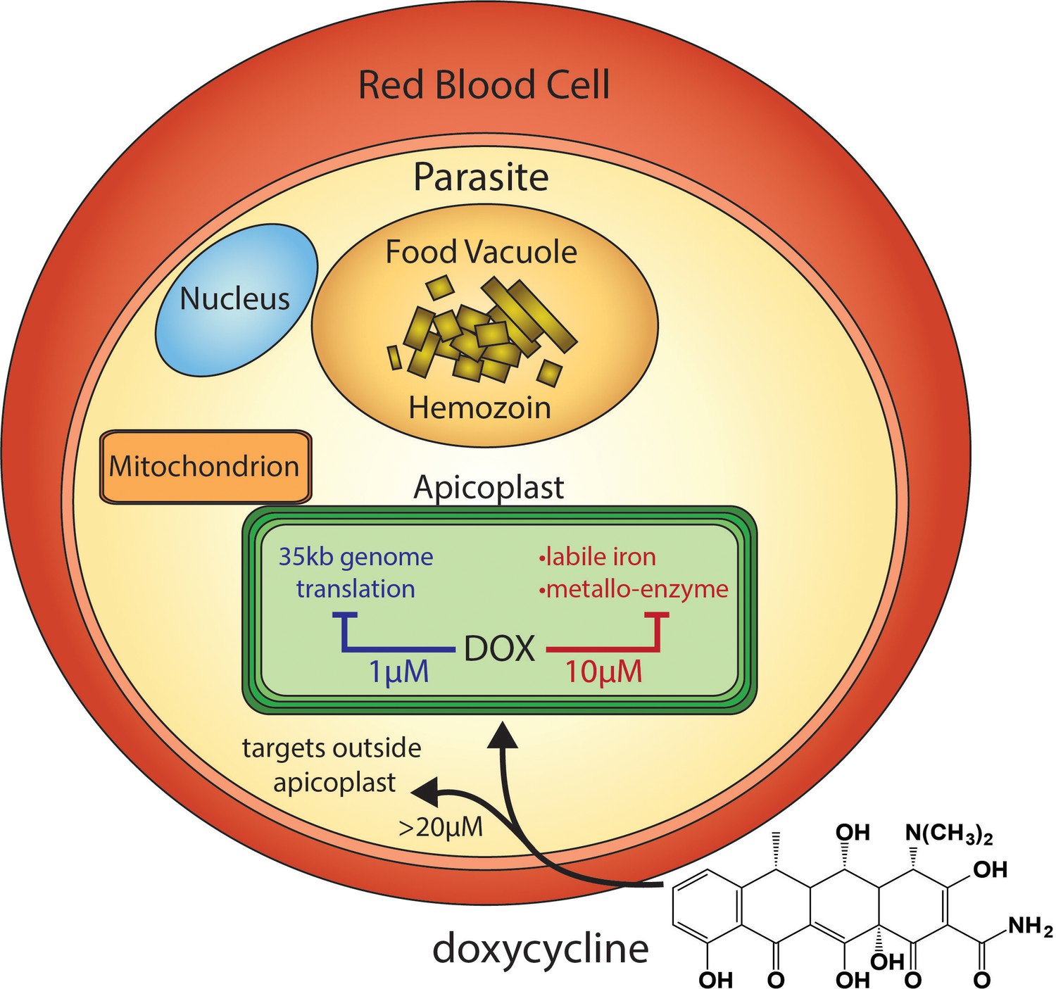
Scheme of intraerythrocytic P. falciparum parasite depicting doxycycline, its canonical delayed-death mechanism at 1 µM inhibiting apicoplast genome translation, the novel metal-dependent mechanism(s) in the apicoplast explored herein at 10 µM, and off-target activity outside the apicoplast at >20 µM.
Results
First-cycle activity by 10 µM DOX has an apicoplast-specific mechanism
Prior studies have shown that 200 µM isopentenyl pyrophosphate (IPP), an essential apicoplast product, rescues parasites from the delayed-death activity of 1–3 µM DOX, confirming an apicoplast-specific target (Yeh and DeRisi, 2011). To provide a baseline for comparison, we first used continuous-growth and 48 hr growth-inhibition assays to confirm that IPP rescued parasites from 1 µM DOX (Figure 2A) and that DOX concentrations > 5 µM killed parasites with first-cycle activity (Figure 2A-C and Figure 2—figure supplement 1) as previously reported (Dahl et al., 2006). To test the apicoplast specificity of first-cycle DOX activity, we next asked whether 200 µM IPP could rescue parasites from DOX concentrations > 5 µM. We observed that IPP shifted the 48 hr EC50 value of DOX from 5 ± 1 to 12 ± 2 µM (average ± SD of five independent assays, p=0.001 by two-tailed unpaired t-test) (Figure 2C and Figure 2—figure supplement 1), suggesting that first-cycle growth defects from 5 to 10 µM DOX reflect an apicoplast-specific mechanism but that DOX concentrations > 10 µM cause off-target defects outside this organelle. We further tested this conclusion using continuous growth assays performed at constant DOX concentrations. We observed that IPP fully or nearly fully rescued parasites from first-cycle growth inhibition by 10 µM but not 20 or 40 µM Dox (Figure 2A and D and Figure 2—figure supplement 1). On the basis of IPP rescue, we conclude that 10 µM DOX kills P. falciparum with first-cycle activity by an apicoplast-specific mechanism.

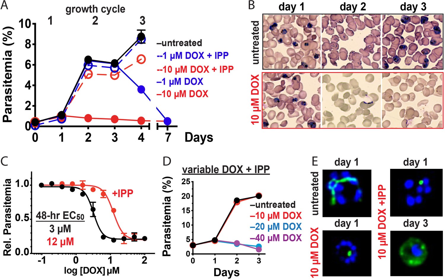
10 µM doxycycline kills P. falciparum with first-cycle, apicoplast-specific activity.
(A) Continuous growth assay of synchronized Dd2 parasites treated with 1 or 10 µM DOX ±200 µM IPP with (B) Giemsa-stained blood smears for days 1–3. (C) 48 hr growth-inhibition curve for DOX-treated Dd2 parasites ± 200 µM IPP. (D) Continuous growth assay of synchronized Dd2 parasites treated with 10–40 µM DOX and 200 µM IPP. (E) Epifluorescence images of synchronized parasites treated as rings with 10 µM DOX ±200 µM IPP and imaged 36 or 65 hr later (green = ACPL GFP, blue = nuclear Hoechst stain). Data points in growth assays are the average ± SD of two to four biological replicates. All growth assays were independently repeated two to four times using different batches of blood (shown in Figure 2—figure supplement 1).
10 µM DOX blocks apicoplast biogenesis in the first cycle
Inhibition of apicoplast biogenesis in the second intraerythrocytic cycle is a hallmark of 1–3 µM DOX-treated P. falciparum, resulting in unviable parasite progeny that fail to inherit the organelle (Dahl et al., 2006). IPP rescues parasite viability after the second cycle without rescuing apicoplast inheritance, such that third-cycle daughter parasites lack the organelle and accumulate apicoplast-targeted proteins in cytoplasmic vesicles (Yeh and DeRisi, 2011). We treated synchronized ring-stage D10 (Waller et al., 2000) or NF54 (Swift et al., 2020) parasites expressing the acyl carrier protein leader sequence fused to GFP (ACPL-GFP) with 10 µM DOX and assessed apicoplast morphology 30–36 hr later in first-cycle schizonts. In contrast to the second-cycle effects of 1–3 µM DOX, the apicoplast in 10 µM DOX-treated parasites failed to elongate in the first cycle. Rescue by 200 µM IPP produced second-cycle parasite progeny with a dispersed GFP signal indicative of apicoplast loss (Figure 2E and Figure 2—figure supplement 2). We conclude that 10 µM DOX blocks apicoplast biogenesis in the first cycle.
First- and second-cycle effects of DOX on the apicoplast are due to distinct mechanisms
What is the molecular mechanism of faster apicoplast-specific activity by 10 µM DOX? We first considered the model that both 1 and 10 µM DOX inhibit apicoplast translation but that 10 µM DOX kills parasites faster due to more stringent translation inhibition at higher drug concentrations. This model predicts that treating parasites simultaneously with multiple distinct apicoplast-translation inhibitors, each added at a delayed death-inducing concentration, will produce additive, accelerated activity that kills parasites in the first cycle. To test this model, we treated synchronized D10 parasites with combinatorial doses of 2 µM DOX, 2 µM clindamycin, and 500 nM azithromycin and monitored growth over three intraerythrocytic cycles. Treatment with each antibiotic alone produced major growth defects at the end of the second cycle, as expected for delayed-death activity at these concentrations (Dahl and Rosenthal, 2007). Two- and three-way drug combinations caused growth defects that were indistinguishable from individual treatments and provided no evidence for additive, first-cycle activity (Figure 3A and Figure 3—figure supplement 1). These results contradict a simple model that 1 and 10 µM DOX act via a common translation-blocking mechanism and suggest that the first-cycle activity of 10 µM DOX is due to a distinct mechanism.

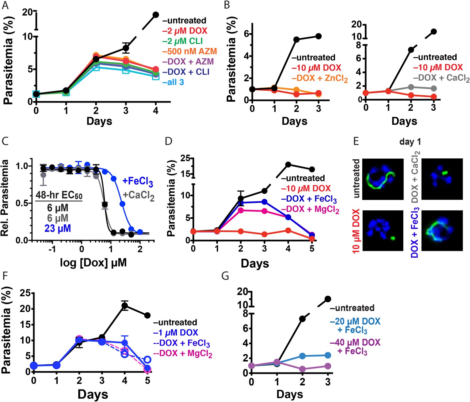
10 µM DOX kills P. falciparum with a first-cycle, metal-dependent mechanism.
Continuous growth assays of synchronized Dd2 parasites treated with (A) DOX, clindamycin (CLI), and/or azithromycin (AZM) and (B) 10 µM DOX and 10 µM ZnCl2 or 500 µM CaCl2. (C) 48 hr growth inhibition assay of D10 parasites treated with DOX without or with 500 µM FeCl3 or CaCl2. (D) Continuous growth assay of synchronized Dd2 parasites treated with 10 µM DOX and 500 µM FeCl3 or MgCl2. (E) Epifluorescence images of synchronized parasites treated as rings with 10 µM DOX ±500 µM FeCl3 or CaCl2 and imaged 36 hr later (green = ACPL GFP, blue = nuclear Hoechst stain). (F) Continuous growth assay of synchronized Dd2 parasites treated with 1 µM DOX and 500 µM FeCl3 or MgCl2. (G) Continuous-growth assay of synchronized Dd2 parasites treated with 20 or 40 µM DOX and 500 µM FeCl3. Data points in growth assays are the average ± SD of two to four biological replicates. All growth assays were independently repeated using different batches of blood (shown in Figure 3—figure supplement 1).
Exogenous iron rescues parasites from first- but not second-cycle effects of 10 µM DOX
Tetracycline antibiotics like DOX tightly chelate a wide variety of di- and trivalent metal ions via their siderophore-like arrangement of exocyclic hydroxyl and carbonyl oxygen atoms (Figure 1), with a reported affinity series of Fe3+>Fe2+>Zn2+>Mg2+>Ca2+ (Albert and Rees, 1956; Nelson, 1998). Indeed, tetracycline interactions with Ca2+ and Mg2+ ions mediate cellular uptake and binding to biomolecular targets such as the tetracycline repressor and 16S rRNA (Nelson, 1998; Orth et al., 2000). We next considered a model that first-cycle effects of 10 µM DOX reflect a metal-dependent mechanism distinct from ribosomal inhibition causing second-cycle death. To test this model, we investigated whether exogenous metals rescued parasites from 10 µM DOX. We failed to observe growth rescue by 10 µM ZnCl2 (toxicity limit [Marvin et al., 2012]) or 500 µM CaCl2 in continuous-growth (Figure 3B and Figure 3—figure supplement 1) or 48 hr growth-inhibition assays (Figure 3C). In contrast, 500 µM FeCl3 (and to a lesser extent 500 µM MgCl2) fully or nearly fully rescued parasites from first-cycle growth inhibition by 10 µM DOX (Figure 3C and D), although partial rescue was observed at FeCl3 concentrations as low as 50 µM (Figure 3—figure supplement 1). However, parasites treated with 10 µM DOX and 500 µM FeCl3 still succumbed to second-cycle, delayed death (Figure 3D and Figure 3—figure supplement 1), as expected for distinct mechanisms of first- and second-cycle DOX activity.
We also observed that 500 µM FeCl3 but not CaCl2 rescued first-cycle apicoplast-branching in 10 µM DOX (Figure 3E and Figure 3—figure supplement 2). These observations contrast with IPP, which rescued parasite viability in 10 µM DOX but did not restore apicoplast branching (Figure 2E). We further noted that FeCl3 selectively rescued parasites from the apicoplast-specific, first-cycle growth effects of 10 µM DOX but did not rescue parasites from the second-cycle effects of 1 µM DOX (Figure 3F) or the off-target effects of 20–40 µM DOX (Figure 3G and Figure 3—figure supplement 1). We conclude that 10 µM DOX kills parasites via a metal-dependent, first-cycle mechanism that blocks apicoplast biogenesis and is distinct from the second-cycle, delayed-death mechanism of 1 µM DOX.
Discussion
Metal-dependent mechanisms of first-cycle activity by 10 µM DOX
What is the metal-dependent mechanism of 10 µM DOX, and why is there preferential rescue of parasite growth by FeCl3? Tetracyclines bind iron more tightly than other metals, with an equilibrium association constant of 1010 M−1 for 1:1 chelation of Fe3+ versus 104 M−1 for Mg2+ (Albert and Rees, 1956). Although the 500 µM concentration of exogenous FeCl3 required for maximal rescue of parasite growth in 10 µM DOX is large relative to the ~1 µM labile iron concentration estimated for the parasite cytoplasm (Scholl et al., 2005), the intracellular iron concentration achieved by exogenous addition of 500 µM FeCl3 remains unclear. Indeed, mechanisms of iron uptake and trafficking by blood-stage P. falciparum remain sparsely understood (Scholl et al., 2005; Mabeza et al., 1999), especially uptake across the four membranes that surround the apicoplast.
We first considered whether exogenous FeCl3 might selectively rescue parasites from 10 µM DOX by blocking or reducing its uptake into the parasite apicoplast, since metal chelation has been reported to influence the cellular uptake of tetracycline antibiotics in other organisms (Nelson, 1998). However, 500 µM FeCl3 or MgCl2 did not rescue second-cycle parasite death in continuous growth assays with 10 µM (Figure 3D) or 1 µM DOX (Figure 3F). Furthermore, exogenous iron resulted in only a small, 1.5 µM shift in EC50 value from 0.5 to 2 µM in a 96 hr growth inhibition assay, in contrast to the 10.5 µM shift provided by IPP (Figure 3—figure supplement 1). These results strongly suggest that DOX uptake into the apicoplast is not substantially perturbed by exogenous iron. The inability of 500 µM FeCl3 to rescue parasites from first-cycle activity by ≥20 µM DOX (Figure 3G) further suggests that general uptake of DOX into parasites is not substantially affected by exogenous iron.
We propose two distinct models to explain the metal-dependent effects of 10 µM DOX, both of which could contribute to apicoplast-specific activity. First, DOX could directly bind and sequester labile iron within the apicoplast, reducing its bioavailability for Fe-S cluster biogenesis and other essential iron-dependent processes in this organelle. Indeed, prior work has shown that apicoplast biogenesis requires Fe-S cluster synthesis apart from known essential roles in isoprenoid biosynthesis (Gisselberg et al., 2013). In this first model, rescue by exogenous FeCl3 would be due to restoration of iron bioavailability, while modest rescue by 500 µM MgCl2 may reflect competitive displacement of DOX-bound iron to restore iron bioavailability. RPMI growth medium already contains ~400 µM Mg2+ prior to supplementation with an addition 500 µM MgCl2, and thus Mg2+ availability is unlikely to be directly limited by 10 µM DOX. Consistent with a general mechanism that labile-iron chelation can block apicoplast biogenesis, we observed in preliminary studies that Plasmodium growth inhibited by the highly specific iron chelator, deferoxamine (DFO) (Mabeza et al., 1999), could be partially rescued by IPP, fully rescued by exogenous FeCl3, and involved a first-cycle defect in apicoplast elongation (Figure 3—figure supplement 3). Development of targeted and incisive probes of labile iron within subcellular compartments remains an ongoing challenge in biology (Breuer et al., 2008), especially in the Plasmodium apicoplast where iron uptake, concentration, and utilization remain sparsely understood. To more broadly evaluate the impact of DOX on iron availability in the apicoplast, we are developing protein-based probes of lipoic acid and isoprenoid biosynthesis, as these two apicoplast-dependent processes require Fe/S-cluster biosynthesis for activity (Gisselberg et al., 2013).
In a second model, DOX could bind to additional macromolecular targets within the apicoplast (e.g. a metalloenzyme) via metal-dependent interactions that inhibit essential functions required for organelle biogenesis. Exogenous 500 µM Fe3+ would then rescue parasites by disrupting these inhibitory interactions via competitive binding to DOX. This second model would be mechanistically akin to diketo acid inhibitors of HIV integrase like raltegravir that bind to active site Mg2+ ions to inhibit integrase activity but are displaced by exogenous metals (Grobler et al., 2002; Hare et al., 2010). To test this model, we are developing a DOX-affinity reagent to identify apicoplast targets that interact with doxycycline and whose inhibition may contribute to first-cycle DOX activity.
Conclusions and implications
These results critically expand the paradigm for understanding the fundamental mechanisms of DOX activity against P. falciparum malaria parasites. These mechanisms include a delayed, second-cycle defect at 1–3 µM DOX that likely reflects inhibition of 70S apicoplast ribosomes, a first-cycle iron-dependent defect within the apicoplast that uniquely operates at 8–10 µM DOX, and a first-cycle iron-independent mechanism outside the apicoplast at ≥20 µM DOX (Figure 1). Pharmacokinetic studies indicate that current 100–200 mg doses of DOX achieve peak human serum concentrations of 6–8 µM over the first six hours which then decrease to 1–2 µM over 24 hr (Newton et al., 2005). Although current DOX treatment regimens result in delayed parasite clearance in vivo, both apicoplast-specific mechanisms of DOX likely operate over this concentration range and contribute to parasite death (Dahl et al., 2006). These multiple mechanisms of DOX, together with limited antimalarial use of DOX in the field, may explain why parasites with stable DOX resistance have not emerged (Conrad and Rosenthal, 2019; Gaillard et al., 2015).
There has been a prevailing view in the literature that delayed-death activity is a fundamental limitation of antibiotics like DOX that block apicoplast maintenance (Ramya et al., 2007; Kennedy et al., 2019). Our results emphasize that DOX is not an intrinsically slow-acting antimalarial drug and support the emerging paradigm (Boucher and Yeh, 2019; Amberg-Johnson et al., 2017; Uddin et al., 2018) that inhibition of apicoplast biogenesis can defy the delayed-death phenotype to kill parasites on a faster time-scale. The first-cycle, iron-dependent impacts of 10 µM DOX or 15 µM DFO on apicoplast biogenesis also suggest that this organelle may be especially susceptible to therapeutic strategies that interfere with acquisition and utilization of iron, perhaps due to limited uptake of exogenous iron and/or limited iron storage mechanisms in the apicoplast.
Finally, this work suggests the possibility of repurposing DOX as a faster-acting antiparasitic treatment at higher dosing, whose multiple mechanisms would be expected to limit parasite resistance. Prior studies indicate that 500–600 mg doses in humans achieve sustained serum DOX concentrations ≥ 5 µM for 24–48 hr with little or no increase in adverse effects (Marlin and Cheng, 1979; Adadevoh et al., 1976). DOX is currently contraindicated for long-term prophylaxis in pregnant women and young children, two of the major at-risk populations for malaria, due to concerns about impacts on fetal development and infant tooth discoloration, respectively, based on observed toxicities for other tetracyclines (Gaillard et al., 2018). Recent studies suggest that these effects are not associated with short-term DOX use (Gaillard et al., 2018; Todd et al., 2015; Cross et al., 2016), and additional tests can define the safety parameters that would govern short-term use of DOX for treatment in these populations. Recent development of tetracycline derivatives with improved activities may provide another option to deploy this important class of antibiotics for antimalarial treatment (Draper et al., 2013).
Materials and methods
Reagent type (species) or resource | Designation | Source or reference | Identifiers | Additional information |
---|---|---|---|---|
Cell line (Plasmodium falciparum) | Dd2 | PMID:1970614 | ||
Cell line (Plasmodium falciparum) | D10 ACPL-GFP | PMID:10775264 | ||
Cell line (Plasmodium falciparum) | NF54-PfMev ACPL-GFP | PMID:32059044 | ||
Software, algorithm | Prism 8 | GraphPad | RRID:SCR_002798 | |
Chemical compound, drug | Doxycycline | Sigma-Aldrich | Cat. No. D3447 | |
Chemical compound, drug | Isopentenyl pyrophosphate | Isoprenoids | Cat. No. IPP001 | |
Chemical compound, drug | Ferric chloride | Sigma-Aldrich | Cat. No. 236489 | |
Chemical compound, drug | Clindamycin | Sigma-Aldrich | Cat. No. C6427 | |
Chemical compound, drug | Azithromycin | Sigma-Aldrich | Cat. No. 75199 | |
Chemical compound, drug | Deferoxamine | Sigma-Aldrich | Cat. No. D9533 |
Materials
All reagents were cell-culture grade and/or of the highest purity available.
Parasite culture
Request a detailed protocolAll experiments were performed using Plasmodium falciparum Dd2 (Wellems et al., 1990), ACPL-GFP D10 (Waller et al., 2000), or ACPL-GFP PfMev NF54 (Swift et al., 2020) parasite strains, which were obtained from colleagues and verified by confirming their expected drug sensitivity and/or sequencing strain-specific genetic markers. Parasite culturing was performed as previously described (Sigala et al., 2015) in Roswell Park Memorial Institute medium (RPMI-1640, Thermo Fisher 23400021) supplemented with 2.5 g/L Albumax I Lipid-Rich BSA (Thermo Fisher 11020039), 15 mg/L hypoxanthine (Sigma H9636), 110 mg/L sodium pyruvate (Sigma P5280), 1.19 g/L HEPES (Sigma H4034), 2.52 g/L sodium bicarbonate (Sigma S5761), 2 g/L glucose (Sigma G7021), and 10 mg/L gentamicin (Invitrogen Life Technologies 15750060). Cultures were maintained at 2% hematocrit in human erythrocytes obtained from the University of Utah Hospital blood bank, at 37°C, and at 5% O2, 5% CO2, 90% N2. Cultures were mycoplasma-free by PCR test.
Parasite growth assays
Request a detailed protocolAll growth assays were performed with two to four biological replicates (defined according to Blainey et al., 2014) in distinct sample wells that were set-up and monitored in parallel. Parasites were synchronized to the ring stage either by treatment with 5% D-sorbitol (Sigma S7900) or by first magnet-purifying schizonts and then incubating them with uninfected erythrocytes for 5 hr followed by treatment with 5% D-sorbitol. Results from growth assays using either of these synchronization methods were indistinguishable within error, and 5% sorbitol was used for synchronization unless stated otherwise.
For continuous growth assays, parasite growth was monitored by diluting sorbitol-synchronized parasites to ~0.5% starting parasitemia, adding additional treatments (antibiotics, IPP, and/or metal salts) at assay initiation, and allowing culture expansion over several days with daily media changes. Growth assays with doxycycline (Sigma D3447), clindamycin (Sigma C6427), and azithromycin (Sigma 75199) were conducted at 0.2% DMSO at the indicated final drug concentration. Growth assays with ZnCl2 (Sigma 208086), CaCl2 (Sigma C4901), MgCl2 (M8266), FeCl3 (Sigma 236489), deferoxamine (Sigma D9533), and/or IPP (NH4+ salt, Isoprenoids IPP001) were conducted at the indicated final concentrations. Parasitemia was monitored daily by flow cytometry by diluting 10 µl of each parasite culture well from 2 to 3 biological replicates into 200 µl of 1.0 µg/ml acridine orange (Invitrogen Life Technologies A3568) in phosphate buffered saline (PBS) and analysis on a BD FACSCelesta system monitoring SSC-A, FSC-A, PE-A, FITC-A, and PerCP-Cy5-5-A channels.
For EC50 determinations via dose-response assay, synchronous ring-stage parasites were diluted to 1% parasitemia and incubated with variable (serially twofold diluted) DOX concentrations ± 200 µM IPP,±50 µM mevalonate (Cayman 20348), ±500 µM FeCl3, or ±500 µM CaCl2 for 48–120 hr without media changes. Parasitemia was determined by flow cytometry for two to four biological replicates for each untreated or drug-treated condition, normalized to the parasitemia in the absence of drug, plotted as the average ± SD of biological replicates as a function of the log of the drug concentration (in µM), and fit to a four-parameter dose-response model using GraphPad Prism 8.0. All growth assays were independently repeated two to five times on different weeks and in different batches of blood. The 48 hr EC50 values determined from five independent assays for DOX ±IPP were averaged and analyzed by unpaired t-test using GraphPad Prism 8.0.
Fluorescence microscopy
Request a detailed protocolFor live-cell experiments, parasites samples were collected at 30–36 or 65 hr after synchronization with magnet purification plus sorbitol treatment (see above). Imaging experiments were independently repeated twice. Parasite nuclei were visualized by incubating samples with 1–2 µg/ml Hoechst 33342 (Thermo Scientific Pierce 62249) for 10–20 min at room temperature. The parasite apicoplast was visualized in D10 (Waller et al., 2000) or NF54 mevalonate-bypass (Swift et al., 2020) cells using the ACPleader-GFP expressed by both lines. Images were taken on DIC/brightfield, DAPI, and GFP channels using either a Zeiss Axio Imager or an EVOS M5000 imaging system. Fiji/ImageJ was used to process and analyze images. All image adjustments, including contrast and brightness, were made on a linear scale. For indicated conditions, apicoplast morphologies in 20–40 total parasites were scored as elongated, focal, or dispersed; counted; and plotted by histogram as the fractional population with the indicated morphology. Analysis of replicate samples indicated standard errors of the mean that were ≤15% for all samples in the percentage of parasites displaying a given apicoplast morphology in a given condition. Two-tailed unpaired t-test analysis using GraphPad Prism was used to evaluate the significance of observed population differences.
Data availability
All data reported or described in this manuscript are available and included in the main and supplemental figures and in the microscopy source data file.
References
-
Intracellular labile ironThe International Journal of Biochemistry & Cell Biology 40:350–354.https://doi.org/10.1016/j.biocel.2007.03.010
-
Antimalarial drug resistance in Africa: the calm before the storm?The Lancet Infectious Diseases 19:e338–e351.https://doi.org/10.1016/S1473-3099(19)30261-0
-
Revisiting doxycycline in pregnancy and early childhood--time to rebuild its reputation?Expert Opinion on Drug Safety 15:367–382.https://doi.org/10.1517/14740338.2016.1133584
-
Tetracyclines specifically target the apicoplast of the malaria parasite Plasmodium falciparumAntimicrobial Agents and Chemotherapy 50:3124–3131.https://doi.org/10.1128/AAC.00394-06
-
Multiple antibiotics exert delayed effects against the Plasmodium falciparum apicoplastAntimicrobial Agents and Chemotherapy 51:3485–3490.https://doi.org/10.1128/AAC.00527-07
-
In vitro and in vivo antimalarial efficacies of optimized tetracyclinesAntimicrobial Agents and Chemotherapy 57:3131–3136.https://doi.org/10.1128/AAC.00451-13
-
Delayed death by plastid inhibition in apicomplexan parasitesTrends in Parasitology 35:747–759.https://doi.org/10.1016/j.pt.2019.07.010
-
Iron chelation therapy for malaria: a reviewPharmacology & Therapeutics 81:53–75.https://doi.org/10.1016/S0163-7258(98)00037-0
-
Pharmacokinetics and tolerability of a single oral 600-mg dose of doxycyclineMedical Journal of Australia 1:575–576.https://doi.org/10.5694/j.1326-5377.1979.tb127087.x
-
Chemical and biological dynamics of tetracyclinesAdvances in Dental Research 12:5–11.https://doi.org/10.1177/08959374980120011901
-
Pharmacokinetics of oral doxycycline during combination treatment of severe Falciparum malariaAntimicrobial Agents and Chemotherapy 49:1622–1625.https://doi.org/10.1128/AAC.49.4.1622-1625.2005
-
Structural basis of gene regulation by the tetracycline inducible tet repressor-operator systemNature Structural Biology 7:215–219.https://doi.org/10.1038/73324
-
Inhibitors of nonhousekeeping functions of the apicoplast defy delayed death in Plasmodium falciparumAntimicrobial Agents and Chemotherapy 51:307–316.https://doi.org/10.1128/AAC.00808-06
-
Bioavailable iron and heme metabolism in Plasmodium falciparumCurrent Topics in Microbiology and Immunology 295:293–324.https://doi.org/10.1007/3-540-29088-5_12
-
No visible dental staining in children treated with doxycycline for suspected rocky mountain spotted feverThe Journal of Pediatrics 166:1246–1251.https://doi.org/10.1016/j.jpeds.2015.02.015
-
Validation of putative Apicoplast-Targeting drugs using a chemical supplementation assay in cultured human malaria parasitesAntimicrobial Agents and Chemotherapy 62:e01161-17.https://doi.org/10.1128/AAC.01161-17
Decision letter
-
Jon ClardyReviewing Editor; Harvard Medical School, United States
-
Dominique Soldati-FavreSenior Editor; University of Geneva, Switzerland
-
Jon ClardyReviewer; Harvard Medical School, United States
-
Christopher D GoodmanReviewer; University of Melbourne, Australia
In the interests of transparency, eLife publishes the most substantive revision requests and the accompanying author responses.
Acceptance summary:
This short report provides an example of a potentially better way to deploy a clinically used antimalarial drug. At 1-3 µM serum concentration, doxycycline kills parasites in their second cycle however this delayed parasite clearance is overcome at 10µM. At this higher concentration the parasite are kills in their first cycle by blocking apicoplast biogenesis via a Fe-sensitive target. This important finding could in principle be rapidly incorporated into current practice.
Decision letter after peer review:
[Editors’ note: the authors submitted for reconsideration following the decision after peer review. What follows is the decision letter after the first round of review.]
Thank you for submitting your work entitled "Doxycycline has Distinct Apicoplast-Specific Mechanisms of Antimalarial Activity" for consideration by eLife. Your article has been reviewed by three peer reviewers, including Jon Clardy as the Reviewing Editor and Reviewer #1, and the evaluation has been overseen by a Senior Editor. The following individuals involved in review of your submission have agreed to reveal their identity: Christopher Goodman (Reviewer #2).
Our decision has been reached after consultation between the reviewers. Based on these discussions and the individual reviews below, we regret to inform you that your work will not be considered further for publication in eLife.
The reviewers agreed that the manuscript reported interesting and potentially significant findings, but that in its current state it did not meet the journal's standards. Shortcomings in the mechanistic analysis in what is known to be a complex system, lack of biological repeats, and quantification were primary concerns.
Reviewer #1:
In many fields of medicine improved therapeutic outcomes have resulted more from better ways to deploy clinically used drugs rather than the introduction of brand new drugs. This short report provides another example in the field of malaria: higher doses of doxycycline (DOX), an approved antimalarial drug, avoid its main known drawback, delayed parasite clearance. DOX at 1-3 µM serum concentration dosing – the current standard – kills parasites in their second cycle while 8-10µM dosing kills parasites in their first cycle. The authors report a change in mechanism – emergence of an Fe-sensitive target – as underlying the effect. There are two issues: faster parasite clearance and responsible mechanism. The faster killing data appeared to be quite solid and might have had some precedents in earlier publications, so that part of the report seems solid.
Identifying a mechanism rigorously requires eliminating plausible alternatives, and the authors began by ruling out increased effects at the known ribosomal target. Again, these experiments seemed convincing. DOX, like other tetracycline analogs, bind metals, and the authors add rather large quantities of metals, including Fe at 500 µM, and this result, along with some controls, establishes that a metal-dependent mechanism is likely. It does, at least for me, establish that it's not the same mechanism as lower DOX dose clearance.
Under normal circumstances, I would expect more for an eLife paper – a clearer notion of mechanism, something less drastic than a super high metal concentration, and more guidance for future experiments. But these are not normal circumstances, and the potential of this finding to quickly translate into improved therapeutic approaches argues for publication essentially as is.
Reviewer #2:
Summary:
The identification of a fast-acting, apicoplast-related mode of action for doxycycline is significant. It is supported by two independent lines of evidence and helps to explain the absence of doxycycline resistance in the field. The role of metal ions in doxycycline function and bioavailability is complex. Dissecting the effects and proving specific activity requires multiple lines of evidence and robust data. Unfortunately, the data presented does not reach that level. Overall, the quality of the data presented throughout the paper (as outlined in detail below) is not of publishable standard in terms of replication and statistical analysis. The work has real potential but requires more robust data to support the claims.
Essential revisions:
1) The experiments are looking at differences in relatively narrow time frames, but the experimental description provided doesn't give sufficient information about the method of synchronization to assess the experiments. A single sorbitol synchronization produces parasites with an age range from 0 to ~18 hours post invasion, and the distribution of stages can vary between cultures. This large window of parasite age can strongly impact the effects of doxycycline (Dahl et al., 2007). Clarification of this procedure is essential to understanding the experimental results.
2) The absence of replication in the drug trials is not consistent with levels of data expected for publication. The minimal expectation is for three independent biological replicates in each drug trial which allows for statistical comparison. The drug assays presented were only done once (technical replicates…subsection “Parasite Growth Assays:”) which is not sufficient. The small differences seen in effective concentrations require robust statistical analysis. This is highlighted by the 2-fold difference in EC50 for two independent experiments for the Dd2 line (Figure 2—figure supplement 1B and C) which is similar in magnitude to the differences seen with IPP supplementation.
3) The data from the epi-fluorescence imaging requires clarification as to whether all images are from the same experiment and how the synchronization was done. Again, there should be data from multiple replicates, the phenotype needs to be quantified and the results statistically analyzed.
4) The presentation of data for the growth trials is confusing. 2-4 replicates were done but it seems that only one replicate is presented in the text and some others (but not all?) are presented in the supplementary figures. Is it possible to include a single figure with all the replicates and include a statistical analysis?
5) The arguments that the impact of iron chloride is not related to iron sequestering and/or oxidizing Doxycyline, or to a change in function related to altering the metals metals bound to the drug, would be improved by the inclusion of dose-response curves comparing effective concentrations with and without metal supplements. While not definitive they would go a long way to distinguishing between the various possibilities. The single experiment showing little change in growth of parasites under 1µM Doxycycline does not seem enough to address this point.
Reviewer #3:
Doxycycline is an established antimalarial with a fairly well-defined, but complex mechanism of action. The authors carefully studied effects of intermediate concentrations of doxycycline to tease out apicoplast-specific and other effects of the compound. Their results are nicely demonstrated and well-described. The results mostly recapitulate what is already known, but they add the observation that iron rescues parasites from rapid killing by intermediate concentrations of doxycline. This offers what appears to be a valid, but incremental advance in our understanding of the antimalarial action of doxycycline. Some specific concerns are below.
1) Abstract. As mentioned in the text, but not noted in the Abstract, the first cycle activity of doxycycline has already been clearly described (e.g. Dahl et al., 2016; Yeh and DeRis, 2011; Wilson, et al., 2015). Readers of the Abstract will assume incorrectly that the authors are describing a novel result.
2) Abstract. The presentation of a "new paradigm" in the Abstract and Discussion section seems overstated. The MS offers only adds a potential explanation of antimalarial effects of intermediate concentrations of doxycycline; impacts of lower and higher concentrations have already been characterized. Also related to this sentence (and another in the discussion), lack of selection of stable resistance to doxycycline is probably explained by limited use for malaria, as seen with atovaquone/proguanil, another drug that selects readily for resistance in vitro, but has only been used for malaria prophylaxis, and for which resistance is uncommon. The suggestion that the lack of resistance selection is due to a unique mechanism of action is only conjecture, and should probably be omitted from the Abstract.
3) Results section. The sentence is confusing. "…rescues the delayed-death activity…" should be changed to "…rescues parasites from the delayed-death activity…". A similar change is needed in the last paragraph of the Results section.
4) Figure 1 is attractive, but not very helpful for this manuscript, as it shows aspects of plasmodial biology not relevant for this discussion and doesn't clearly inform regarding doxycycline action.
5) Most of Figure 2 represents repeats (albeit nicely performed repeats) and variations of experiments that have been published by other groups.
6) Figure 3. The drug combination experiments are interesting, but interpretation is not as simple as implied. For example, clindamycin appears to lack the high dose rapid killing effects of doxycycline (Dahl and Rosenthal, 2007), so it is difficult to interpret rapid effects of combinations of doxycycline and clindamycin.
7) Discussion section. Mention of repurposing of doxycycline might also refer to studies of analogues, many with far-improved potency, as antimalarials (e.g. PMID: 23629719).
https://doi.org/10.7554/eLife.60246.sa1Author response
[Editors’ note: The authors appealed the original decision. What follows is the authors’ response to the first round of review.]
The reviewers agreed that the manuscript reported interesting and potentially significant findings, but that in its current state it did not meet the journal's standards. Shortcomings in the mechanistic analysis in what is known to be a complex system, lack of biological repeats, and quantification were primary concerns.
We thank the reviewers for their careful evaluation and detailed comments. All three reviewers thought our study was interesting and important but raised specific critiques and concerns. We have addressed each of the concerns raised by the reviewers in the revised manuscript and in point-by-point responses to each concern below.
Mechanistic analysis: We have added additional data and analyses to rule out non-specific impacts of metals on DOX uptake or activity, addressed the concern about use of high metal concentrations via additional data and discussion, and provided new data and discussion on unraveling and understanding DOX mechanisms.
Lack of biological repeats: We regret in the prior submission that we mistakenly referred to the parallel samples used in growth assays as “technical replicates” when in fact these replicate samples analyzed in parallel were “biological replicates”. We have clarified in the text and methods that all growth assays data points were based on 2-4 biological replicates (distinct sample wells) but that all assays were also independently repeated on different days and with different blood batches.
Quantification: We have performed additional analyses of the microscopy and growth-assay data to evaluate and establish statistical significance of observed differences.
Reviewer #1:
In many fields of medicine improved therapeutic outcomes have resulted more from better ways to deploy clinically used drugs rather than the introduction of brand new drugs. This short report provides another example in the field of malaria: higher doses of doxycycline (DOX), an approved antimalarial drug, avoid its main known drawback, delayed parasite clearance. DOX at 1-3 µM serum concentration dosing – the current standard – kills parasites in their second cycle while 8-10µM dosing kills parasites in their first cycle. The authors report a change in mechanism – emergence of an Fe-sensitive target – as underlying the effect. There are two issues: faster parasite clearance and responsible mechanism. The faster killing data appeared to be quite solid and might have had some precedents in earlier publications, so that part of the report seems solid.
Identifying a mechanism rigorously requires eliminating plausible alternatives, and the authors began by ruling out increased effects at the known ribosomal target. Again, these experiments seemed convincing. DOX, like other tetracycline analogs, bind metals, and the authors add rather large quantities of metals, including Fe at 500 µM, and this result, along with some controls, establishes that a metal-dependent mechanism is likely. It does, at least for me, establish that it's not the same mechanism as lower DOX dose clearance.
We thank the reviewer for this feedback and helpful perspective. In our view, the most important finding in our manuscript is that higher-dose DOX can kill parasites quickly (i.e., in the first intraerythrocytic cycle) by an apicoplast-specific mechanism that is distinct from the canonical second-cycle mechanism ascribed to apicoplast ribosome inhibition. The apicoplast-specific effects of DOX have been assumed to be intrinsically slow-acting due to a single mechanism. Our results critically expand this paradigm to clearly establish that higher-dose DOX can specifically block apicoplast biogenesis to kill parasites in the first cycle by a metal-dependent mechanism that is distinct from the canonical mechanism uniquely observed at lower DOX dose. This conclusion of a second, independent apicoplast-specific mechanism, which does not require knowing the specific metal-dependent mechanism, has important therapeutic implications for targeting apicoplast biogenesis and for guiding clinical use of DOX. The importance and timeliness of this conclusion are what drove our decision to submit this work to eLife as a short report.
Under normal circumstances, I would expect more for an eLife paper – a clearer notion of mechanism, something less drastic than a super high metal concentration, and more guidance for future experiments. But these are not normal circumstances, and the potential of this finding to quickly translate into improved therapeutic approaches argues for publication essentially as is.
500 µM FeCl3 is required for maximal parasite rescue from 10 µM DOX, but we do see partial rescue at iron concentrations as low as 50 µM (Figure 3—figure supplement 1D). While this amount of iron may seem high relative to the 1 µM labile iron estimated in the parasite cytoplasm, it is unclear what effective iron concentration inside the parasite, especially inside the apicoplast, is achieved by exogenous 500 µM iron. Indeed, iron uptake and trafficking mechanisms in bloodstage parasites are very sparsely studied or understood, and exogenous iron would have to traverse 7 membranes (RBC membrane, parasitophorous vacuole membrane, parasite membrane, and 4 apicoplast membranes) to reach the apicoplast matrix.
We agree with the reviewer on the importance and desire to elucidate the specific iron-dependent mechanism of higher-dose DOX in the apicoplast. We have expanded our discussion of possible iron-dependent mechanisms to include preliminary data for studies with the highly-specific iron chelator, deferoxamine (DFO), that support a general mechanism that labile-iron chelation can block apicoplast biogenesis (Figure 3—figure supplement 3). We have on-going studies of DFO and plan to publish a full analysis of these effects elsewhere. We also discuss experimental strategies we are currently using moving forward to test the impact of higher-dose DOX on labile iron availability in the apicoplast as well as to use DOX-affinity reagents to identify apicoplast-specific targets. We have tried to strike a balance on providing some guidance on what approaches we will take to resolve the first-cycle mechanism while not being overly speculative on what the outcomes may be, especially as the core conclusions of this manuscript do not depend critically on knowing the exact metal-dependent mechanism(s).
Although we agree on the importance of understanding mechanism, we also know that demonstration of specific mechanisms can be very challenging and slow. As a point of perspective, we note that apicoplast ribosomal translation inhibition by low-dose DOX has never been formally demonstrated as the specific mechanism of second-cycle parasite death. The observed phenotypes are consistent with this assumed mechanism and the known effects of DOX on bacterial ribosomes but have never been directly shown in Plasmodium parasites. Indeed, the Dahl et al., (2006) study, which is rightly regarded as one of the definitive studies of tetracycline effects on the apicoplast, studied apicoplast transcription, not translation.
Reviewer #2:
Summary:
The identification of a fast-acting, apicoplast-related mode of action for doxycycline is significant. It is supported by two independent lines of evidence and helps to explain the absence of doxycycline resistance in the field. The role of metal ions in doxycycline function and bioavailability is complex. Dissecting the effects and proving specific activity requires multiple lines of evidence and robust data. Unfortunately, the data presented does not reach that level. Overall, the quality of the data presented throughout the paper (as outlined in detail below) is not of publishable standard in terms of replication and statistical analysis. The work has real potential but requires more robust data to support the claims.
We thank the reviewer for this feedback, and we certainly agree on the importance of robust data and analyses. As explained below, we regret in the prior submission that we mistakenly referred to independent, replicate culture wells in growth assays as “technical replicates”, which gave the misimpression that we measured parasitemia values multiple times for a single culture well. In revising the manuscript, we have followed the convention of Blainey et al., (2014) in defining biological replicates as biologically distinct sample wells set-up and monitored in parallel. We have clarified in the text and methods that all growth assays involved 2-4 biological replicate samples that were averaged and plotted ± standard deviation values. Because biological replicates involve growth in the same batch of blood, we have also independently repeated all growth assays on different days and in different batches of blood. We show these independent assay replicates in the supplemental figures to emphasize that our observations were robust across multiple independent experiments.
For microscopy experiments, we have performed the analysis suggested by the reviewer by analyzing 20-40 total parasites in replicate experiments in each condition and using two-tailed unpaired t-tests to analyze and establish the significance of observed population differences. These analyses are now included as supplemental figures.
Essential revisions:
1) The experiments are looking at differences in relatively narrow time frames, but the experimental description provided doesn't give sufficient information about the method of synchronization to assess the experiments. A single sorbitol synchronization produces parasites with an age range from 0 to ~18 hours post invasion, and the distribution of stages can vary between cultures. This large window of parasite age can strongly impact the effects of doxycycline (Dahl et al., 2007). Clarification of this procedure is essential to understanding the experimental results.
The Dahl et al., study mentioned by the reviewer used pulsed, 12-hour drug treatments initiated at different delays after culture synchronization. The effects of these short pulses are much more susceptible to variations in culture synchrony.
Our study used continuous drug exposure (i.e., drugs were added immediately after synchronization and maintained continuously) whose effects are expected to be less dependent on culture synchrony. Our initial experiments used a single treatment with 5% D-sorbitol to synchronize parasite, which results in a ~15-hour synchrony window. Culture synchrony and transition between stages were monitored by flow cytometry and blood smear (e.g., Figure 2B). To test the dependence of growth effects from continuous DOX treatment on culture synchrony, we repeated 48- and 96-hour dose-response assays with DOX, IPP, FeCl3, and CaCl2 using a tighter 5-hour synchrony window obtained by magnet-purifying schizonts, incubating schizonts with fresh, uninfected erythrocytes for 5 hours, and then treating with 5% D-sorbitol (to kill remaining schizonts). The results obtained from synchronization with sorbitol alone versus magnet + sorbitol were indistinguishable within error (see 48-hour EC50 curves for Dd2 sychronized by sorbitol alone and D10 synchronized by magnet + sorbitol).
We have revised the text and Materials and methods section to clearly indicate how parasites were synchronized in growth assays and microscopy experiments.
2) The absence of replication in the drug trials is not consistent with levels of data expected for publication. The minimal expectation is for three independent biological replicates in each drug trial which allows for statistical comparison. The drug assays presented were only done once (technical replicates…subsection “Parasite Growth Assays:”) which is not sufficient. The small differences seen in effective concentrations require robust statistical analysis. This is highlighted by the 2-fold difference in EC50 for two independent experiments for the Dd2 line (Figure 2—figure supplement 1B and C) which is similar in magnitude to the differences seen with IPP supplementation.
We regret in the prior submission that we mistakenly referred to independent, replicate culture wells in growth assays as “technical replicates”, which gave the misimpression that we measured parasitemia values multiple times for a single culture well. We have clarified in the text and Materials and methods section that all growth assays involved 2-4 biological replicate samples that were averaged and plotted ± standard deviation values. Because biological replicates involve growth in the same batch of blood, we have repeated all growth assays on different days and in different batches of blood. We show these independent assay replicates in the supplemental figures to emphasize that our observations were robust across multiple independent experiments.
We determined 48-hour EC50 values for DOX ±IPP in 4-5 independent experiments (using separate batches of blood and performed on different days). Although the reviewer is correct that the DOX EC50 values varied by up to 2-fold between assays, in every individual assay IPP reproducibly shifted the EC50 value ≥2-fold to higher value. To evaluate the significance of these differences, we average the EC50 values for all assays, calculated a standard deviation, and used an unpaired t-test to evaluate statistical significance. This scatter-plot analysis, pasted below, revealed that 200 µM IPP shifted the DOX EC50 value from 5 ±1 µM to 12 ±2 µM, with a P value of 0.001, which strongly supports the significance of this shift. We state this analysis in the Results section and graphically display all values in Figure 2—figure supplement 1C.
3) The data from the epi-fluorescence imaging requires clarification as to whether all images are from the same experiment and how the synchronization was done. Again, there should be data from multiple replicates, the phenotype needs to be quantified and the results statistically analyzed.
The microscopy data is based on 2 independent experiments performed on different days with different blood batches with D10 parasites expressing ACPL-GFP and synchronized by magnet purification, reinvasion, and 5% D-sorbitol. We have revised the Materials and methods section to indicate these details. We have performed the analysis suggested by the reviewer by analyzing 20-40 total parasites in replicate experiments in each condition and using two-tailed unpaired t-test to analyze the significance of observed population differences. These analyses are now included as Figure 2—figure supplement 2 and Figure 3—figure supplement 2.
4) The presentation of data for the growth trials is confusing. 2-4 replicates were done but it seems that only one replicate is presented in the text and some others (but not all?) are presented in the supplementary figures. Is it possible to include a single figure with all the replicates and include a statistical analysis?
As explained above, we have clarified in the text and methods that all growth assays involved 2-4 biological replicate samples that were averaged and plotted ± standard deviation values in individual growth assays. Because even biological replicates involve growth in the same batch of blood, we have repeated all growth assays on different days and in different batches of blood. We show these independent assay replicates in the supplemental figures to emphasize that our observations were robust across multiple independent experiments. Because continuous-growth assays performed on different days involved different starting parasitemia values, it is difficult to combine the data into one single representation. Instead, we have included these independent experiments as supplemental figures to demonstrate the reproducibility of our results in different blood batches and in some cases with different parasite strains.
5) The arguments that the impact of iron chloride is not related to iron sequestering and/or oxidizing Doxycyline, or to a change in function related to altering the metals metals bound to the drug, would be improved by the inclusion of dose-response curves comparing effective concentrations with and without metal supplements. While not definitive they would go a long way to distinguishing between the various possibilities. The single experiment showing little change in growth of parasites under 1µM Doxycycline does not seem enough to address this point.
We have performed the experiment suggested by the reviewer and now include 48- and 96-hour dose-response curves for DOX ± 500 µM FeCl3 or CaCl2 as Figure 3C and Figure 3—figure supplement 1E, along with pertinent discussion of the results. Both experiments fully support the conclusion based on continuous growth assays at 1 or 10 µM DOX that FeCl3 but not CaCl2 substantially rescues first- but not second-cycle parasite growth in 1 or 10 µM DOX. Iron shifted the 48-hour EC50 by 17 µM (which is greater than the 7 µM shift by IPP), but only shifted the 96-hour EC50 by 1.5 µM (which is much less than the 10.5 µM shift by IPP).
As discussed in the manuscript, our observation that exogenous iron does not appreciably rescue parasites from the second-cycle effects of 1 or 10 µM DOX strongly suggests that the DOX uptake and availability has not been substantially reduced by iron.
Reviewer #3:
Doxycycline is an established antimalarial with a fairly well-defined, but complex mechanism of action. The authors carefully studied effects of intermediate concentrations of doxycycline to tease out apicoplast-specific and other effects of the compound. Their results are nicely demonstrated and well-described. The results mostly recapitulate what is already known, but they add the observation that iron rescues parasites from rapid killing by intermediate concentrations of doxycline. This offers what appears to be a valid, but incremental advance in our understanding of the antimalarial action of doxycycline. Some specific concerns are below.
We thank the reviewer for this feedback. As we note below, the prevailing view in the literature (e.g., Ramya et al., 2007; Kennedy, Crisafulli and Ralph, 2019) is that DOX and other antibiotics that target apicoplast maintenance are fundamentally slow-acting drugs that cause delayed parasite death. The major contribution of our manuscript is our discovery that intermediate concentrations of DOX kill parasites quickly (i.e., in the first cycle) by apicoplast-specific effects that involve a novel iron-dependent mechanism, which has never been shown before. Our study thus critically expands the paradigm (see further explanation below) for understanding the fundamental antimalarial mechanisms of DOX. In our view, this new insight goes well beyond an “incremental advance in our understanding”. We feel strongly that this new understanding is exciting and important, can inform and impact clinical use of DOX and development of new therapeutic strategies that target apicoplast biogenesis, and will be of strong interest to the parasitology, microbial pathogenesis, and cell biology communities that read eLife.
1) Abstract. As mentioned in the text, but not noted in the Abstract, the first cycle activity of doxycycline has already been clearly described (e.g. Dahl et al., 2016; Yeh and DeRis, 2011; Wilson, et al., 2015). Readers of the Abstract will assume incorrectly that the authors are describing a novel result.
We have revised the Abstract and manuscript text to clearly indicate that first-cycle activity by DOX was previously known and to focus attention on the novelty of our finding that this faster activity is rescued by exogenous IPP and iron, which has not previously been shown.
2) Abstract. The presentation of a "new paradigm" in the Abstract and Discussion section seems overstated. The manuscript offers only adds a potential explanation of antimalarial effects of intermediate concentrations of doxycycline; impacts of lower and higher concentrations have already been characterized.
We thank the reviewer for this helpful perspective. We have revised the Abstract and Discussion section to state that our results “expand the paradigm for understanding the fundamental antiparasitic mechanisms of DOX.” The prevailing view in the literature (e.g., Ramya et al., 2007; Kennedy, Crisafulli and Ralph, 2019) is that DOX and other antibiotics that target apicoplast maintenance are fundamentally slow-acting drugs that cause delayed parasite death. Our results clarify that apicoplast-specific effects of 10 µM DOX are not intrinsically slow acting and that blocking apicoplast biogenesis can lead to first-cycle parasite death. We agree that the existing paradigm for understanding the action of 1 µM DOX on the apicoplast remains valid. However, our results indicate that this paradigm must be expanded to explain the apicoplast-specific action of 10 µM DOX.
Also related to this sentence (and another in the Discussion section), lack of selection of stable resistance to doxycycline is probably explained by limited use for malaria, as seen with atovaquone/proguanil, another drug that selects readily for resistance in vitro, but has only been used for malaria prophylaxis, and for which resistance is uncommon. The suggestion that the lack of resistance selection is due to a unique mechanism of action is only conjecture, and should probably be omitted from the Abstract.
We agree with the reviewer that limited use of DOX for treatment likely contributes substantially to the lack of parasite DOX resistance. We have removed this statement from the Abstract and have revised the subsection “Conclusions and implications” to state that “multiple mechanisms of DOX, together with limited antimalarial use of DOX in the field, may explain why parasites with stable DOX resistance have not emerged.”
3) Results section. The sentence is confusing. "…rescues the delayed-death activity…" should be changed to "…rescues parasites from the delayed-death activity…". A similar change is needed in the last paragraph of the Results section.
We thank the reviewer for this suggestion, which we agree enhances clarity. We have revised these and other uses in the manuscript to refer to rescue of parasites or parasite growth.
4) Figure 1 is attractive, but not very helpful for this manuscript, as it shows aspects of plasmodial biology not relevant for this discussion and doesn't clearly inform regarding doxycycline action.
We thank the reviewer for this feedback. We have revised Figure 1 to only depict general features of blood-stage P. falciparum biology and to graphically summarize the distinct mechanisms of doxycycline elucidated in our study that operate in discrete concentration ranges. Colleagues who reviewed drafts of our manuscript consistently mentioned that a graphical summary of proposed mechanisms was helpful and clarifying. We anticipate that this figure will also be helpful to general readers who may be unfamiliar with blood-stage P. falciparum, the apicoplast, and the many membranes that separate the extracellular space from the apicoplast matrix.
5) Most of Figure 2 represents repeats (albeit nicely performed repeats) and variations of experiments that have been published by other groups.
Previous studies have reported second-cycle parasite death by 1 µM DOX, its rescue by IPP, and first-cycle death by DOX concentrations >5 µM. We have clarified in the text that these results were already known. We have repeated these studies in Figure 2 to provide an experimental baseline from which to test the ability of IPP and metals to rescue parasite growth in 10 µM DOX and to show that iron does not rescue second-cycle growth inhibition by 1 or 10 µM DOX. IPP and iron rescue of first-cycle parasite growth in 10 µM DOX has never been shown before our study. We feel strongly that repeating the prior studies is an important control in order to conclude that IPP and iron can selectively rescue parasite growth.
6) Figure 3. The drug combination experiments are interesting, but interpretation is not as simple as implied. For example, clindamycin appears to lack the high dose rapid killing effects of doxycycline (Dahl and Rosenthal, 2007), so it is difficult to interpret rapid effects of combinations of doxycycline and clindamycin.
We would like to clarify that the experimental test in Figure 3A does not require rapid killing by clindamycin in order to test the model that faster parasite death by higher dose DOX is due to more stringent translation inhibition at higher drug combination. These drugs are all proposed to bind to apicoplast ribosomes (CLI and AZM to the 50S subunit, DOX to the 30S subunit) and inhibit protein translation. All of the drug concentrations used in this experiment (2 µM DOX, 2 µM clindamycin, and 500 nM azithromycin) cause second-cycle parasite death when used individually. If delayed, second-cycle death by 1 µM DOX is due to incomplete inhibition of ribosomal translation (and faster action by 10 µM DOX due to more stringent ribosome inhibition), then combination with other ribosome inhibitors (all used at concentrations that cause slow death via presumed ribosome binding) would be predicted to increase the stringency of ribosome inhibition in an additive fashion (akin to increasing DOX concentration alone). Thus, if incomplete ribosome inhibition is the cause of delayed death, then increasing the stringency of inhibition would be predicted to accelerate activity into the first cycle.
Our observation that combination of all three inhibitors at these concentrations does not lead to first-cycle parasite death strongly suggests that increased ribosome inhibition at higher DOX concentration is not the mechanism of faster, first-cycle activity at 10 µM DOX. The additional observations in Figure 3 that iron rescues parasites from first- but not second-cycle death in 1 or 10 µM DOX strongly support our conclusion that first-cycle activity by 10 µM reflects a distinct mechanism from ribosomal inhibition ascribed to 1 µM DOX.
7) Discussion section. Mention of repurposing of doxycycline might also refer to studies of analogues, many with far-improved potency, as antimalarials (e.g. PMID: 23629719).
We thank the reviewer for bringing this study to our attention. We now cite this paper at the end of the subsection “Conclusions and Implications”.
https://doi.org/10.7554/eLife.60246.sa2Article and author information
Author details
Funding
National Institute of Diabetes and Digestive and Kidney Diseases (T32DK007115)
- Megan Okada
National Heart and Lung Institute (R25HL108828)
- Shai-anne Nalder
National Institute of Diabetes and Digestive and Kidney Diseases (U54DK110858)
- Paul A Sigala
Burroughs Wellcome Fund (1011969)
- Paul A Sigala
Pew Charitable Trusts (32099)
- Paul A Sigala
National Institute of General Medical Sciences (R35GM133764)
- Paul A Sigala
The funders had no role in study design, data collection and interpretation, or the decision to submit the work for publication.
Acknowledgements
We thank Jeremy Burrows, Dan Goldberg, Don Granger, Daria Hazuda, Jerry Kaplan, Sean Prigge, Dennis Winge and members of the Sigala lab for helpful discussions. PAS holds a Career Award at the Scientific Interface from the Burroughs Wellcome Fund and a Pew Biomedical Scholarship from the Pew Charitable Trusts. Microscopy and flow cytometry were performed using core facilities at the University of Utah.
Senior Editor
- Dominique Soldati-Favre, University of Geneva, Switzerland
Reviewing Editor
- Jon Clardy, Harvard Medical School, United States
Reviewers
- Jon Clardy, Harvard Medical School, United States
- Christopher D Goodman, University of Melbourne, Australia
Publication history
- Received: June 21, 2020
- Accepted: November 1, 2020
- Accepted Manuscript published: November 2, 2020 (version 1)
- Version of Record published: November 16, 2020 (version 2)
Copyright
© 2020, Okada et al.
This article is distributed under the terms of the Creative Commons Attribution License, which permits unrestricted use and redistribution provided that the original author and source are credited.
Metrics
-
- 3,292
- Page views
-
- 256
- Downloads
-
- 9
- Citations
Article citation count generated by polling the highest count across the following sources: Crossref, PubMed Central, Scopus.
Download links
Downloads (link to download the article as PDF)
Open citations (links to open the citations from this article in various online reference manager services)
Cite this article (links to download the citations from this article in formats compatible with various reference manager tools)
Further reading
-
- Genetics and Genomics
- Microbiology and Infectious Disease
Staphylococcus aureus infections are associated with high mortality rates. Often considered an extracellular pathogen, S. aureus can persist and replicate within host cells, evading immune responses, and causing host cell death. Classical methods for assessing S. aureus cytotoxicity are limited by testing culture supernatants and endpoint measurements that do not capture the phenotypic diversity of intracellular bacteria. Using a well-established epithelial cell line model, we have developed a platform called InToxSa (intracellular toxicity of S. aureus) to quantify intracellular cytotoxic S. aureus phenotypes. Studying a panel of 387 S. aureus bacteraemia isolates, and combined with comparative, statistical, and functional genomics, our platform identified mutations in S. aureus clinical isolates that reduced bacterial cytotoxicity and promoted intracellular persistence. In addition to numerous convergent mutations in the Agr quorum sensing system, our approach detected mutations in other loci that also impacted cytotoxicity and intracellular persistence. We discovered that clinical mutations in ausA, encoding the aureusimine non-ribosomal peptide synthetase, reduced S. aureus cytotoxicity, and increased intracellular persistence. InToxSa is a versatile, high-throughput cell-based phenomics platform and we showcase its utility by identifying clinically relevant S. aureus pathoadaptive mutations that promote intracellular residency.
-
- Microbiology and Infectious Disease
The amoeba-resistant bacterium Legionella pneumophila causes Legionnaires’ disease and employs a type IV secretion system (T4SS) to replicate in the unique, ER-associated Legionella-containing vacuole (LCV). The large fusion GTPase Sey1/atlastin is implicated in ER dynamics, ER-derived lipid droplet (LD) formation, and LCV maturation. Here, we employ cryo-electron tomography, confocal microscopy, proteomics, and isotopologue profiling to analyze LCV-LD interactions in the genetically tractable amoeba Dictyostelium discoideum. Dually fluorescence-labeled D. discoideum producing LCV and LD markers revealed that Sey1 as well as the L. pneumophila T4SS and the Ran GTPase activator LegG1 promote LCV-LD interactions. In vitro reconstitution using purified LCVs and LDs from parental or Δsey1 mutant D. discoideum indicated that Sey1 and GTP promote this process. Sey1 and the L. pneumophila fatty acid transporter FadL were implicated in palmitate catabolism and palmitate-dependent intracellular growth. Taken together, our results reveal that Sey1 and LegG1 mediate LD- and FadL-dependent fatty acid metabolism of intracellular L. pneumophila.