DDK/Hsk1 phosphorylates and targets fission yeast histone deacetylase Hst4 for degradation to stabilize stalled DNA replication forks
- Laboratory of Chromatin Biology and Epigenetics, Centre for DNA Fingerprinting and Diagnostics, India
- Graduate Studies, Manipal Academy of Higher Education, India
Figures
Figure 1 with 1 supplement
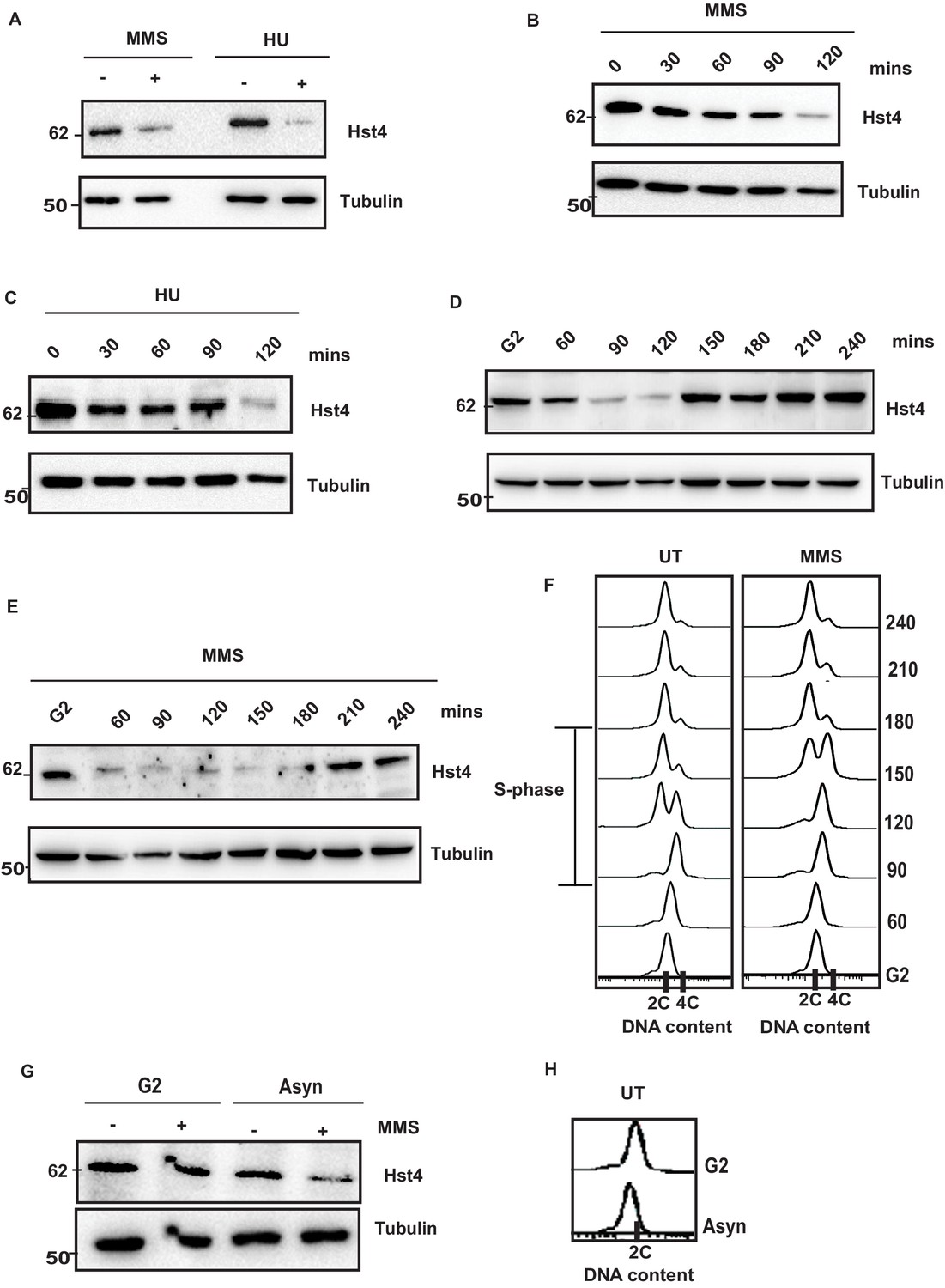
Replication stress leads to downregulation of Hst4.
(A) Wild-type (wt) strain (ROP238) containing TAP-tagged Hst4 was grown to 0.5 O.D., treated with 0.015 % MMS and 10 mM HU for 2 hr, respectively, whole cell extracts were prepared and immunoblotted with anti-PAP antibody to detect TAP-tagged Hst4. (B) (C) wt strain (ROP238) containing TAP-tagged Hst4 was grown to 0.5 O.D., treated with 0.015 % MMS and 10 mM HU, extracts were prepared from cells collected at indicated time points and western blot was performed. (D, E) The cdc25-22 cells expressing TAP-tagged Hst4 (DHP38) were first synchronized at G2 phase and then released into the S phase in the absence or presence of 0.015 % MMS, cells were collected at indicated time points and western blot was performed. (F) Flow cytometry profile of experiments in (D and E). (G) The cdc25-22 cells (DHP38) were synchronized at G2 phase by growing cells at 36 °C, after 2 hr 0.015 % MMS was added and grown for another 2 hr, asynchronous cells grown at 25 degrees were used for this experiment. (H) Flow cytometry profile of experiment in (G).
-
Figure 1—source data 1
Uncropped western blot images for Figure 1A– E and G.
- https://cdn.elifesciences.org/articles/70787/elife-70787-fig1-data1-v2.zip
Figure 1—figure supplement 1
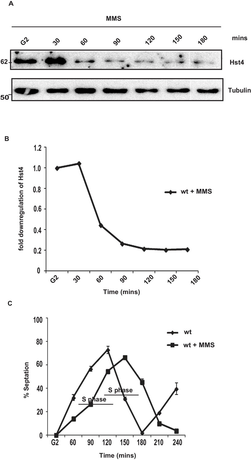
S-phase dependent downregulation of Hst4 upon MMS treatment.
(A) The cdc25-22 cells (DHP38) were first synchronized at G2 phase and then released into the S phase in the presence of 0.015 % MMS, time points were collected and western blot was performed. (B) Quantitation for the data shown in A. (C) Septation index for corresponding cell cycle arrest in Figure 1F showing percentage of cells with septa at each time point after release from G2 arrest. Calcofluor was used to stain the septa and DAPI was used to localize the nucleus.
Figure 2 with 1 supplement

The C terminus of Hst4 is required for proteasome dependent degradation of Hst4 upon replication stress.
(A) Half-life of Hst4 was measured by growing wt (ROP238) cells to O.D 0.5 and treating with cycloheximide (CHX) at100 µg/ml in the presence and absence of 0.015 % MMS. Cells were collected at indicated timepoints and western blot was performed. (B) wt strain (ROP238) containing TAP-tagged Hst4 and mts2-1 strain (DHP37) were grown to 0.5 O.D and treated with 0.015 % MMS for 2 hr and whole cell extracts were prepared and immunoblotted with anti-PAP antibody. The treatment was performed at 25 °C. (C) mts2-1 strain (DHP37) was grown to 0.5 O.D and cycloheximide (CHX) treatment at 100 µg/ml in the presence and absence of 0.015 % MMS was done, cells were collected at indicated timepoints and western blot was performed. The treatment was performed at non-permissive temperature (36 °C). (D) mts2-1 strain expressing pREP-6XHis-Ub or empty vector were grown to OD 0.5 and shifted to 36 °C for 1 hr followed by treatment with 0.015 % MMS for 2 hr or kept untreated. Ubiquitinated proteins were pulled down using Ni-NTA beads and western blot was performed and probed with anti-PAP antibody and anti-His antibody. (E) The hst4∆ strain (ROP57) transformed with GFP-Fl-Hst4-pSGP573 and GFP-C∆-Hst4-pSGP573 were grown to mid-log phase in EMM-Ura medium and treated with 0.015 % MMS and whole cell extracts were prepared and western blot was performed. (F) Quantification of data shown in (E). (G) The hst4∆ strain (ROP57) transformed with GFP-Fl-Hst4 and GFP-C∆-Hst4 were grown to mid-log phase in EMM-Ura medium, treated with 0.015 % MMS for 2 hr and live cell microscopy was performed and imaged under confocal microscope. Hoechst was used to stain the DNA.
-
Figure 2—source data 1
Uncropped western blot images for Figure 2A–E.
- https://cdn.elifesciences.org/articles/70787/elife-70787-fig2-data1-v2.zip
Figure 2—figure supplement 1
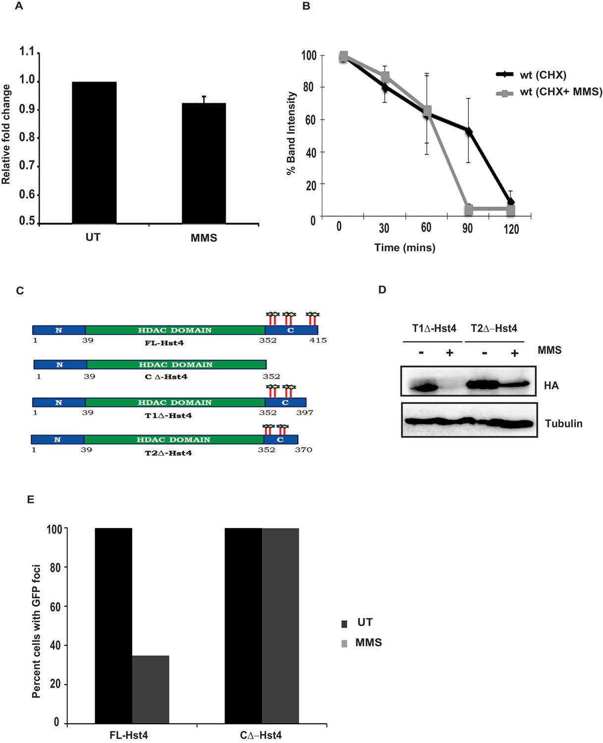
Hst4 is post-translationally regulated and minimum region required for Hst4 degradation lies at the C-terminus.
(A) Wild-type strain of Hst4 (ROP238) was grown in absence and presence of 0.015 % MMS and RNA was extracted and RT-qPCR was performed. The qPCR data are shown as expression fold changes after normalization against the actin mRNA control, and represent the mean ± SEM (n = 3). (B) Quantification of data shown in Figure 2A. Error bars represent SEM, n = 3. (C) The domain diagram of Hst4 showing different truncations containing potential phosphorylation sites at the C-terminus. (D) The hst4Δ strain (ROP57) transformed with T1Δ-Hst4-pSLF272 and T2Δ-Hst4-pSLF272 were grown to mid-log phase in EMM-Ura medium and treated with 0.015 % MMS for 2 hr and whole cell extracts were prepared and western blot was performed. (E) Quantification of immunofluorescence data shown in Figure 2G.
-
Figure 2—figure supplement 1—source data 1
Uncropped western blot images for Figure 2—figure supplement 1D.
- https://cdn.elifesciences.org/articles/70787/elife-70787-fig2-figsupp1-data1-v2.pdf
Figure 3 with 1 supplement
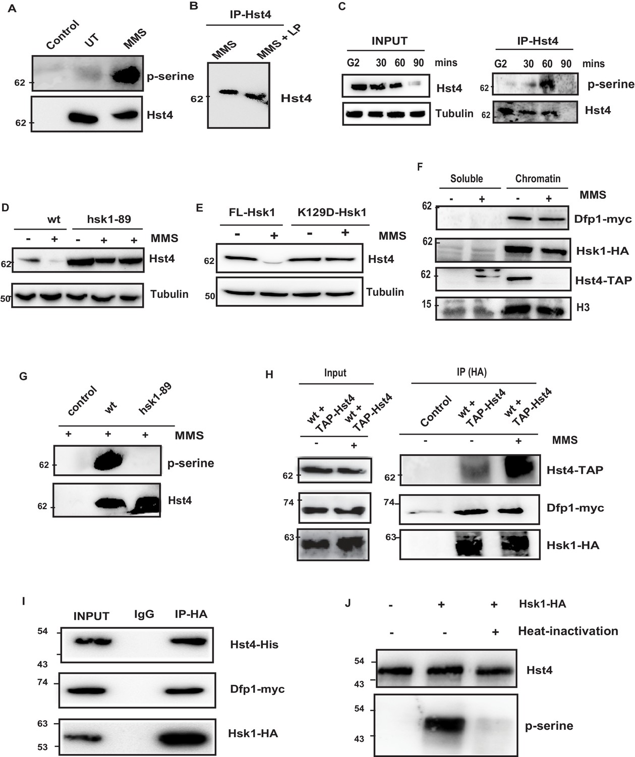
Replication-dependent regulation of Hst4 via phosphorylation by DDK/Hsk1.
(A) wt strain (ROP238) containing TAP-tagged Hst4 was grown to 0.5 O.D., treated with 0.015 % MMS for 1 hr and samples were immunoprecipitated using IgG-sepharose beads and western blot was performed with phosphoserine antibody. Extracts from untagged strain (ROP191) were used as control. (B) wt strain (ROP238) were treated with 0.015 % MMS, harvested, whole cell extracts were prepared and Hst4 was immunoprecipitated using IgG sepharose beads. Half of the immunoprecipitated sample were kept untreated and other half was treated with lamda phosphatase for 30 min. The IP samples were boiled in 2 X SDS loading buffer and ran on 8 % SDS Polyacrylamide gel and immunoblotted. (C) The cdc25-22 cells (DHP38) were first synchronized at G2 phase and then released into the S phase and cells were collected at indicated timepoints. Hst4 was immunoprecipitated using IgG-sepharose beads and western blot was performed with phosphoserine antibody. (D) wt (ROP238) and hsk1-89 (DHP78) strains containing TAP-tagged Hst4 were grown to 0.5 O.D. and treated with 0.015 % MMS for indicated time points and whole cell extracts were prepared and immunoblotted with anti-PAP antibody. The hsk1-89 strain was grown at 25 °C till O.D. 0.5 and then shifted to 30 °C for inactivation of kinase activity for 1 hr and then treated with MMS for 2 hr and 4 hr. (E) The complementation of hsk1-89 strain with FL-Hsk1 (pSLF272) or kinase dead K129D-Hsk1 (pSLF272) plasmid constructs. The transformants were grown in EMM-Ura at 25 °C and treated with 0.015 % MMS for 2 hr after shifting to 30 °C, whole cell extracts were prepared and immunoblotted with anti-PAP antibody. (F) wt (ROP238) and DHP115 strains was grown to OD 0.5 and treated with 0.015 % MMS for 2 hr. The chromatin fractionation was performed as mentioned in the methods followed by western blot to detect indicated proteins. (G) wt (ROP238) and hsk1-89 (DHP78) strains were grown and treated with 0.015 % MMS for 1 hr and immunoprecipitation of Hst4 was done. The hsk1-89 strain was shifted to 30 °C for inactivation of kinase activity and then treated with MMS. Extracts from untagged strain (ROP191) were used as control. (H) The DHP115 strain containing hsk1-HA and dfp1-myc was transformed with TAP-tagged Hst4- pREP81 vector was grown to O.D. 0.5, treated with 0.015 % MMS for 1 hr and crosslinking was performed by treating with 1 % formaldehyde for 30 min. Whole cell extracts were prepared and immunoprecipitated using anti-HA antibody and western blot was performed. (I) Recombinant FL-Hst4 (pET28a) was expressed in BL21 cells and purified by Ni-NTA beads. The Hsk1-HA was purified from fission yeast strain containing hsk1-HA and dfp1-myc (DHP115) and bound to protein A/G beads after immunoprecipitation. Beads enriched with Hsk1 were incubated with recombinant Hst4, washed twice with lysis buffer. IgG was used as control. Western blot was performed with anti-His, anti-HA and anti-myc antibodies. (J) Recombinant FL-Hst4 (pET28a) was expressed in BL21 cells and purified by Ni-NTA beads. The Hsk1-HA was purified from fission yeast strain (DHP115) and in vitro kinase assay was performed as described in materials and methods.
-
Figure 3—source data 1
Uncropped western blot images for Figure 3B–J.
- https://cdn.elifesciences.org/articles/70787/elife-70787-fig3-data1-v2.zip
-
Figure 3—source data 2
- https://cdn.elifesciences.org/articles/70787/elife-70787-fig3-data2-v2.zip
Figure 3—figure supplement 1
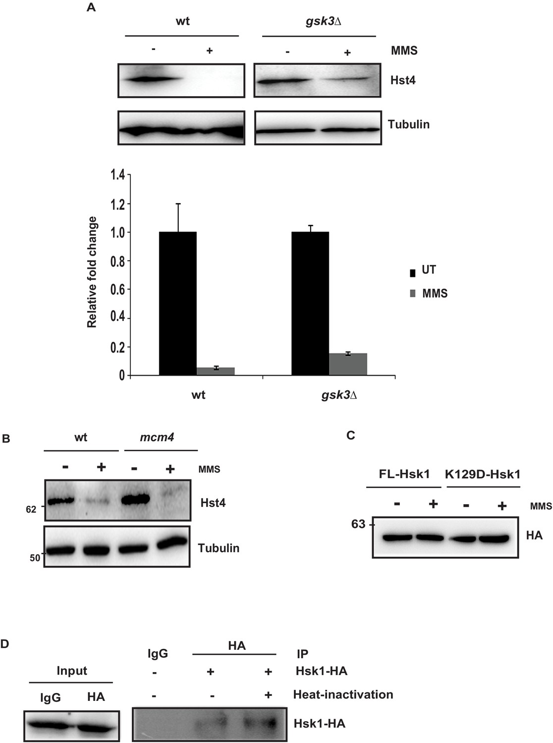
Gsk3-beta and Mcm4 are not required for downregulation of Hst4.
(A) The wt (ROP238) and gsk3∆ strain (DHP77) were grown to mid-log phase in YES medium, treated with 0.015 % MMS for 2 hr, whole cell extracts were prepared and western blot was performed. Quantitation of the blot is shown. (B) wt strain (ROP238) and mcm4 (DHP172) containing TAP-Hst4 was grown to 0.5 O.D. and treated with 0.015 % MMS for 2 hr immunoblotted with anti-PAP antibody. The mcm4 strain was shifted to 36 °C for 2 hr and then treated with MMS. (C) The complementation of hsk1-89 strain with FL-Hsk1 (pSLF272) or kinase dead K129D-Hsk1 (pSLF272) plasmid constructs. The transformants were grown in EMM-Ura at 25 °C and treated with 0.015 % MMS for 2 hr after shifting to 30 °C, whole cell extracts were prepared and immunoblotted with anti-HA antibody to check the expression of Hsk1. (D) Western blot showing the immunoprecipitated Hsk1 for the experiment in Figure 3J. IgG was used as control for IP.
-
Figure 3—figure supplement 1—source data 1
Uncropped western blot images for Figure 3—figure supplement 1A, B, C.
- https://cdn.elifesciences.org/articles/70787/elife-70787-fig3-figsupp1-data1-v2.pdf
Figure 4 with 1 supplement
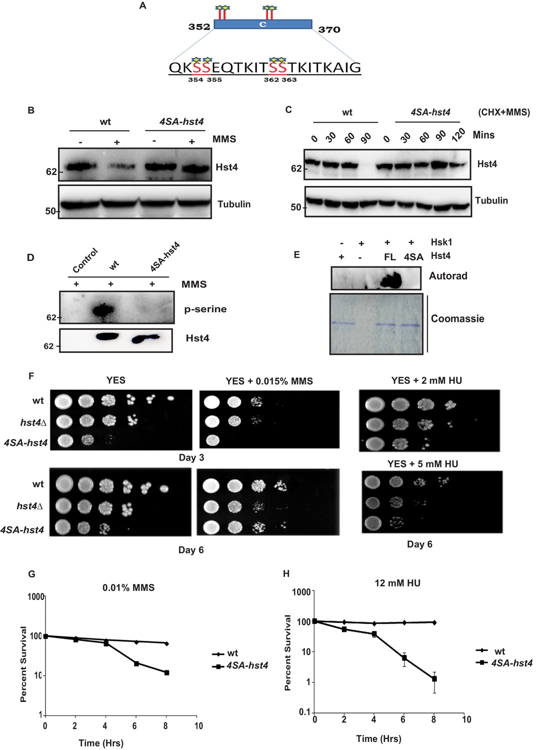
Phosphorylation of Hst4 at serine residues 354, 355, 362, and 363 is required for cell survival upon replication stress.
(A) The C-terminal residues 352–370 required for degradation of Hst4 are shown. The serine residues marked with star, which are putative consensus DDK sites were mutated to alanines by site-directed mutagenesis. (B) wt strain (ROP238) and 4SA-hst4 strain (DHP146) was grown to 0.5 O.D., treated with 0.015 % MMS for 2 hr, whole cell extracts were prepared and immunoblotted with indicated antibodies. (C) wt (ROP238) and 4SA-hst4 (DHP146) strains were grown to 0.5 O.D, cycloheximide (CHX) treatment at 100 µg/ml in the presence and absence of 0.015 % MMS was done, cells were collected at indicated timepoints and western blot was performed. (D) wt strain (ROP238) and 4SA-hst4 strain (DHP146) was grown to 0.5 O.D., treated with 0.015 % MMS for 1 hr and samples were immunoprecipitated using IgG-sepharose beads and western blot was performed with phosphoserine antibody. TAP-tagged Hst4 was probed with anti-PAP antibody. Extracts from untagged strain (ROP191) were used as control. (E) Hsk1-HA was purified from DHP115 strain using anti-HA antibody. The recombinant FL-Hst4 and 4SA-Hst4 proteins were purified from BL21 cells and in vitro kinase assay was performed. The image shows autoradiogram and coomassie staining is shown for protein expression. (F) Spot assay showing sensitivity of non-degradable Hst4 in shown concentrations of HU and MMS containing plates. Indicated strains were grown to OD one and fivefold serial dilutions were prepared and plated onto YES, YES + HU and YES + MMS plates or EMM-Ura plates and incubated at 30 °C for 3–6 days. (G) (H) Mid-log phase cultures of wild-type and 4SA-hst4 cells were treated with 0.015 % MMS and 12 mM HU (time 0). Samples were taken at the indicated timepoints, washed free of MMS and HU, and viability was determined by colony formation on YES plates for 4 days at 30 °C. Error bars represent SEM, n = 3, and are normalized to untreated cells.
-
Figure 4—source data 1
Uncropped western blot images for Figure 4C–E.
- https://cdn.elifesciences.org/articles/70787/elife-70787-fig4-data1-v2.zip
Figure 4—figure supplement 1
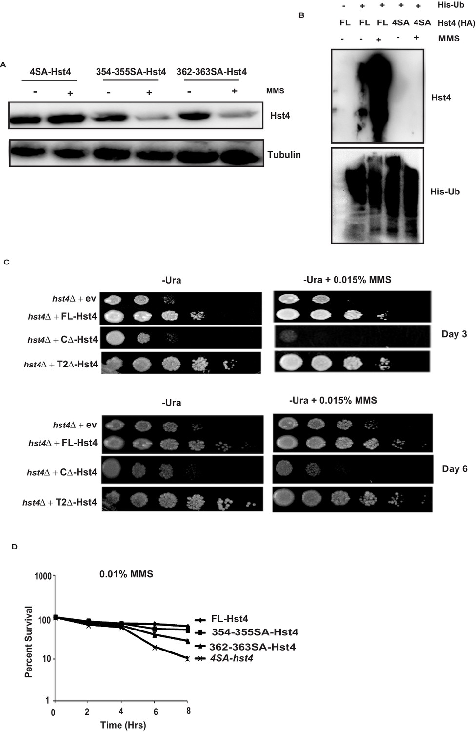
Stability and sensitivity of Hst4 C-terminal domain deletion and serine to alanine mutants in replication stress.
(A) The hst4∆ strain (ROP57) transformed with 4SA-Hst4-pSLF273 (354,355,362,363), 354-355SA-Hst4-pSLF273, and 362-363SA-Hst4-pSLF273 were grown to mid-log phase in EMM-Ura medium, treated with 0.015 % MMS for 2 hr and whole cell extracts were prepared and western blot was performed. (B) mts2-1 strain expressing pREP-6XHis-Ub or empty vector along with FL-Hst4 (pSLF273) or 4SA-Hst4 (pSLF273) were grown to OD 0.5 and shifted to 36 °C for 1.5 hr followed by treatment with 0.015 % MMS for 2 hr or kept untreated. Ubiquitinated proteins were pulled down using Ni-NTA beads and western blot was performed and probed with anti-HA antibody and anti-His antibody. (C) Spot assay showing sensitivity of truncation constructs CΔ-Hst4-pSLF273 and T1Δ-Hst4-pSLF273 in shown concentrations of MMS containing plates. Indicated strains were grown to OD one and fivefold serial dilutions were prepared and plated onto EMM-Ura plates with or without MMS and incubated at 30 °C and photographs taken on day 3 and day 6 are shown. (D) Mid-log phase cultures of hst4∆ strain (ROP57) transformed with FL-Hst4, 354-355SA-Hst4, 362-363SA-Hst4, and 4SA-Hst4 (354,355,362,363SA) cloned in pSLF273 vector, were treated with 0.015 % MMS (time 0). Samples were taken at the indicated timepoints, washed free of MMS and viability was determined by colony formation on YES plates for 4 days at 30 °C. Error bars represent SEM, n = 3, and are normalized to untreated cells.
-
Figure 4—figure supplement 1—source data 1
Uncropped western blot images for Figure 4—figure supplement 1A and B.
- https://cdn.elifesciences.org/articles/70787/elife-70787-fig4-figsupp1-data1-v2.pdf
Figure 5 with 1 supplement
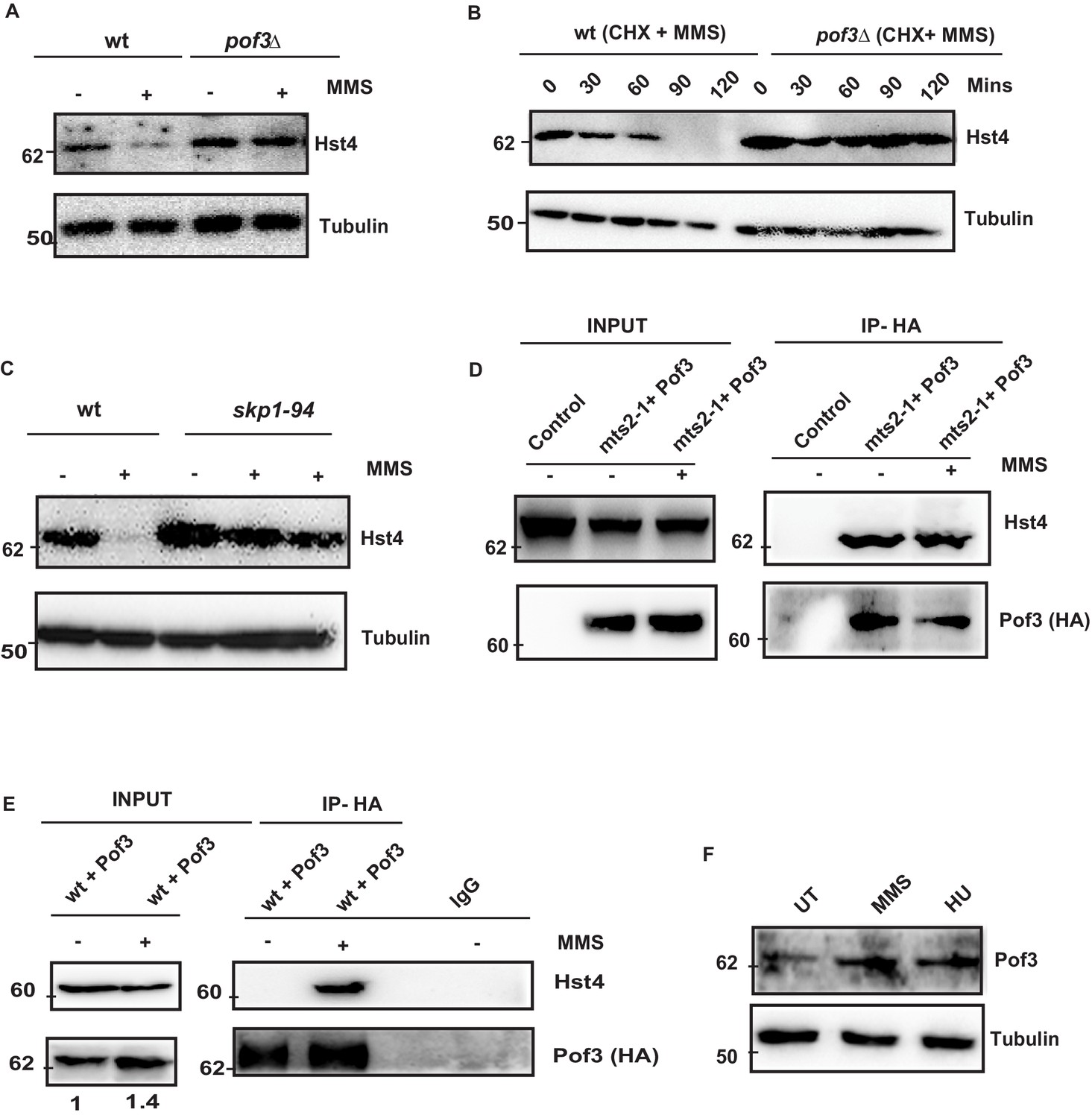
SCFPof3-mediated degradation of Hst4 on replication stress.
(A) wt strain (ROP238) and F box mutant strain pof3Δ (DHP35) containing TAP-tagged Hst4 were grown to 0.5 O.D., treated with 0.015 % MMS for 2 hr and whole cell extracts were prepared and immunoblotted with anti-PAP antibody. (B) wt strain (ROP238) and pof3Δ (DHP35) was grown to 0.5 O.D, treated with cycloheximide (CHX) at 100 µg/ml inpresence and absence of 0.015 % MMS, cells were collected at indicated timepoints and western blot was performed. (C) wt strain (ROP238) and skp1-94 (DHP57) containing TAP-tagged Hst4 were grown to 0.5 O.D. at 25 °C, shifted to 36 °C, after 1 hr, 0.015 % MMS was added, wt cells were collected after 2 hours of treatment and skp1-94 cells were collected after 2 and 3 hr. (D) HA-Pof3 in pSLF272 vector was expressed in mts2-1 strain (DHP37). Cells were grown to midlog phase in EMM-Ura medium at 25 °C, shifted to 36 °C and after 1 hr, 0.015 % MMS was added and treated for 1 hr. Pof3 was immunoprecipitated using anti-HA antibody and western blot was performed using indicated antibodies. The extract from untransformed strain (DHP37) was used as a control. (E) HA-Pof3 in pSLF272 vector was expressed in wt strain (ROP238). Cells were grown to midlog phase in EMM-Ura medium and then treated with 0.015 % MMS for 1 hr. Pof3 was immunoprecipitated using anti-HA antibody and western blot was performed using indicated antibodies. Imunoprecipitation by IgG antibody was used as control. (F) Whole cell levels of Pof3 (ENY3684) upon treatment with 0.015 % MMS and 10 mM HU, respectively were checked by western blot using anti-myc antibody.
-
Figure 5—source data 1
Uncropped western blot images for Figure 5A–C and E.
- https://cdn.elifesciences.org/articles/70787/elife-70787-fig5-data1-v2.zip
Figure 5—figure supplement 1
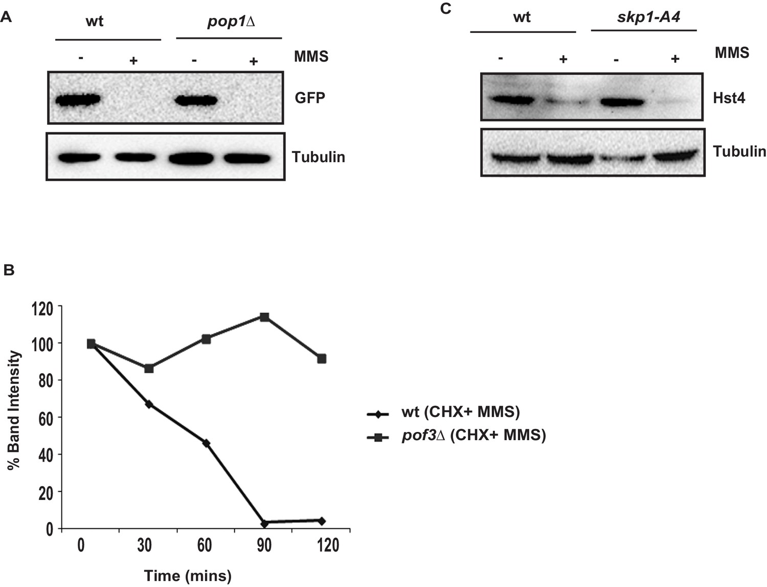
Pop1 is not required for downregulation of Hst4.
(A) The wt (ROP238) and pop1∆ strain transformed with FL-Hst4-pSGP573 were grown to mid-log phase in EMM-Ura medium, treated with 0.015 % MMS for 2 hr, whole cell extracts were prepared and western blot was performed. (B) Quantification of data shown in Figure 5B. (C) The wt (ROP238) and skp1-A4 (DHP56) strains were grown to mid-log phase and treated with 0.015 % MMS for 2 hr, whole cell extracts were prepared and western blot was performed. The skp1-A4 strain was shifted to 36 °C for 2 hr and then treated with MMS.
-
Figure 5—figure supplement 1—source data 1
Uncropped western blot images for Figure 5—figure supplement 1C.
- https://cdn.elifesciences.org/articles/70787/elife-70787-fig5-figsupp1-data1-v2.pdf
Figure 6 with 1 supplement
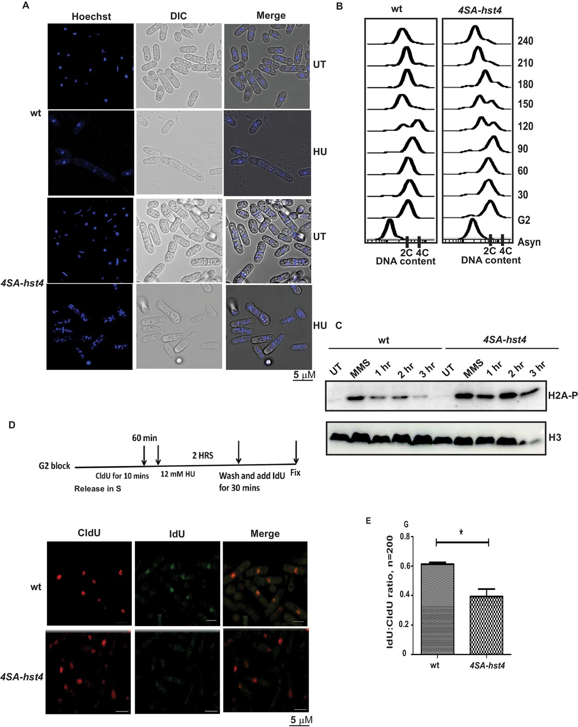
Dynamic regulation of Hst4 is required for stable fork stalling and recovery from replication stress.
(A) Wild-type strain (ROP238) and 4SA-hst4 (DHP146) were grown to OD 0.5 and treated with 12 mM HU for 6 hr and DNA was stained with Hoechst and life cell microscopy was performed. Scale- 5 µM. (B) The cdc25-22 (FY4225) and cdc25-22 4SA-hst4 (DHP148) grown to midlog phase in YES medium at 25 °C, were shifted to 36°C for 4 hr to synchronize at G2 phase, cells were collected every 30 min and cell cycle profile was analyzed by flow cytometry. (C) wt type strain (ROP238) and 4SA-hst4 (DHP146) were grown to OD 0.5 and treated with 0.03 % MMS for 2 hr, cells were washed and allowed to recover till 3 hr in fresh medium. Samples were collected at indicated timepoints and western blot was performed. (D) The cdc25-22 (FY4225) and cdc25-22 4SA-hst4 (DHP148) strains engineered to incorporate halogenated nucleotides were grown to midlog phase in YES medium at 25 °C, were shifted to 36°C for 4 hr to synchronize at G2 phase, released into the cell cycle, 60 min post-release, CldU was added for 10 min, washed off, treated with 12 mM HU for 2 hr, washed off and allowed to recover in the presence of IdU for 30 min. Cells were fixed and immunofluorescence was performed after dual staining with CldU and IdU antibodies as mentioned in materials and methods. (E) The graph showing ratio of IdU to CldU indicating fork restart. A total of 100–200 cells were counted from two independent experiments and statistical analysis was performed. * indicating p < 0.05.
-
Figure 6—source data 1
Uncropped western blot images for Figure 6C.
- https://cdn.elifesciences.org/articles/70787/elife-70787-fig6-data1-v2.zip
Figure 6—figure supplement 1
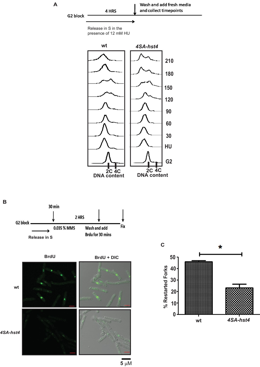
4SA-hst4 is required for DNA replication fork restart post replication stress.
(A) The cdc25-22 (FY4225) and cdc25-22 4SA-hst4 (DHP148) strains engineered to incorporate halogenated nucleotides were grown to midlog phase in YES medium at 25 °C, were shifted to 36°C for 4 hr to synchronize at G2 phase, released into the cell cycle in the presence of 15 mM HU for 4 hr to induce early S-phase arrest, cells were washed off and recovery timepoints were collected, Flow Cytometry was performed on indicated timepoints. (B) The cdc25-22 (FY4225) and cdc25-22 4SA-hst4 (DHP148) strains engineered to incorporate halogenated nucleotides were grown to midlog phase in YES medium at 25 °C, were shifted to 36°C for 4 hr to synchronize at G2 phase, released into the cell cycle, 30 min post-release, treated with 0.035 % MMS for 3 hr, washed off and allowed to recover in the presence of BrdU for 30 min. Cells were fixed and immunofluorescence was performed after staining BrdU antibody as mentioned in methods. (C) The graph showing percent restarted forks where brdU positive cells were counted. 100–200 cells were counted from two independent experiments and statistical analysis was performed. * indicating p < 0.05.
Figure 7 with 1 supplement
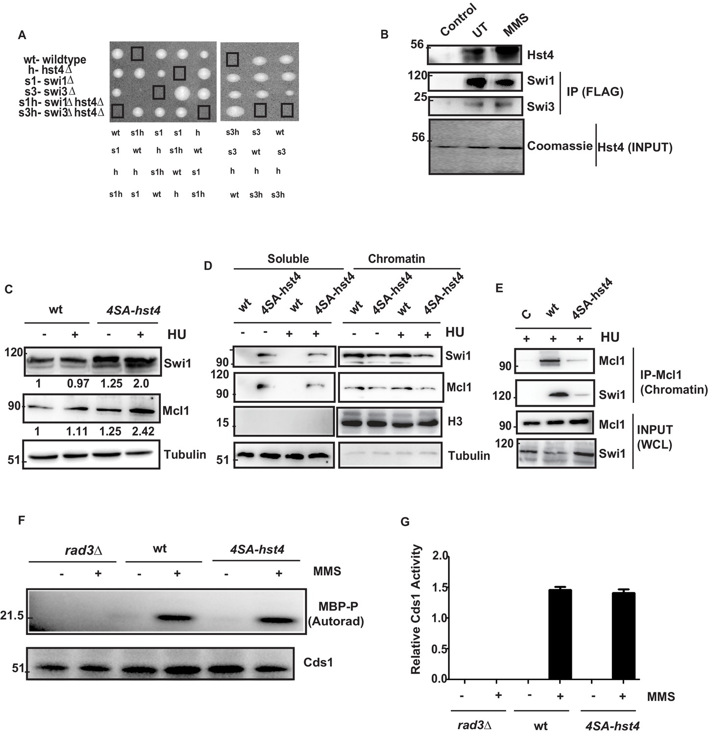
The degradation of Hst4 is required for the stable association of Fork Protection Complex at the replication fork.
(A) Tetrads each from the hst4∆ strain (ROP58) crossed with the swi1∆ (EN3182) and swi3∆ (EN3366) are shown, photographed after 4 days of growth on YES plates. The genotypes were determined by replica plating on selection media are shown. (B) Recombinant FL-Hst4 (pET28a) was expressed in BL21 cells and purified by Ni-NTA beads. Swi1 (EN3381) or Swi3 (EN3382) FLAG-tagged strains were grown, immunoprecipitation was performed using FLAG M2 beads and incubated with recombinant Hst4 to check interaction. IgG was used as control. Western blot was performed with anti-FLAG antibody. Coomasie staining shows the expression of FL-Hst4. (C) wt (DHP143) and 4SA-hst4 (DHP144) with FLAG tagged Swi1 were grown to OD 0.5, treated with 12 mM HU for 2 hr, whole cell extracts were prepared and immunoblotted with indicated antibodies. (D) wt (DHP143) and 4SA-hst4 (DHP144) were grown to OD 0.5 and treated with 12 mM HU for 4 hr. The chromatin fractionation was performed as mentioned in the methods followed by western blot to detect indicated proteins. (E) wt (DHP143) and 4SA-Hst4 (DHP144) were grown to OD 0.5, treated with 12 mM HU for 4 hr. Chromatin fractionation was performed and chromatin associated proteins were immunoprecipitated using anti-And-1 (Mcl1) antibody. Western blot was performed using indicated antibodies. (F) wt (yFS988), rad3∆ (DHP173), and 4SA-hst4 (DHP174) containing Cds1-HA was grown till 0.5 OD and treated with 0.03 % MMS for 2 hr. Cds1 was immunoprecipitated with anti-HA antibody and in vitro Cds1 kinase activity was monitored using MBP as the substrate and γp32 ATP was incorporated. The figure shows autoradiogram after 24 hr of exposure. (G) The graph showing relative kinase activity normalized to total Cds1 from two independent experiments. Error bars represent SEM.
-
Figure 7—source data 1
Uncropped western blot images for Figure 7B–D and F.
- https://cdn.elifesciences.org/articles/70787/elife-70787-fig7-data1-v2.zip
Figure 7—figure supplement 1
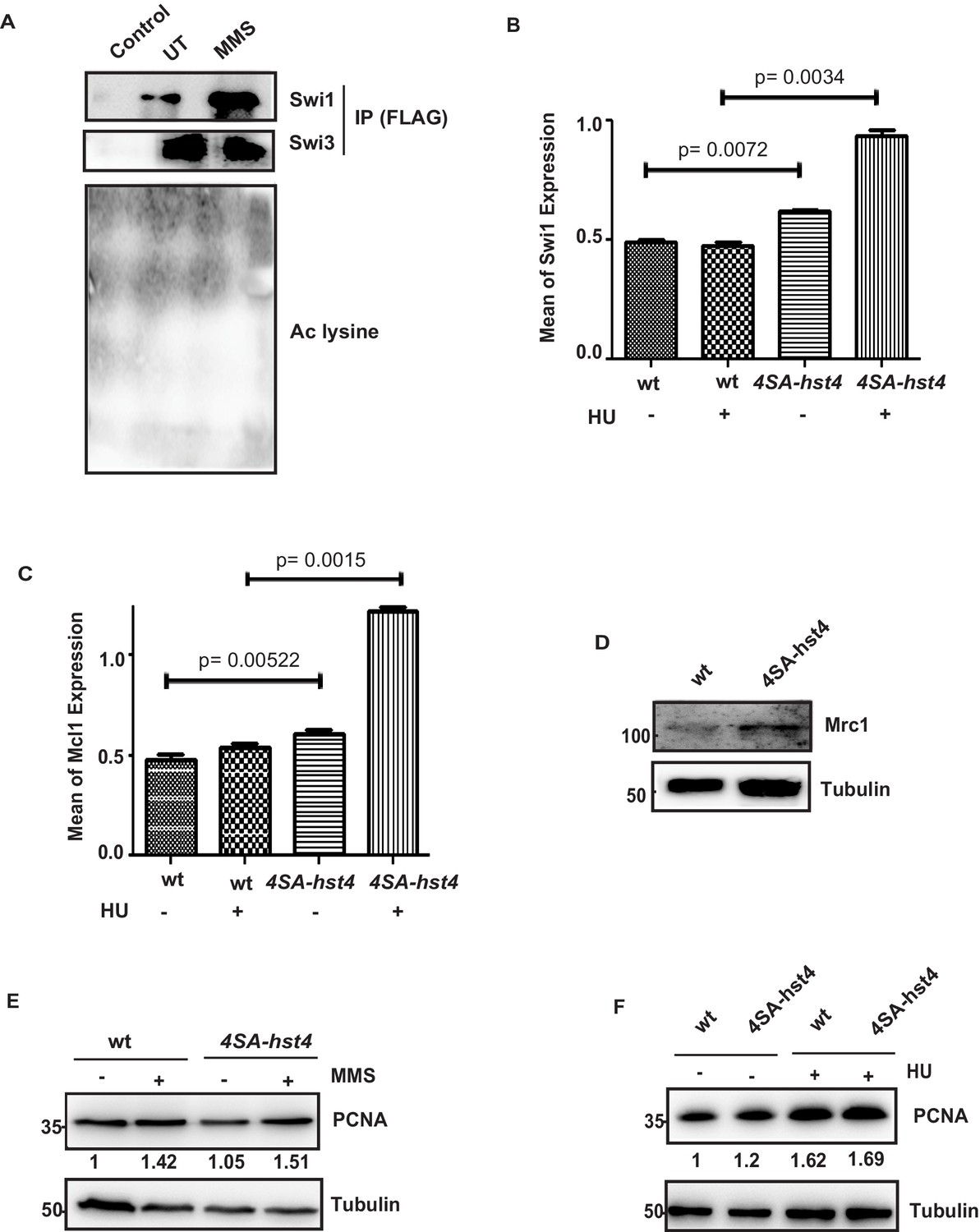
Regulation of replisome components in 4SA-hst4 mutant.
(A) The wt Swi1 (EN3381) or Swi3 (EN3382) FLAG-tagged strains were grown and immunoprecipitation was performed using FLAG M2 beads, followed by western blot and probed with anti-FLAG and ant-acetyl lysine antibodies. (B) Quantitation of the levels of Swi1 from the data shown in Figure 7C. The errors bar represent SEM. The p value less than 0.05 is significant. (C) Quantitation of the levels of Mcl1 from the data shown in Figure 7C. The errors bar represent SEM. The p value less than 0.05 is significant (D) The wt (DHP152) and 4SA-hst4 (DHP177) containing mrc1-myc were grown to mid-log phase, whole cell extracts were prepared and western blot was performed with anti-myc antibody. (E) The wt (ROP238) and 4SA-hst4 strain (DHP146) were grown to mid-log phase and treated with 0.015 % MMS for 2 hr and whole cell extracts were prepared and western blot was performed with anti-PCNA antibody. (F) The wt (ROP238) and 4SA-hst4 strain (DHP146) were grown to mid-log phase and treated with 10 mM HU for 2 hr and whole cell extracts were prepared and western blot was performed with anti-PCNA antibody.
-
Figure 7—figure supplement 1—source data 1
Uncropped western blot images for Figure 7—figure supplement 1D.
- https://cdn.elifesciences.org/articles/70787/elife-70787-fig7-figsupp1-data1-v2.pdf
Figure 8 with 1 supplement
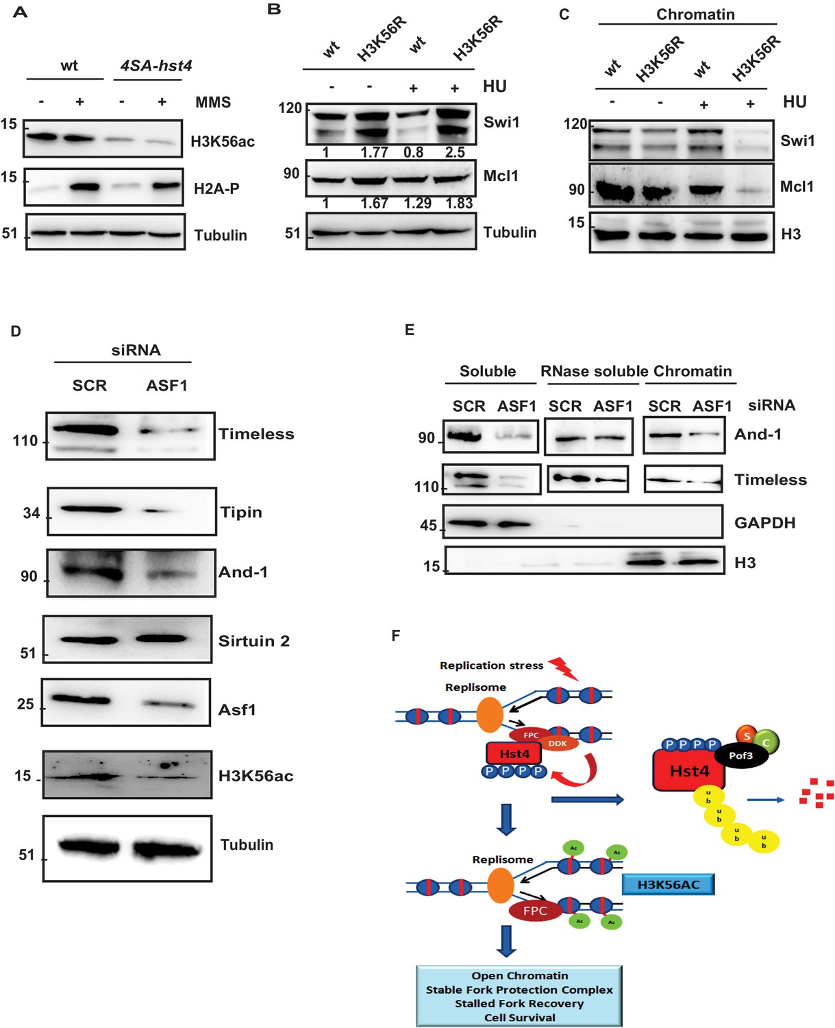
H3K56 acetylation-dependent stabilization of Fork Protection Complex at the chromatin regulates genomic stability.
(A) wt strain (ROP238) and 4SA-hst4 strain (DHP146) were grown to 0.5 O.D., treated with 0.015 % MMS for 2 hr, whole cell extracts were prepared and immunoblotted with indicated antibodies. (B) wt strain and H3K56R strain (DHP169) were grown to 0.5 O.D.treated with 12 mM HU for 2 hr, whole cell extracts were prepared and immunoblotted with indicated antibodies. (C) wt and H3K56R strain (DHP169) was grown to 0.5 O.D. treated with 12 mM HU for 4 hr, chromatin fractionation followed by western blot was performed. (D) U2OS cells were transfected with scramble and Asf1a siRNA. At 48 hr post transfection, whole cell extracts were prepared and western blot was performed to detect indicated proteins. The expression of Asf1a and H3K56ac was detected by respective antibodies. Tubulin was checked as a loading control (E) To study chromatin association of FPC, U2OS cells were transfected with scramble and Asf1a siRNA. At 48 h post transfection, chromatin was extracted as described in materials and methods and western blot was performed to detect FPC components. GAPDH and H3 were Checked as controls for fractionation. (F) Model for the role of dynamic regulation of Hst4 in maintenance of FPC stability via H3K56ac. In S-phase or upon replication stress, DDK/Hsk1 interacts with Hst4 and phosphorylates it at serine residues at the C-terminus. The phosphorylated Hst4 is recognized and ubiquitinated by SCFpof3 ubiquitin ligase and degraded via proteasome. Degradation of Hst4 increases H3K56ac levels leading to stable maintenance of FPC Swi1 and Mcl1 at the chromatin which helps in fork recovery and cell survival.
-
Figure 8—source data 1
Uncropped western blot images for Figure 8A–E.
- https://cdn.elifesciences.org/articles/70787/elife-70787-fig8-data1-v2.zip
-
Figure 8—source data 2
- https://cdn.elifesciences.org/articles/70787/elife-70787-fig8-data2-v2.zip
Figure 8—figure supplement 1
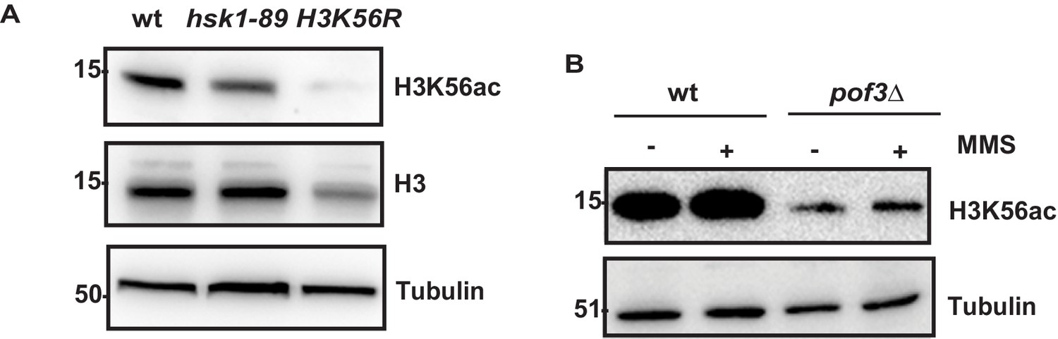
Hypoacetylation of histone H3K56 in hsk1-89 and pof3 mutant cells.
(A) The wt (ROP238), hsk1-89 (DHP78) and H3K56R strain (ROP253) were grown to mid-log phase, whole cell extracts were prepared and western blot was performed. (B) The wt (ROP238) and pof3∆ (DHP35) were grown to mid-log phase, treated with 0.015 % MMS for 2 hr and whole cell extracts were prepared and western blot was performed.
-
Figure 8—figure supplement 1—source data 1
Uncropped western blot images for Figure 8—figure supplement 1A and B.
- https://cdn.elifesciences.org/articles/70787/elife-70787-fig8-figsupp1-data1-v2.pdf
Tables
Key resources table
| Reagent type (species) or resource | Designation | Source or reference | Identifiers | Additional information |
|---|---|---|---|---|
| Antibody | Anti-PAP(Peroxidase Peroxidase Soluble Complex antibody produced in rabbit) | Sigma-Aldrich | Cat#P1291RRID:AB_1079562 | WB (1:5,000) |
| Antibody | Anti-HA(rabbit polyclonal antibody) | Bethyl laboratories | Cat#A190-208ARRID:AB_67466 | WB (1:10,000) |
| Antibody | Anti-HA(rabbit polyclonal antibody) | Abcam | Cat#ab9110RRID:AB_307019 | WB (1:5000)IP- (4 µg) |
| Antibody | Anti-H3K56ac(rabbit polyclonal antibody) | Abcam | Cat#ab71956RRID:AB_10861799 | WB (1:2,000) |
| Antibody | Anti-H3(rabbit polyclonal antibody) | Abcam | Cat#ab1791RRID:AB_302613 | WB (1:5,000) |
| Antibody | Anti-FLAG(mouse monoclonal antibody) | Sigma-Aldrich | Cat#F1804RRID:AB_262044 | WB (1:2,000) |
| Antibody | Anti-myc(mouse monoclonal antibody) | Sigma-Aldrich | Cat#M4439RRID:AB_439694 | WB (1:5,000) |
| Antibody | Anti-And-1 (Mcl1)(mouse monoclonal antibody) | Biolegend | Cat#630,301RRID:AB_2215084 | WB (1:5,000)IP- (2 µg) |
| Antibody | anti-GFP(mouse monoclonal antibody) | Cell Signalling | Cat#2,955RRID:AB_1196614 | WB (1:2,000)IF- (1:250) |
| Antibody | Anti-His(mouse monoclonal antibody) | BD biosciences | Cat#552,565RRID:AB_394432 | WB (1:5,000) |
| Antibody | Anti-Phosphoserine(rabbit polyclonal antibody) | Abcam | Cat#ab9332RRID:AB_307184 | WB (1:3,00) |
| Antibody | H2A-P(rabbit polyclonal antibody) | Abcam | Cat#ab15083RRID:AB_301630 | WB (1:5,000) |
| Antibody | Anti-BrdU(mouse monoclonal antibody) | BD biosciences | Cat#347,580RRID:AB_400326 | IF (1:1,00) |
| Antibody | Anti-CldU(rat monoclonal antibody) | Abcam | Cat#ab6326RRID:AB_305426 | IF (1:1,00) |
| Antibody | Anti-Timeless(rabbit monoclonal antibody) | Abcam | Cat#ab109512RRID:AB_10863023 | WB (1:2000) |
| Antibody | Anti-Tipin | Abcam | Cat#ab229329 | WB (1:2000) |
| Antibody | Anti-Sirt2(rabbit monoclonal antibody) | Cell Signalling | Cat#D4O5ORRID:AB_2716762 | WB (1:2500) |
| Antibody | Anti-GAPDH(rabbit monoclonal antibody) | Cell Signalling | Cat#14C10RRID:AB_561053 | WB (1:5000) |
| Strain, strain background (Escherichia coli) | BL21(DE3) pLysS | Lab stock | Chemically Competent cells | |
| Cell line (Homo-sapiens) | U2OS(Osteosarcoma), cells | ATCC | ATCC ID: HTB-96RRID:CVCL_0042 | Gift from Dr. Rashna Bhandari’s lab |
| Transfected construct (human) | siRNA toAsf1a | Thermo Fisher Scientific | Cat#4392420Assayid- S226044 | |
| Recombinant DNA reagent | pSLF273 | Forsburg Lab | Cloning of FL-Hst4, T1Δ-Hst4, T2Δ-Hst4 and point mutations. | |
| Recombinant DNA reagent | pSLF272 | Forsburg Lab | Cloning of FL-Hsk1, KD-Hsk1 and Pof3 | |
| Recombinant DNA reagent | pET28a | Lab stock | Cloning of FL-Hst4 and 4SA-Hst4 | |
| Recombinant DNA reagent | pSGP573 | Forsburg Lab | Cloning of FL-Hst4 and CΔ-Hst4 | |
| Recombinant DNA reagent | pREP1-His6-ubiquitin | Takashi Toda | ||
| Chemical compound, drug | Methyl methane sulphonate (MMS) | Sigma-Aldrich | Cat#129,925 | |
| Chemical compound, drug | Hydroxyurea (HU) | Sigma-Aldrich | Cat#H8627 | |
| Other | Bisbenzimide Hoechst | Sigma-Aldrich | Cat#B2261 | 10 µg/µl |
| Software, algorithm | ImageJ | ImageJ | https://imagej.net/Fiji/Downloads | Quantification of western blots |
| Software, algorithm | Graphpad Prism 6.0 | Graphpad | https://www.graphpad.com/ scientific-software/prism// | Statistical analysis and graphs representation |
| Software, algorithm | FlowJo | BD Life Sciences | RRID:SCR_008520 |
Table 1
List of strains used in the study.
| Strain | Genotype | Source |
|---|---|---|
| ROP238 | h + ade6 M216 arg3-D4 his3-D1 leu1-32 ura4-D18 hst4-TAP:KanR | Lab stock |
| ROP191 | h_ ade6-210 arg3-D4 his3-D1 leu1-32 ura4-D18 | Lab stock |
| DHP38 | h + cdc25-22 ade6-216 arg3-D4 his3-D1 leu1-32 ura4-D18 hst4-TAP:KanR | This Study |
| DHP37 | h- ade6-216 arg3-D4 his3-D1 leu1-32 ura4-D18 hst4-TAP:KanR mts2-1 | This Study |
| ROP57 | h + ade6 M216 arg3-D4 his3-D1 leu1-32 ura4-D18 hst4∆-his3+ | Lab stock |
| DHP57 | h + skp1-94 his3-D1 arg3-D4 leu1-32 ura4-D18 ade? hst4-TAP:KanR | This Study |
| DHP56b | h- skp1-A4 his3-D1 arg3-D4 leu1-32 ura4-D18 ade? hst4-TAP:KanR | This Study |
| SKP471 | h− leu1-32 ura4-D18 pof3Δ::kan | Takashi Toda |
| TP403-2B | h90 leu1-32 ura4-D18 pop1::ura+ | Takashi Toda |
| DHP35 | h- arg3-D4 his3-D1 leu1-32 ura4-D18 pof3::kanR hst4-TAP:KanR | This Study |
| DHP78 | h + hsk1-89:ura4+ his3-D1arg3-D4 leu1-32 ura4-D18 ade? hst4-TAP:KanR | This Study |
| ROP247 | h + h3.2 K56R h3.1∆/h4.1∆::his3+ h3.3∆ /h4.3∆::arg3+ leu1-32 ura4-D18 his3-D1 arg3-D4 ade6-210 | Lab collection |
| DHP77 | h + ade6-216 arg3-D4 his3-D1 leu1-32 ura4-D18 hst4-TAP:KanR gsk3::ura4+ | This Study |
| ENY3684 | h + pof3-myc:hphMX6 pol2-3FLAG:kanMX6 leu1-32 ura4-D18 | Eishi Noguchi |
| yFS988 | h- ura4-D18 ade-? cdc10-M17 leu1-32::pYJ294(leu1 cds1-6his2HA) | Nick Rhind |
| FY4225 | h- cdc25-22 leu1::hENT-leu1+ his7-366::hsv-tk-his7+ ura4? ade6-M210 | Susan.L.Forsburg |
| FY1077 | h- hsk1HA::ura4+ ura4-D18 leu1-32 ade6-M216 | Susan Forsburg |
| FY1763 | h + leu1:dfp1 +6his3HA leu1+ dfp1-D1 ura4-D18 ade6-M216 | Susan Forsburg |
| DHP146 | h- ade6-216 arg3-D4 his3-D1 leu1-32 ura4-D18 4SA-hst4-TAP:KanR | This Study |
| DHP148 | h- cdc25-22 leu1::hENT-leu1+ his7-366::hsv-tk-his7+ ura4? ade6-M210 4SA-hst4-TAP::KanR | This Study |
| DHP168 | h- h3.1/h4.1Δ::his3+ h3.3/h4.3Δ::arg3+ leu1-32 ura4-D18 arg3-D ade6-M210 swi1-3FLAG::KanR | This Study |
| DHP169 | h + H3.2 K56R h3.1/h4.1Δ::his3+ h3.3/h4.3Δ::arg3+ leu1-32 ura4-D18 arg3-D ade6-M210 swi1-3FLAG::KanR | This Study |
| MS193 | h- leu1-32 ura4-D18 cdc21 | Hisao Masai |
| DHP172 | h + leu1-32 ura4-D18 ade6-M210 cdc21 (mcm4) hst4-TAP::Kan | This Study |
| DHP173 | h + ura4-D18 ade-? leu1-32::pYJ294(leu1 cds1-6his2HA) rad3::ura4+ | This Study |
| DHP174 | h- ura4-D18 ade-? leu1-32::pYJ294(leu1 cds1-6his2HA) 4SA-hst4-TAP::Kan | This Study |
| EN3182 | h− swi1::Kanr leu1-32 ura4-D18 | Eishi Noguchi |
| EN3366 | h− swi3::Kanr leu1-32 ura4-D18 | Eishi Noguchi |
| EN3381 | h− swi1-3FLAG:Kanr leu1-32 ura4-D18 | Eishi Noguchi |
| EN3382 | h− swi3-3FLAG:Kanr leu1-32 ura4-D18 | Eishi Noguchi |
| DHP143 | h + leu1-32 ura4-D18 arg3-D4 ade6-M216 swi1-3FLAG::KanR | This Study |
| DHP144 | h- leu1-32 ura4-D18 arg3-D4 ade6-M216 swi1-3FLAG::KanR 4SA-hst4-TAP::KanR | This Study |
| DHP177 | h- leu1-32 ura4-D18 arg3-D4 ade6-M216 mrc1-13myc::Kan 4SA-hst4-TAP::Kan | This Study |
| DHP115a | h + hsk1HA::ura4+ ura4-D18 leu1-32 ade6-M216 dfp1-13 myc:G418r | This Study |
| DHP152 | h- leu1-32 ura4-D18 mrc1-13myc::Kan | Hisao Masai |
Additional files
-
Transparent reporting form
- https://cdn.elifesciences.org/articles/70787/elife-70787-transrepform1-v2.pdf
-
Source data 1
Source data for all figures.
- https://cdn.elifesciences.org/articles/70787/elife-70787-supp1-v2.pdf
Download links
A two-part list of links to download the article, or parts of the article, in various formats.
Downloads (link to download the article as PDF)
Open citations (links to open the citations from this article in various online reference manager services)
Cite this article (links to download the citations from this article in formats compatible with various reference manager tools)
DDK/Hsk1 phosphorylates and targets fission yeast histone deacetylase Hst4 for degradation to stabilize stalled DNA replication forks
eLife 10:e70787.
https://doi.org/10.7554/eLife.70787


