Lys417 acts as a molecular switch that regulates the conformation of SARS-CoV-2 spike protein
Figures
Figure 1

Identification of residue 417 as a molecular switch that regulates the conformation of SARS-CoV-2 spike.
(A) Structure of trimeric SARS-CoV-2 spike ectodomain in the closed conformation with three receptor-binding domains (RBDs) down (PDB 6VXX). Each monomeric subunit of the spike trimer is colored differently. The RBD contains a core structure (in cyan) and a receptor-binding motif (RBM; in magenta). Lys417 in the RBD is shown as blue sticks. (B) A hydrogen bond is formed between the side chain of Lys417 from one spike subunit and the main chain of Asn370 from another spike subunit, stabilizing the trimeric spike in the closed conformation. (C) Residue 417 is a valine in SARS-CoV-1 spike and has been a mutational hotspot in later SARS-CoV-2 variants.
Figure 2 with 2 supplements
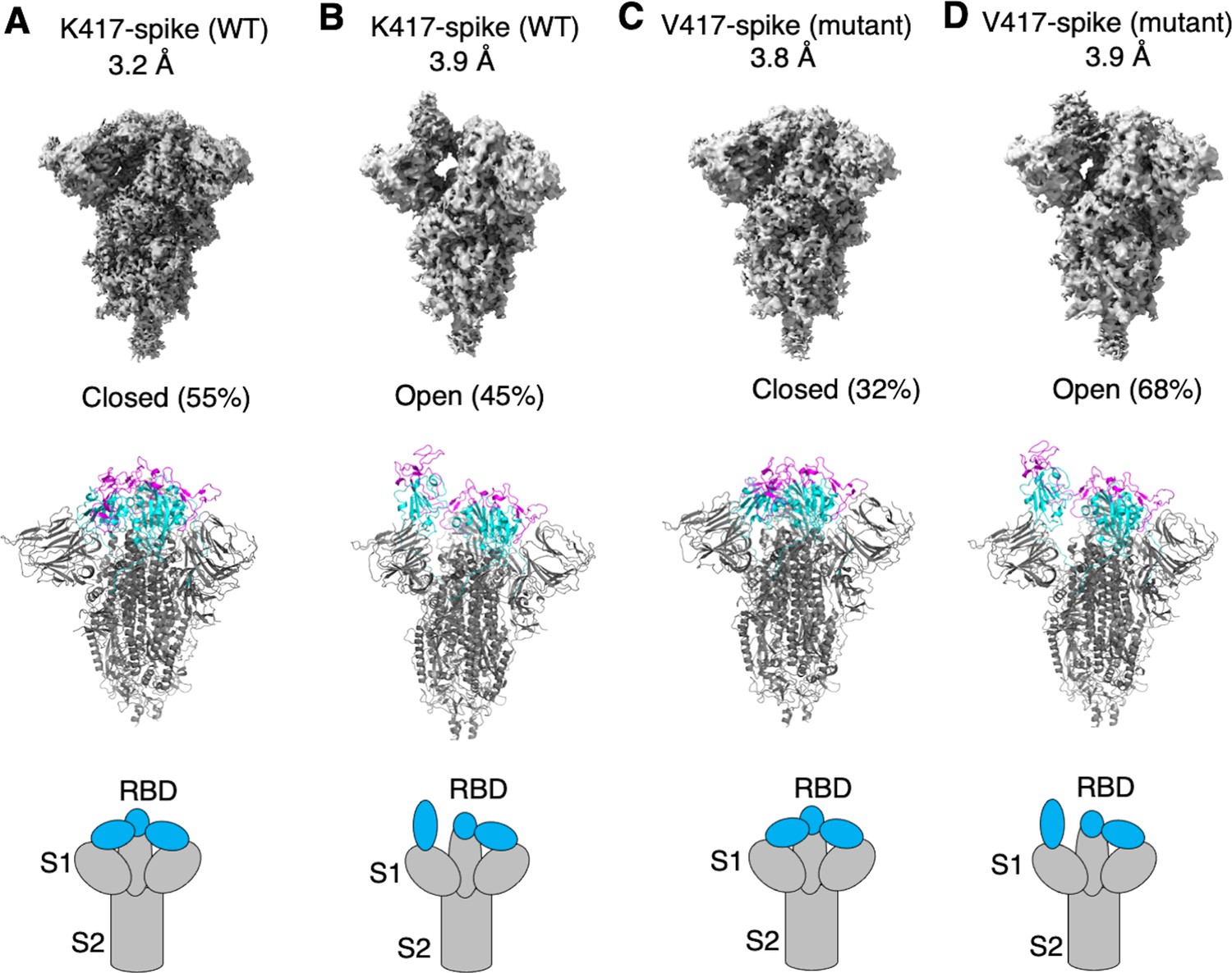
Cryo-EM analyses of residue 417 in regulating the conformation of recombinant SARS-CoV-2 spike ectodomain.
Prototypic SARS-CoV-2 spike ectodomains containing either Lys417 (as in prototypic SARS-CoV-2) or Val417 (as in SARS-CoV-1) were subjected to cryo-EM analyses and the particle distributions for open and closed conformations were calculated. Cryo-EM densities (top), atomic models (middle), and schematic presentations (bottom) for each of the protein classes are shown. (A) K417-spike in the closed conformation. (B) K417-spike in the open conformation. (C) V417-spike in the closed conformation. (D) V417-spike in the open conformation.
Figure 2—figure supplement 1
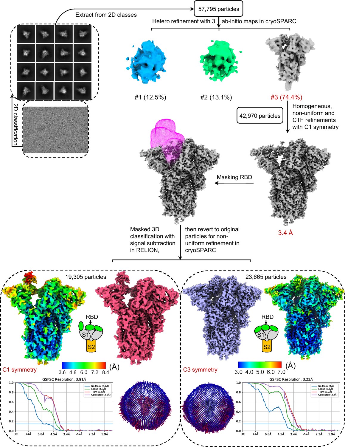
Flow chart of cryo-EM data processing for prototypic SARS-CoV-2 spike ectodomain containing Lys417 (K417-spike).
Representative raw cryo-EM image and 2D classes are presented. Further 3D refinements using the good particles generated a 3.2 Å map for the closed spike and a 3.9 Å map for the open spike. Angular distribution plots, the final maps, half-map Fourier shell correlation (FSC) curves, and accompanying local resolution illustrations are displayed.
Figure 2—figure supplement 2
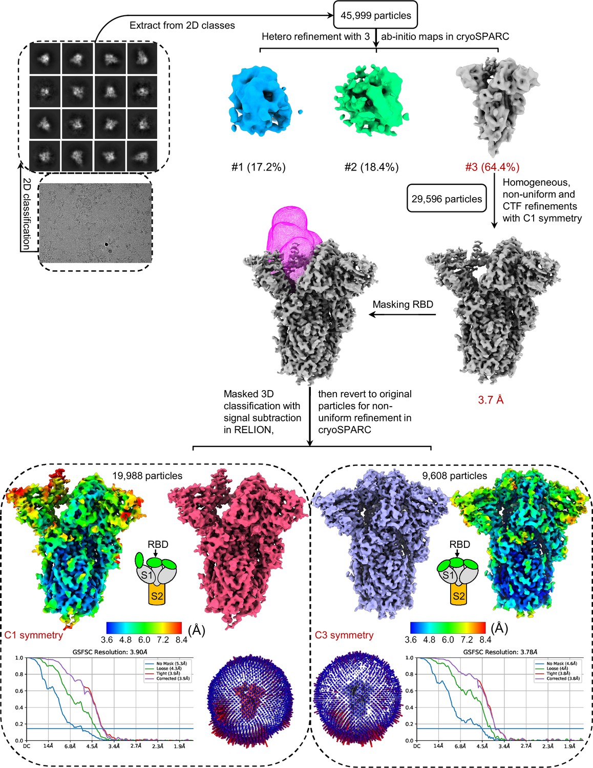
Flow chart of cryo-EM data processing for prototypic SARS-CoV-2 spike ectodomain containing Val417 (V417-spike).
Representative raw cryo-EM image and 2D classes are presented. Further 3D refinements using the good particles generated a 3.8 Å map for the closed spike and a 3.9 Å map for the open spike. Angular distribution plots, the final maps, half-map Fourier shell correlation (FSC) curves, and accompanying local resolution illustrations are displayed.
Figure 3 with 1 supplement

Biochemical analyses of residue 417 in regulating the conformation of membrane-anchored full-length SARS-CoV-2 spike and of the functions of the spike in different conformations.
(A) Protein pull-down assay using recombinant human ACE2 as the bait and cell-surface-anchored full-length SARS-CoV-2 spike as the target. The spike contains either Lys417 (wild-type residue) or Val417 (mutant residue). Top: cell-surface-expressed SARS-CoV-2 spike. Middle: pull-down results using His6-tagged ACE2. Bottom: pull-down results using Fc-tagged ACE2 (Figure 3—source data 1). The expected molecular weights of SARS-CoV-2 spike monomer and S2 monomer are ~180 kDa and ~80 kDa, respectively. (B) Flow cytometry assay to detect the interactions between recombinant human ACE2 and cell-surface-anchored full-length SARS-CoV-2 spike (Figure 3—source data 2). The spike contains either Lys417 (wild-type residue) or Val417 (mutant residue) and contains either intact furin motif or inactivated furin motif. See Figure 3—figure supplement 1 for details of this experiment. Data are mean + SEM. A comparison (two-tailed Student’s t-test) was performed on data between indicated groups (n = 3). ***p<0.001. (C) SARS-CoV-2 pseudovirus entry into human-ACE2-expressing cells. The virus-surface-anchored spike contains either Lys417 (wild-type residue) or Val417 (mutant residue). Top: pseudovirus entry efficiency normalized against the expression level of the spike (see bottom) (Figure 3—source data 3). Bottom: SARS-CoV-2 spike in packaged pseudoviruses (Figure 3—source data 4). Data are mean + SEM. A comparison (two-tailed Student’s t-test) was performed on data between indicated groups (n = 8). ***p<0.001. All experiments in this figure were repeated independently three times with similar results.
-
Figure 3—source data 1
Raw image for Figure 3A.
- https://cdn.elifesciences.org/articles/74060/elife-74060-fig3-data1-v3.pptx
-
Figure 3—source data 2
Numerical data for Figure 3B.
- https://cdn.elifesciences.org/articles/74060/elife-74060-fig3-data2-v3.xlsx
-
Figure 3—source data 3
Numerical data for Figure 3C.
- https://cdn.elifesciences.org/articles/74060/elife-74060-fig3-data3-v3.pptx
-
Figure 3—source data 4
Raw image for Figure 3C.
- https://cdn.elifesciences.org/articles/74060/elife-74060-fig3-data4-v3.xlsx
Figure 3—figure supplement 1
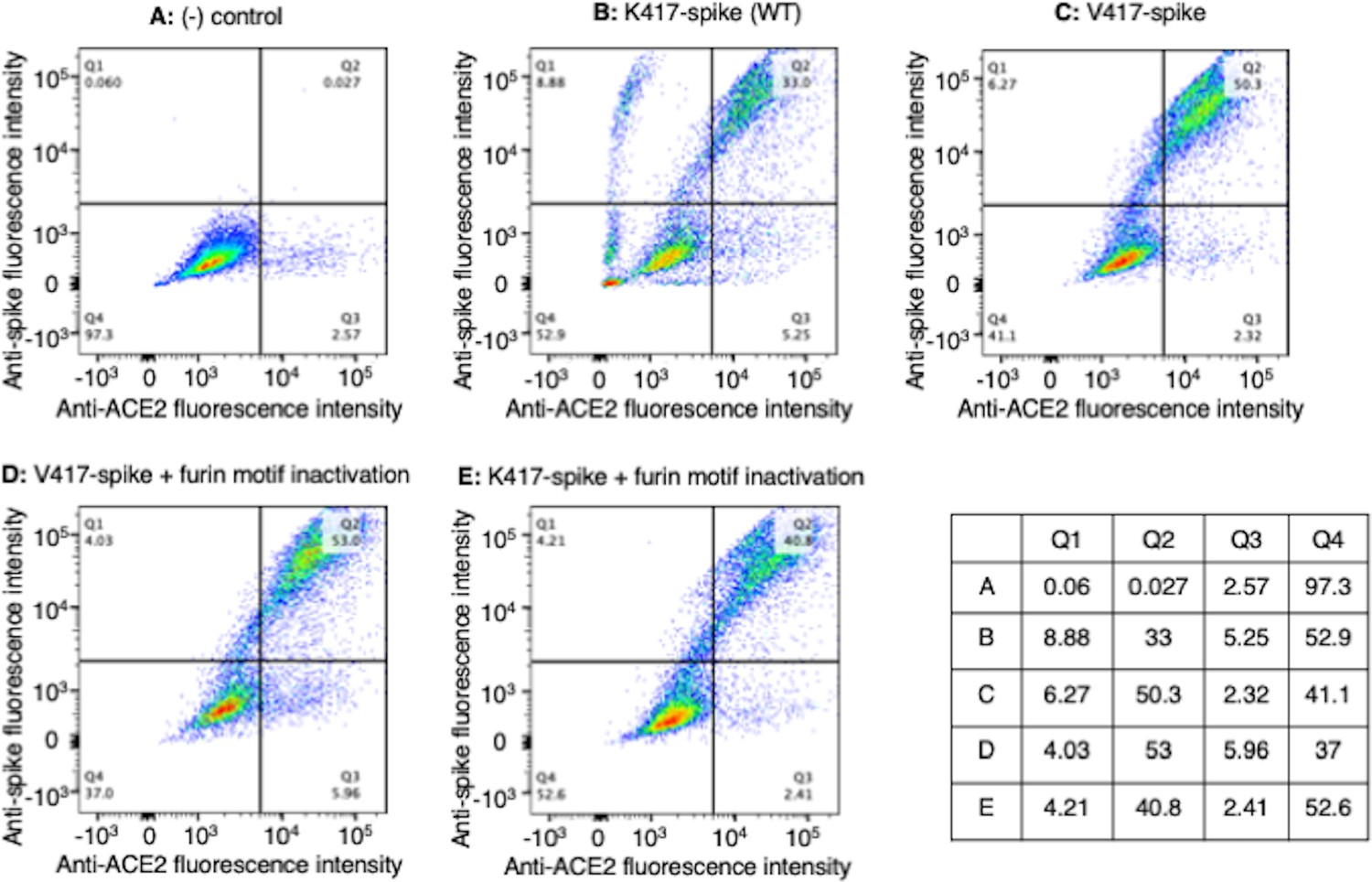
Flow cytometry assay on the interactions between cell-surface-anchored full-length trimeric spike and recombinant human ACE2.
HEK293T cells expressing full-length C9-tagged prototypic SARS-CoV-2 spike (containing either Lys417 or Val417 and containing either intact furin motif or inactivated furin motif) were incubated with recombinant Fc-tagged human ACE2. A fluorescence-labeled anti-Fc-tag antibody and a fluorescence-labeled anti-C9-tag antibody were used to label cell-bound ACE2 and cell-surface-expressed spike, respectively. The dot plots display fluorescence intensities indicative of the interactions between one of the spikes and ACE2. The accompanying table shows the percentage of labeled cells in quadrants Q1, Q2, Q3, and Q4 for each of the conditions. (A) (-) control - cells transfected with the vector only. (B) K417-spike. (C) V417-spike. (D) V417-spike containing an inactivated furin motif. (E) K417-spike containing an inactivated furin motif.
Figure 4

Analyses of epitopes on SARS-CoV-2 receptor-binding domain (RBD) that are accessible to neutralizing antibodies or nanobodies when the spike adopts different conformations.
(A) Epitopes targeted by neutralizing conventional antibodies. Numerous RBD-targeting human antibodies have been discovered individually from COVID-19 patients. They bind to five groups of epitopes. Only one representative antibody for each group is shown. The PDB codes are 7N4L for group 1, 7M7W for group 2, 7M6G for group 3, 7AKD for group 4, and 7M7W for group 5. Three epitopes out of the five groups are accessible to conventional antibodies only when the spike adopts the open conformation. (B) Epitopes targeted by neutralizing nanobodies (single-domain antibodies). We previously discovered four RBD-targeting nanobodies from camelid animals. They bind to three groups of epitopes. The PBD codes are 7KM5 for Nanosota-1, 8G72 for Nanosota-2, 8G74 for Nanosota-3, and 8G75 for Nanosota-4. The Nanosota-2 epitope is accessible to the nanobody only when the spike adopts the open conformation.
Figure 5 with 1 supplement
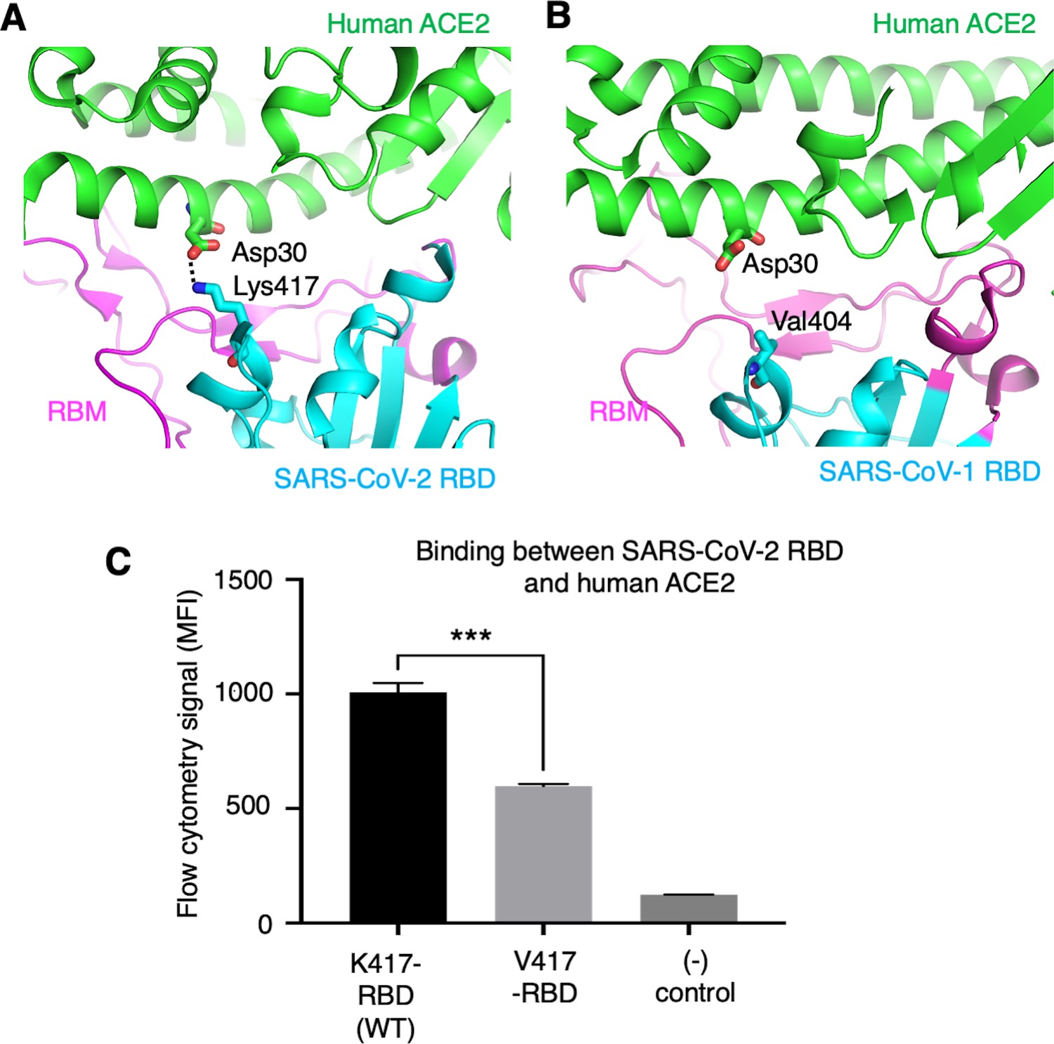
Role of residue 417 in direct interaction with ACE2 receptor.
(A) Lys417 in SARS-CoV-2 receptor-binding domain (RBD) forms a favorable salt bridge with Asp30 in human ACE2. PDB code: 6M0J. (B) Val404 in SARS-CoV-1 RBD (whose position is equivalent to residue 417 in SARS-CoV-2 RBD) does not form any direct interaction with human ACE2. PDB code: 2AJF. (C) Flow cytometry assay to detect the interactions between recombinant SARS-CoV-2 RBD and cell-surface-anchored human ACE2 (Figure 5—source data 1). The RBD contains either Lys417 (wild-type residue) or Val417 (mutant residue). MFI: median fluorescence intensity. See Figure 5—figure supplement 1 for details of this experiment. Data are mean + SEM. A comparison (two-tailed Student’s t-test) was performed on data between indicated groups (n = 3). ***p<0.001. This experiment was repeated independently three times with similar results.
-
Figure 5—source data 1
Numerical data for Figure 5C.
- https://cdn.elifesciences.org/articles/74060/elife-74060-fig5-data1-v3.zip
Figure 5—figure supplement 1
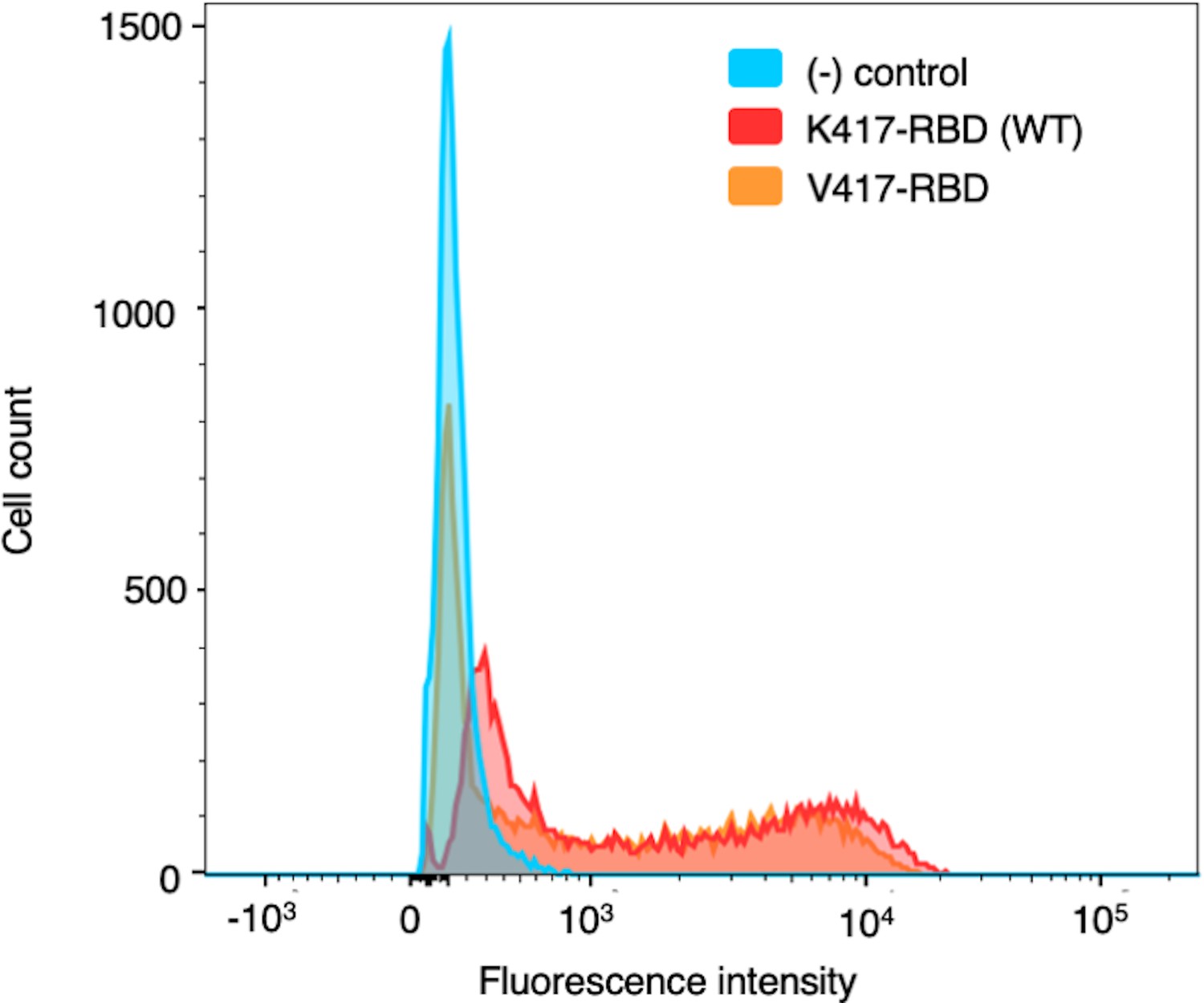
Flow cytometry assay on the interactions between cell-surface-anchored human ACE2 and recombinant SARS-CoV-2 receptor-binding domain (RBD).
Human-ACE2-expressing HEK293T cells were incubated with recombinant His-tagged SARS-CoV-2 RBD (containing either Lys417 or Val417) or buffer only. A fluorescence-labeled anti-His-tag antibody was used to detect cell-bound RBD. The histogram displays the differential fluorescence intensity, indicating the interactions between one of the RBDs and cell-surface ACE2. The (-) control represents cells treated with buffer only.
Additional files
-
Supplementary file 1
Cryo-EM data collection, refinement, and validation statistics.
- https://cdn.elifesciences.org/articles/74060/elife-74060-supp1-v3.docx
-
Transparent reporting form
- https://cdn.elifesciences.org/articles/74060/elife-74060-transrepform1-v3.docx
Download links
A two-part list of links to download the article, or parts of the article, in various formats.
Downloads (link to download the article as PDF)
Open citations (links to open the citations from this article in various online reference manager services)
Cite this article (links to download the citations from this article in formats compatible with various reference manager tools)
Lys417 acts as a molecular switch that regulates the conformation of SARS-CoV-2 spike protein
eLife 12:e74060.
https://doi.org/10.7554/eLife.74060


