A new early branching armored dinosaur from the Lower Jurassic of southwestern China
- Centre for Vertebrate Evolutionary Biology, Yunnan University, China
- Department of Earth Sciences, Natural History Museum, United Kingdom
- Yimen Administration of Cultural Heritage, China
- Key Laboratory of Evolutionary Systematics of Vertebrates, Institute of Vertebrate Paleontology and Paleoanthropology, Chinese Academy of Sciences, China
- Center for Excellence in Life and Paleoenvironment, China
- Department of Biology, Indiana University of Pennsylvania, United States
Figures
Figure 1

Geographical and stratigraphic location of Yuxisaurus kopchicki CVEB21701.
(A) Location of the quarry yielding Yuxisaurus kopchicki, with a red star indicating the locality. (B) Sediments of the Fengjiahe Formation at the quarry site. (C) Stratigraphic column of the Fengjiahe Formation in the Jiaojiadian area (modified from Bai, 1999).
Figure 2
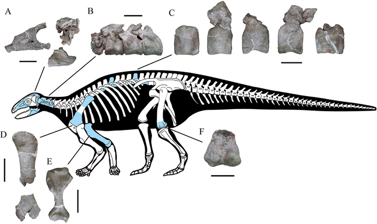
Skeletal reconstruction of Yuxisaurus kopchicki showing some of the main preserved elements from the holotype (highlighted in blue), with details of the skull bones (A), cervical vertebrae (B), dorsal vertebrae (C), left scapula (D), right humerus (E), and left femur (F).
Scale bars equal 5 cm (A–C) or 10 cm (D–F). The facial region and distal scapula are mirrored. Osteoderms have been omitted for convenience.
Figure 3

Right maxilla of Yuxisaurus kopchicki in (A) lateral, (B) medial, (C) dorsal, and (D) ventral views.
Maxillary tooth row in (E) lingual view with the last tooth in (F) lingual view. Abbreviations: af, antorbital fenestra; afo, antorbital fossa; fenestra; aso, anterior supraorbital; ju, jugal; mrl, maxillary ramus of the lacrimal; mso, mesosupraorbital; orb, orbital; aso anterior supraorbital; prf, prefrontal. Scale bar equals 5 cm.
Figure 4
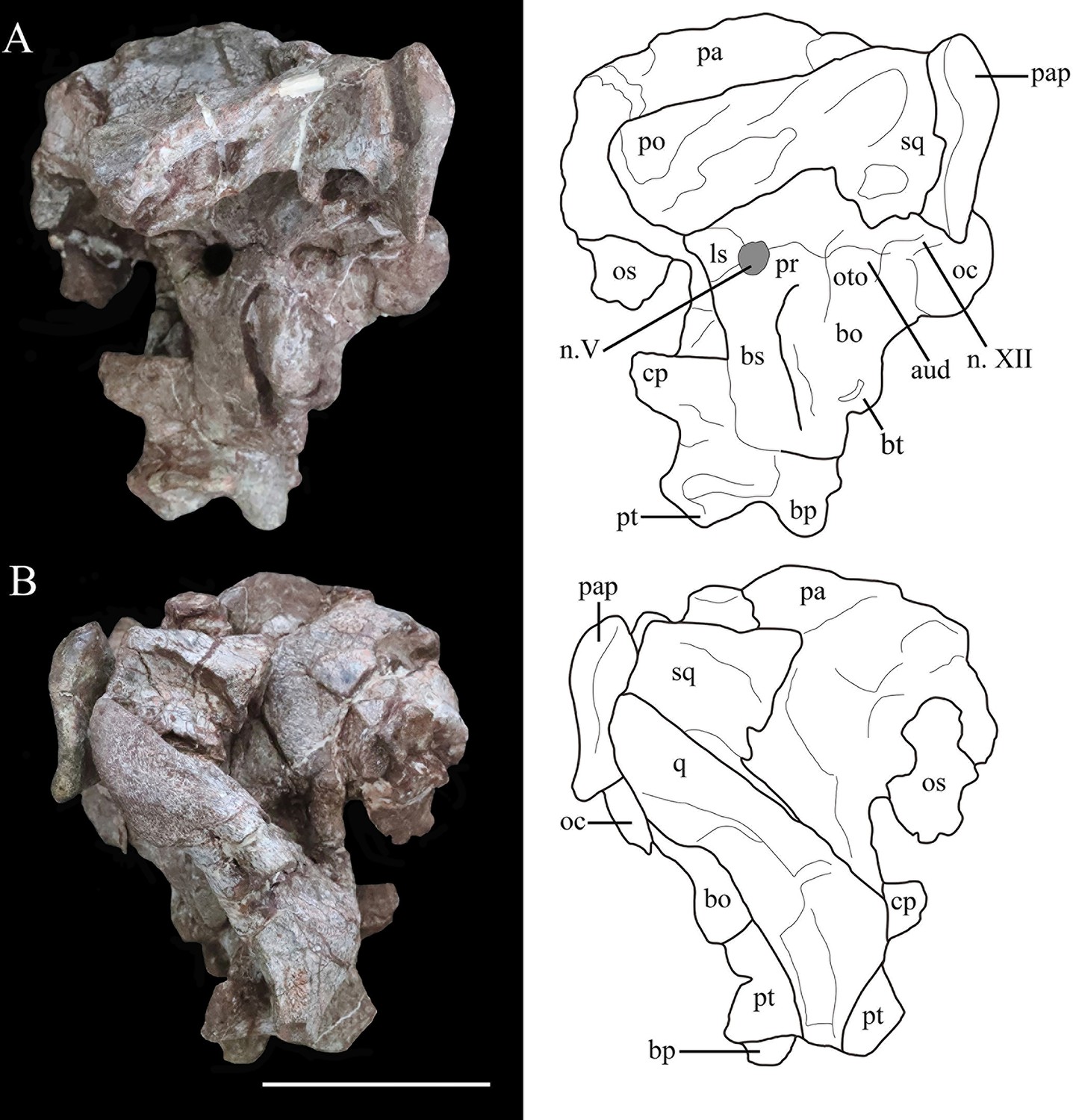
Photographs (left) and line drawings (right) of the braincase and partial skull roof of Yuxisaurus kopchicki in left lateral (A) and right lateral (B) views.
Abbreviations: aud, auditory recess; bo, basioccipital; bp, basipterygoid process; bs, basisphenoid; cp, cultriform process (parasphenoid rostrum); fm, foramen magnum; ls, laterosphenoid; n. V, exit of trigeminal nerve; n. XII, exit of cranial nerve XII; oc, occipital condyle; os, orbitosphenoid; oto, otoccipital; pa, parietal; pap, paroccipital process; po, postorbital; pr, prootic; pt, pterygoid; q, quadrate; sq, squamosal. Scale bar equals 5 cm.
Figure 5
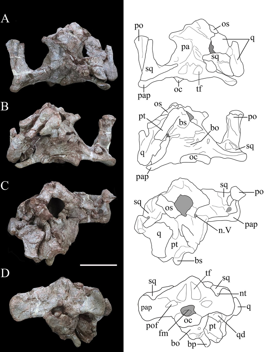
Photographs (left) and line drawings (right) of the braincase of Yuxisaurus kopchicki in (A) dorsal, (B) ventral, (C) anterior, and (D) posterior views.
Abbreviations: bo, basioccipital; bp, basipterygoid process; bs, basisphenoid; cp, cultriform process (parasphenoid rostrum); fm, foramen magnum; nt, ‘V’-shaped notch; n. V, exit of trigeminal nerve; oc, occipital condyle; os, orbitosphenoid; pa, parietal; pap, paroccipital process; po, postorbital; pt, pterygoid; q, quadrate; qd, quadrate depression; sq, squamosal. Scale bar equals 5 cm.
Figure 6

Possible skull roof fragment of Yuxisaurus kopchicki in (A) dorsal, (B) lateral, and (C) ventral views.
Abbreviations: cd, channel-like depression; d, dome. Scale bar equals 5 cm.
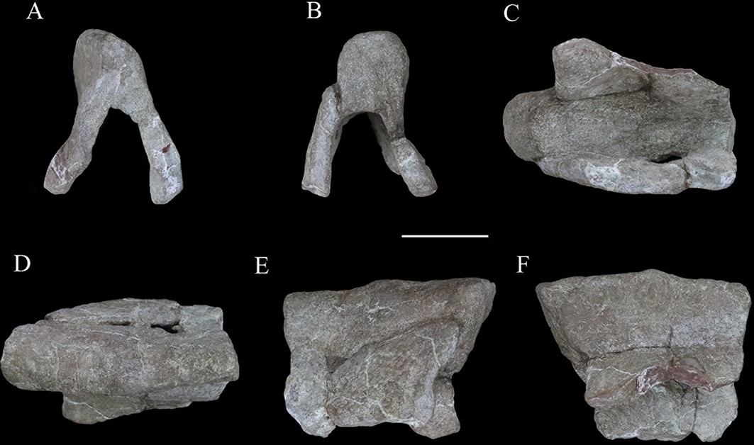
Figure 7
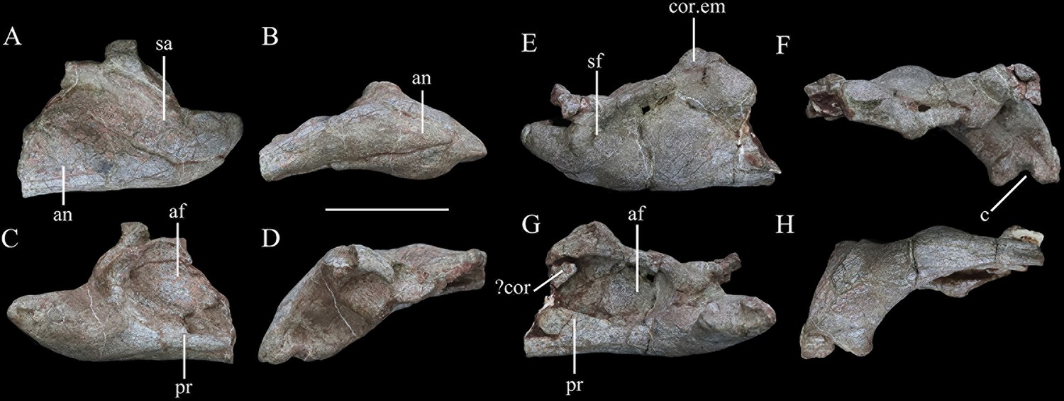
Mandibular remains of Yuxisaurus kopchicki.
Posterior part of left hemimandible in (A) lateral, (B) ventral, (C) medial, and (D) dorsal views. Posterior part of right hemimandible in (E) lateral, (F) ventral, (G) medial, and (H) dorsal views. Abbreviations: af, adductor fossa; an, angular; c, concavity; cor, coronoid; cor.em, coronoid eminence; pr, prearticular; sa, surangular; sf, surangular foramen. Scale bar equals 5 cm.
Figure 8
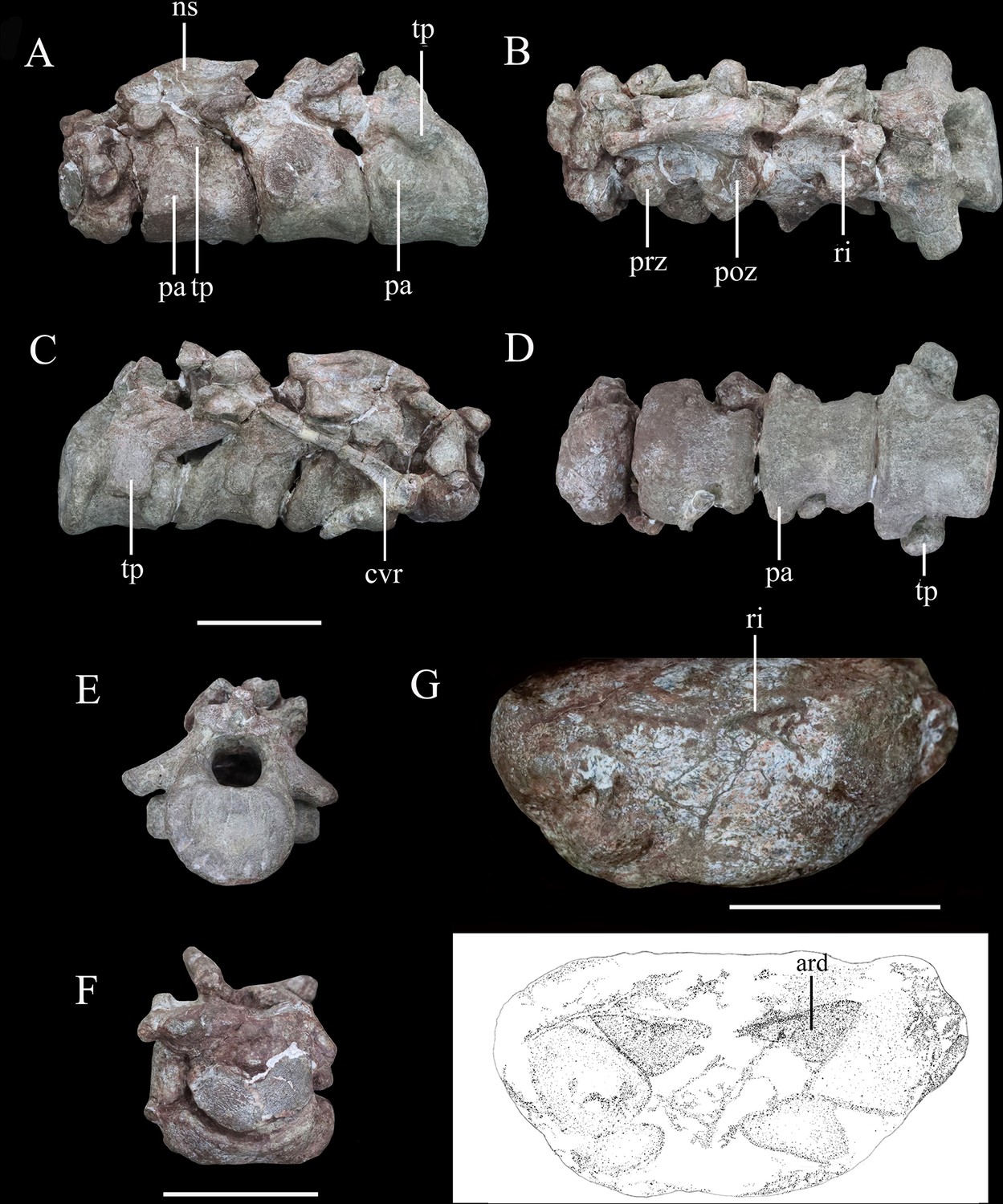
Articulated series of the anterior-most cervical vertebrae (atlas, axis, and cervicals 3 and 4) of Yuxisaurus kopchicki in (A) left lateral, (B) dorsal, (C) right lateral, (D) ventral, (E) posterior, and (F) anterior views.
Atlas in (G) ventral view with interpretative diagram beneath. Abbreviations: ard, arrow-like depression; cvr, cervical rib; ns, neural spine; pap, parapophysis; poz, postzygapophysis; prz, prezygapophysis; ri, ridge; tp, transverse process. Scale bar equals 5 cm.
Figure 9
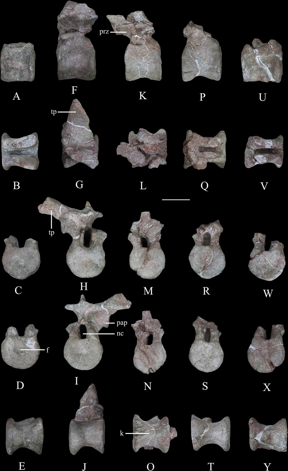
Dorsal vertebrae of Yuxisaurus kopchicki.
D1 in (A) left lateral, (B) dorsal, (C) anterior, (D) posterior, and (E) ventral views. D2 in (F) left lateral, (G) dorsal, (H) anterior, (I) posterior, and (J) ventral views. D3 in (K) left lateral, (L) dorsal, (M) anterior, (N) posterior, and (O) ventral views. D4 in (P) left lateral, (Q) dorsal, (R) anterior, (S) posterior, and (T) ventral views. D5 in (U) left lateral, (V) dorsal, (W) anterior, (X) posterior, and (Y) ventral views. Abbreviations: f, fossa; k, keel; nc, neural canal; pap, parapophysis; prz, prezygapophysis; tp, transverse process. Scale bar equals 5 cm.
Figure 10
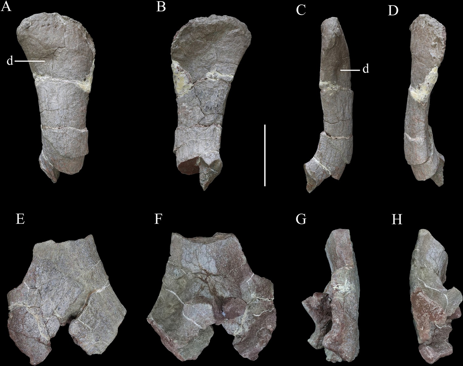
Scapulae of Yuxisaurus kopchicki.
Distal part of right scapula in (A) lateral, (B) medial, (C) ventral, and (D) dorsal views. Proximal part of left scapula in (E) lateral, (F) medial, (G) ventral, and (H) dorsal views. Abbreviation: d, depression. Scale bar equals 10 cm.
Figure 11
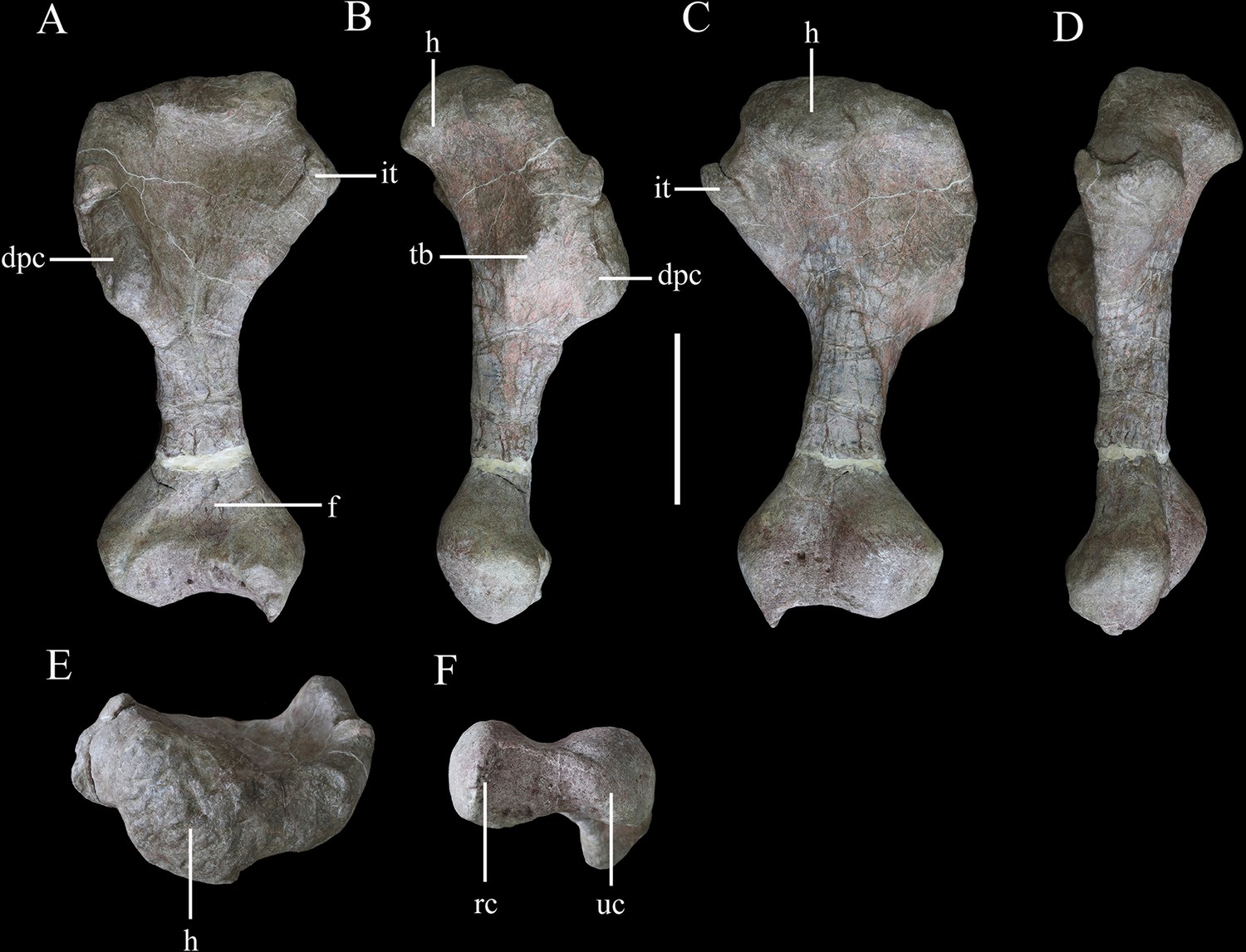
Right humerus of Yuxisaurus kopchicki in (A) anterior, (B) lateral, (C) posterior, (D) medial, (E) proximal, and (F) distal views.
Abbreviations: dpc, deltopectoral crest; f, fossa; h, humeral head; it, internal tuberosity; rc, radial condyle; tb, tubercle; uc, ulnar condyle. Scale bar equals 10 cm.
Figure 12
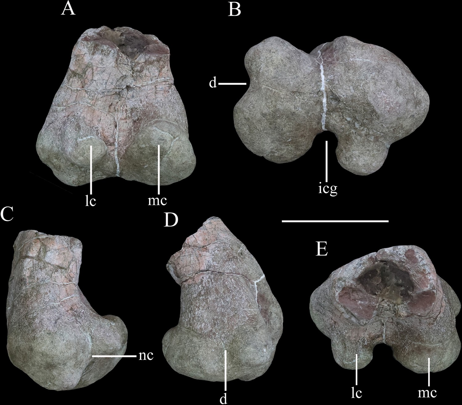
Distal end of right femur of Yuxisaurus kopchicki in (A) posterior, (B) ventral, (C) lateral, (D) medial, and (E) dorsal views.
Abbreviations: d, depression; icg, intercondylar groove; lc, lateral condyle; mc, medial condyle; nc, notch. Scale bar equals 10 cm.
Figure 13
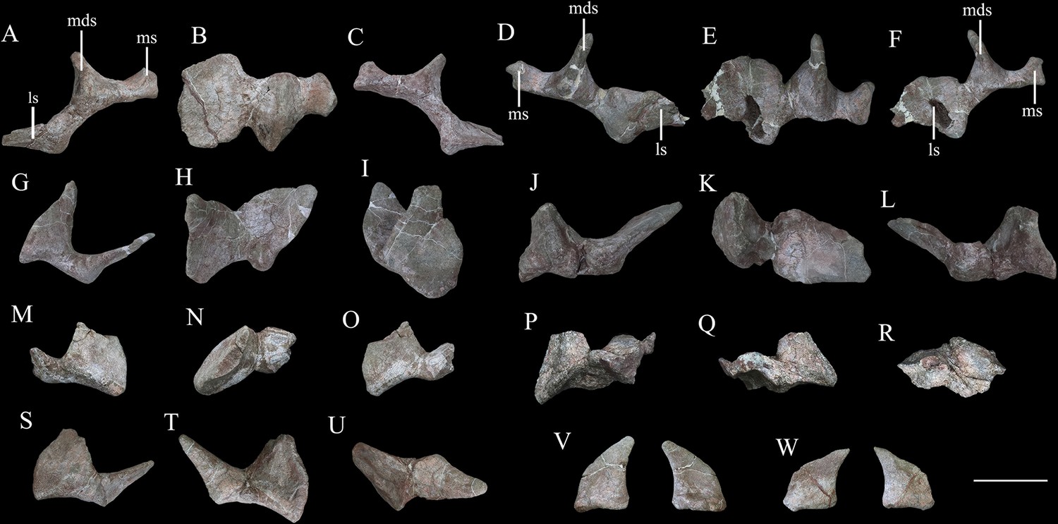
Cervical and pectoral osteoderms of Yuxisaurus kopchicki.
Tripartite compound osteoderm (TPO) 1 in (A) anterior, (B) dorsal, and (C) posterior views. TPO 2 in (D) anterior, (E) dorsal, and (F) posterior views. Bipartite osteoderm (BPO) 1 in (G) anterior, (H) dorsal, and (I) medial views; BPO 2 in (J) anterior, (K) dorsal, and (L) posterior views; BPO 3 in (M) anterior, (N) dorsal, and (O) posterior views; BPO 4 in (P) anterior, (Q) posterior, and (R) dorsal views; and BPO 5 in (S) anterior, (T) posterior, and (U) dorsal views. Blade-like cervical spines in anterior and posterior views (V, W). Abbreviations: ls, lateral spine; mds, middle scute; ms, medial scute. Scale bar equals 10 cm.
Figure 14
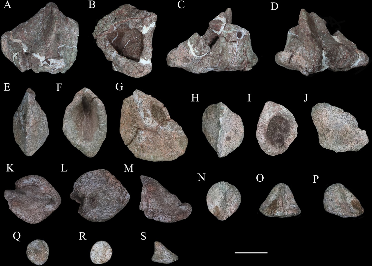
Six selected individual osteoderms of Yuxisaurus kopchicki.
Osteoderm 1 in (A) dorsal, (B) ventral, (C) posterior, and (D) anterior views; osteoderm 2 in (E) dorsal, (F) ventral, and (G) lateral views; osteoderm 3 in (H) dorsal, (I) ventral, and (J) lateral views; osteoderm 4 in (K) dorsal, (L) ventral, and (N) lateral views; osteoderm 5 in (N) dorsal, (O) anterior, and (P) lateral views; osteoderm 6 in (Q) dorsal, (R) ventral, and (S) lateral views. Scale bar equals 5 cm.
Figure 15
‘Pup tent’-shaped osteoderm of Yuxisaurus kopchicki in (A) posterior, (B) anterior, (C) ventral, (D) dorsal, and (E, F) side views.
Scale bar equals 10 cm.
Figure 16
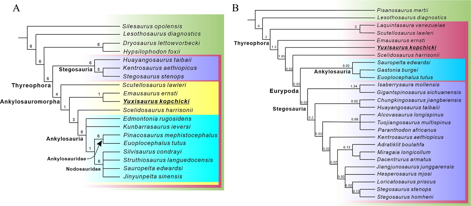
Phylogenetic relationships of Yuxisaurus within Thyreophora.
(A) Strict consensus of the six most parsimonious trees (MPTs) recovered from analysis of the modified Norman, 2021 data set. (B) Strict consensus of the two MPTs recovered from analysis of the modified Maidment et al., 2020 data set. Bremer support values are shown adjacent to the nodes.
Figure 17
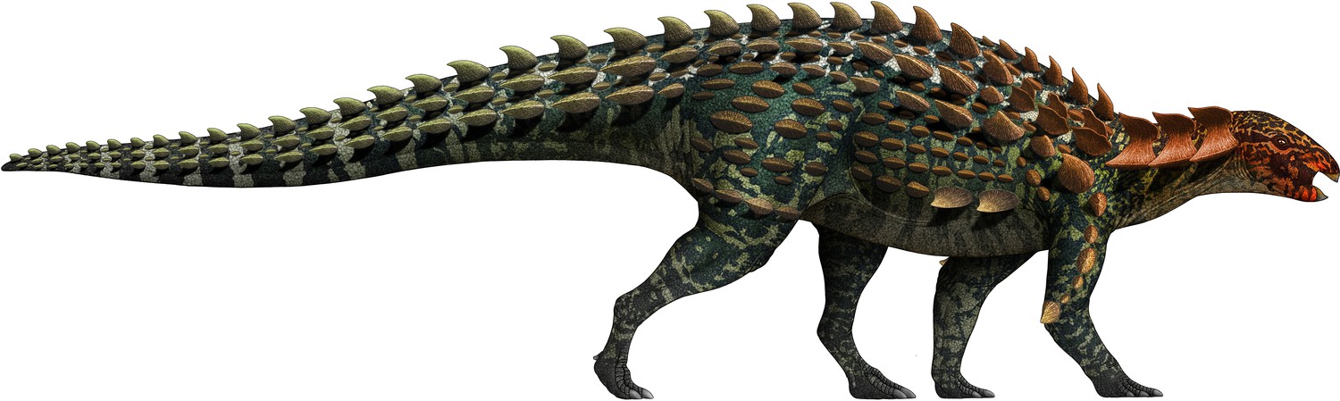
Life restoration of Yuxisaurus kopchicki.
The osteoderm arrangement is hypothetical but that includes many of the types of armor found with the skeleton.
Additional files
-
Supplementary file 1
Data matrix modified from Norman, 2021 used in the phylogenetic analysis (in txt format).
- https://cdn.elifesciences.org/articles/75248/elife-75248-supp1-v2.txt
-
Supplementary file 2
Data matrix modified from Maidment et al., 2020 used in the phylogenetic analysis (in txt format).
- https://cdn.elifesciences.org/articles/75248/elife-75248-supp2-v2.txt
-
Transparent reporting form
- https://cdn.elifesciences.org/articles/75248/elife-75248-transrepform1-v2.docx
Download links
A two-part list of links to download the article, or parts of the article, in various formats.
Downloads (link to download the article as PDF)
Open citations (links to open the citations from this article in various online reference manager services)
Cite this article (links to download the citations from this article in formats compatible with various reference manager tools)
A new early branching armored dinosaur from the Lower Jurassic of southwestern China
eLife 11:e75248.
https://doi.org/10.7554/eLife.75248


