Digital restoration of the pectoral girdles of two Early Cretaceous birds and implications for early-flight evolution
- Key Laboratory of Vertebrate Evolution and Human Origins, Institute of Vertebrate Paleontology and Paleoanthropology, Chinese Academy of Sciences, China
- CAS Center for Excellence in Life and Paleoenvironment, China
- University of Chinese Academy of Sciences, China
- University of Alberta, Canada
- Shenyang Normal University, China
- Philip J. Currie Dinosaur Museum, Canada
- Centre for Vertebrate Evolutionary Biology, Yunnan University, China
Figures
Figure 1
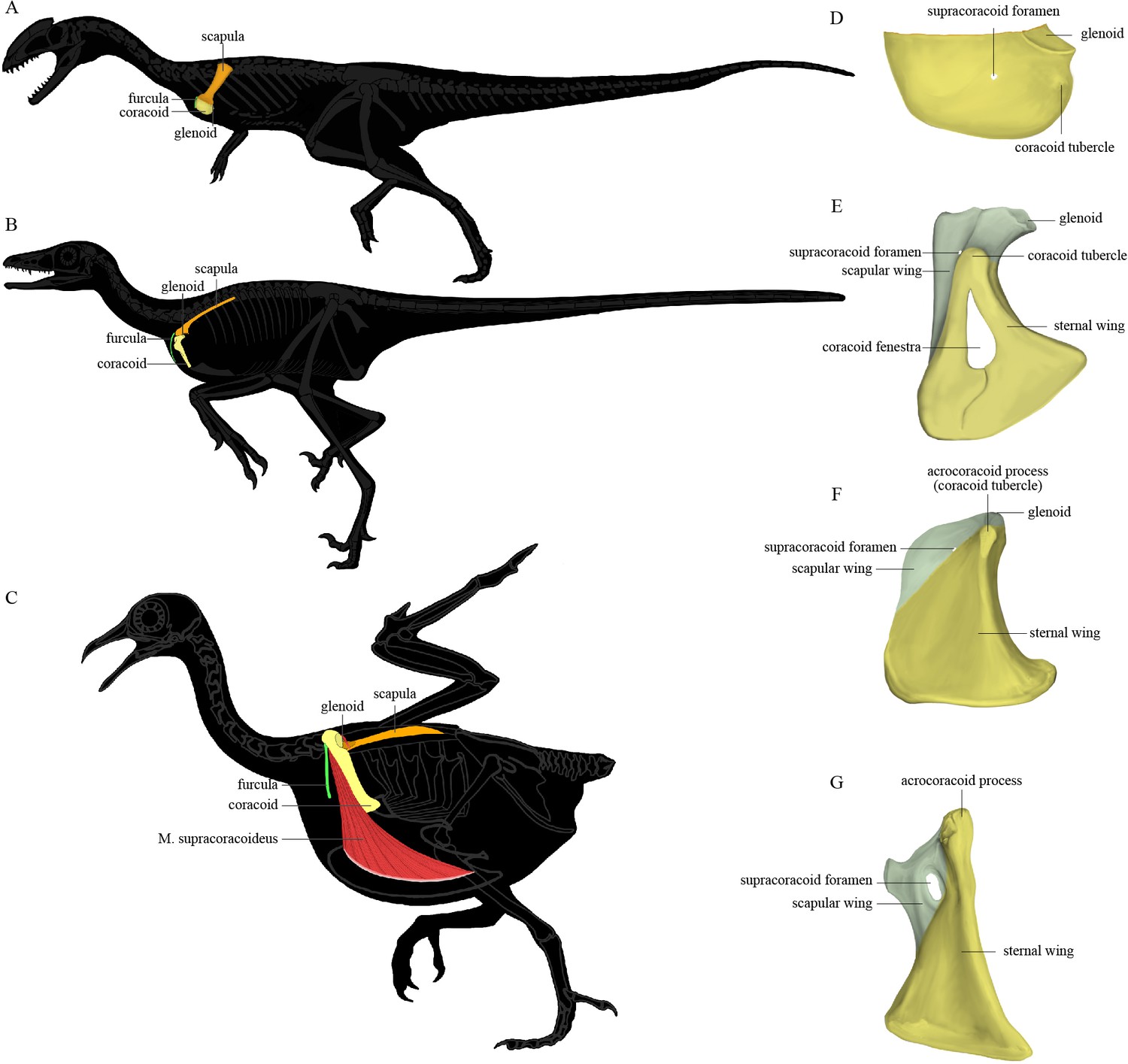
The position of the pectoral girdle and the form of the coracoid in different theropod groups.
(A–C) Skeletal silhouettes showing the anatomical position of the pectoral girdle in (A) the early-diverging theropod Coelophysis, (B) the early-diverging pennaraptoran Microraptor, and (C) the modern bird Columba. The M. supracoracoideus is illustrated in (C) but typically covered by the M. pectoralis, which is not illustrated. (D–G) Illustrations of the left coracoids of (D) Coelophysis (modified from Tykoski, 1998), (E) the early-diverging pennaraptoran Sinornithosaurus (modified from Xu et al., 1999), (F) the early-diverging avialan Archaeopteryx (modified from Wellnhofer et al., 2009), and (G) the early-diverging avialan Jeholornis (based on STM 2-49 and IVPP V 13886). Coracoid of Coelophysis in lateral view, coracoids of other taxa in ventral view.
Figure 2

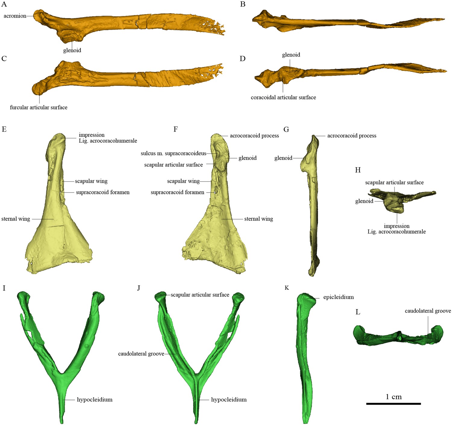
Pectoral girdle bones of Sapeornis chaoyangensis PMoL-AB00015.
(A–D) Left scapula in lateral, dorsal, medial (costal), and ventral views. (E–H) Left coracoid in ventral, dorsal, lateral, and cranial views. (I–L) Furcula in cranial, caudal, lateral, and ventral views. The black arrows in (J) and (L) indicate the concave surface for the tendon of M. supracoracoideus.
Figure 3
Pectoral girdle bones of Piscivorenantiornis inusitatus IVPP V 22582.
(A–D) Left scapula in lateral, dorsal, medial (costal), and ventral views. (E–H) Right coracoid in ventral, dorsal, lateral, and cranial views. (I–L) Furcula in cranial, caudal, left, and ventral views.
Figure 4
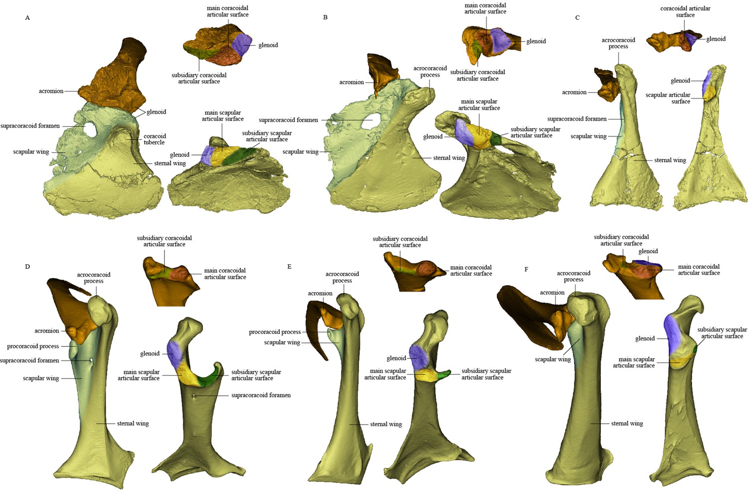
Comparison of scapula and coracoid morphology across various paravian taxa.
Each panel shows articulated left scapula and coracoid in ventral view (on left) and opposing articular surfaces of left scapula and coracoid (on right, with cranial direction toward top of figure for both scapula and coracoid). (A) Sinovenator changii (mirrored), (B) Sapeornis chaoyangensis, (C) Piscivorenantiornis inusitatus, (D) Tyto alba, (E) Egretta garzetta, and (F) Pavo muticus.
Figure 5
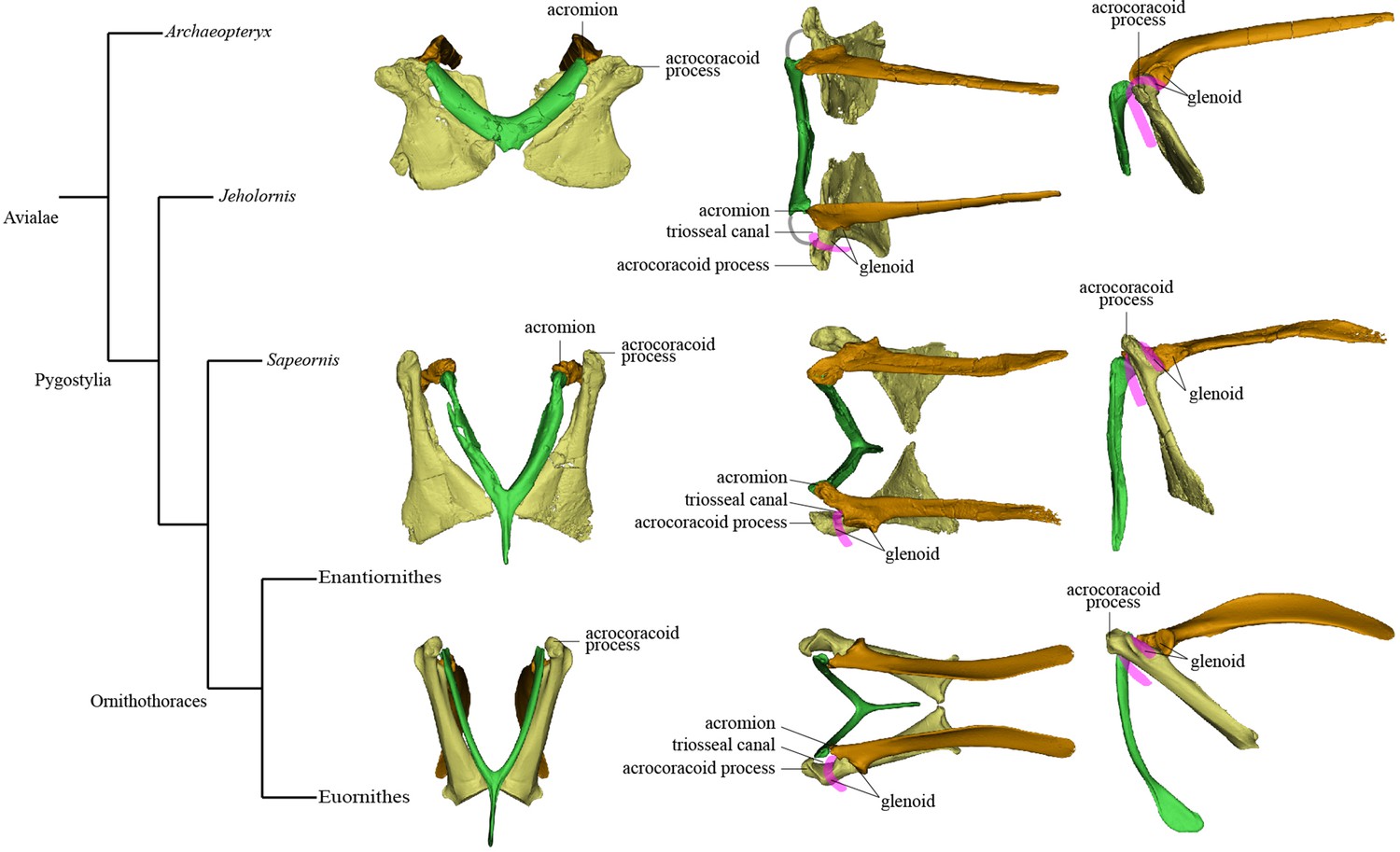
Simplified phylogeny with hypothetical steps in pectoral girdle evolution.
The pectoral girdles of Sapeornis chaoyangensis, Piscivorenantiornis inusitatus, and Pavo muticus (from top to bottom) are shown in cranial, dorsal, and left lateral views. The pink lines in the dorsal and lateral views represent the tendon of M. supracoracoideus, and the gray line in the dorsal view of the Sapeornis rendering represents the coracoclavicular ligament that connects the coracoid and furcula. Phylogenetic framework following Wang et al., 2018a.
Additional files
Download links
A two-part list of links to download the article, or parts of the article, in various formats.
Downloads (link to download the article as PDF)
Open citations (links to open the citations from this article in various online reference manager services)
Cite this article (links to download the citations from this article in formats compatible with various reference manager tools)
Digital restoration of the pectoral girdles of two Early Cretaceous birds and implications for early-flight evolution
eLife 11:e76086.
https://doi.org/10.7554/eLife.76086


