Female fertility and the zona pellucida
- Department Cell, Developmental, and Regenerative Biology Icahn School of Medicine at Mount Sinai One Gustave L. Levy Place, United States
Figures
Figure 1
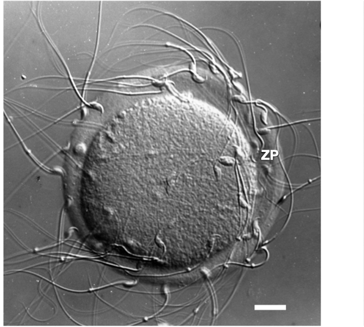
Photographic image of a light micrograph (Nomarski differential interference contrast) of an unfertilized mouse egg incubated in the presence of free-swimming sperm.
Sperm are shown bound to the zona pellucida. Scale bar (≃1 cm) = ≃14 µm.
Figure 2
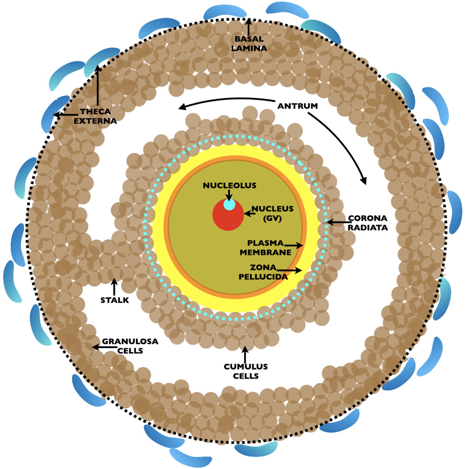
Drawing of an ovarian Graafian follicle prior to ovulation of an unfertilized egg.
Shown is a fully grown oocyte (pale green) containing a nucleus/germinal vesicle (GV) (red), nucleolus (turquoise), plasma membrane (orange), and zona pellucida (yellow). Two-to-three layers of cumulus cells (brown) surround the oocyte and the innermost cumulus cells, the corona radiata, are indicated by a dashed turquoise circle. The oocyte is connected to the granulosa cells (brown) by a stalk and is located in a fluid filled cavity, the antrum. At the outermost region of the Graafian follicle is a basal lamina (dashed black circle) and outside of this are the theca externa cells (blue). At ovulation, oocytes arrested in metaphase II of meiosis are expelled from the Graafian follicle surrounded by two-to-three layers of cumulus cells. Oocytes resume meiosis, complete the first meiotic reductive division, called meiotic maturation, with separation of homologous chromosomes and emission of the first polar body, and become unfertilized eggs. At fertilization, eggs complete meiosis with separation of chromatids and emission of a second polar body. The sperm’s genome restores the fertilized egg to a diploid state.
Figure 3

Graph depicting changes in the width of the mouse oocyte’s zona pellucida during growth of the oocyte.
Mouse oocytes grow from ≃12 µm (nongrowing oocytes) to ≃80 µm in diameter (fully grown oocyte) over 2–3 weeks; this corresponds to more than a 300-fold increase in oocyte volume, from ≃0.9 to ≃300 pl, during oocyte growth. Concomitant with oocyte growth, the zona pellucida increases in width from zero for nongrowing oocytes to ≃6.2 ± 1.9 µm for fully grown oocytes.
Figure 4
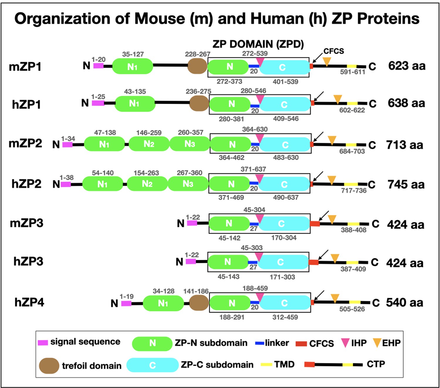
Schematic representation of the organization of mouse zona pellucida proteins, mZP1–3 (623, 713, and 424 amino acids, respectively), and human zona pellucida proteins, hZP1–4 (638, 745, 424, and 540 amino acids, respectively).
In each case, the polypeptide contains a signal sequence (SS) at the N-terminus (pink), a ZP domain (ZPD; black box) consisting of ZP-N (green) and ZP-C (turquoise) subdomains and a short linker region (blue), and a consensus furin cleavage site (CFCS; arrow), transmembrane domain (TMD; yellow), and C-terminal propeptide (CTP). mZP1, hZP1, and hZP4 also have a trefoil domain (brown) adjacent to the ZPD. mZP1, mZP2, hZP1, hZP2, and hZP4 have one or three extra copies of the ZP-N subdomain (green) between the N-terminus of the polypeptides and the ZPD. The positions of the internal (IHP) and external (EHP) hydrophobic patches are indicated by red and orange triangles, respectively. The amino acid numbers for each region of the mouse and human zona pellucida polypeptides are indicated above and below the drawings of the polypeptides.
Figure 5
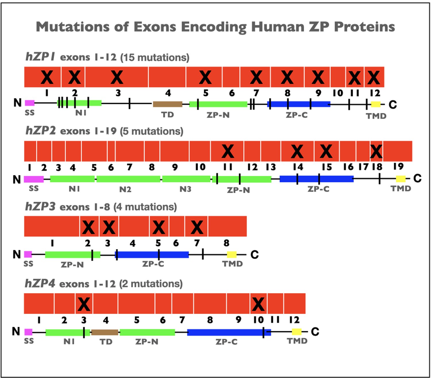
Schematic representations of mutations in exons encoding human zona pellucida proteins, hZP1–4.
Shown is the organization of exons (red) for hZP1 (12 exons), hZP2 (19 exons), hZP3 (8 exons), and hZP4 (12 exons). Exons subject to mutations that caused infertility are marked by an X. Also shown are schematic representations of hZP1–4 polypeptides with the signal sequence (SS; red), trefoil domain (TD; brown), ZP domain (ZPD) consisting of ZP-N (green) and ZP-C (blue) subdomains, transmembrane domain (TMD; yellow), and extra copies of ZP-N subdomain (green) between the N-terminus of the polypeptides and the ZPD. The sites of mutations in the polypeptides are indicated by black vertical lines.
Tables
Table 1
Phenotypes of ZP1, 2, 3 null female mice.
| Genotype | Fertility | Zona Pellucida | References |
|---|---|---|---|
| ZP1–3 wild-type | Reduced fertile | Normal | |
| ZP1 homozygous-null | Reduced | Abnormal | Rankin et al., 1999 |
| ZP2 homozygous-null | Infertile | None | Rankin et al., 2001 |
| ZP3 homozygous-null | Infertile | None | Rankin et al., 1996; Liu et al., 1996 |
| ZP3 heterozygous-null | Fertile | Thin | Wassarman et al., 1997 |
Download links
A two-part list of links to download the article, or parts of the article, in various formats.
Downloads (link to download the article as PDF)
Open citations (links to open the citations from this article in various online reference manager services)
Cite this article (links to download the citations from this article in formats compatible with various reference manager tools)
Female fertility and the zona pellucida
eLife 11:e76106.
https://doi.org/10.7554/eLife.76106


