Functional gradients in the human lateral prefrontal cortex revealed by a comprehensive coordinate-based meta-analysis
- MIND team, Inria, CEA, Université Paris-Saclay, France
- NeuroSpin, CEA, Université Paris-Saclay, France
Figures
Figure 1 with 5 supplements
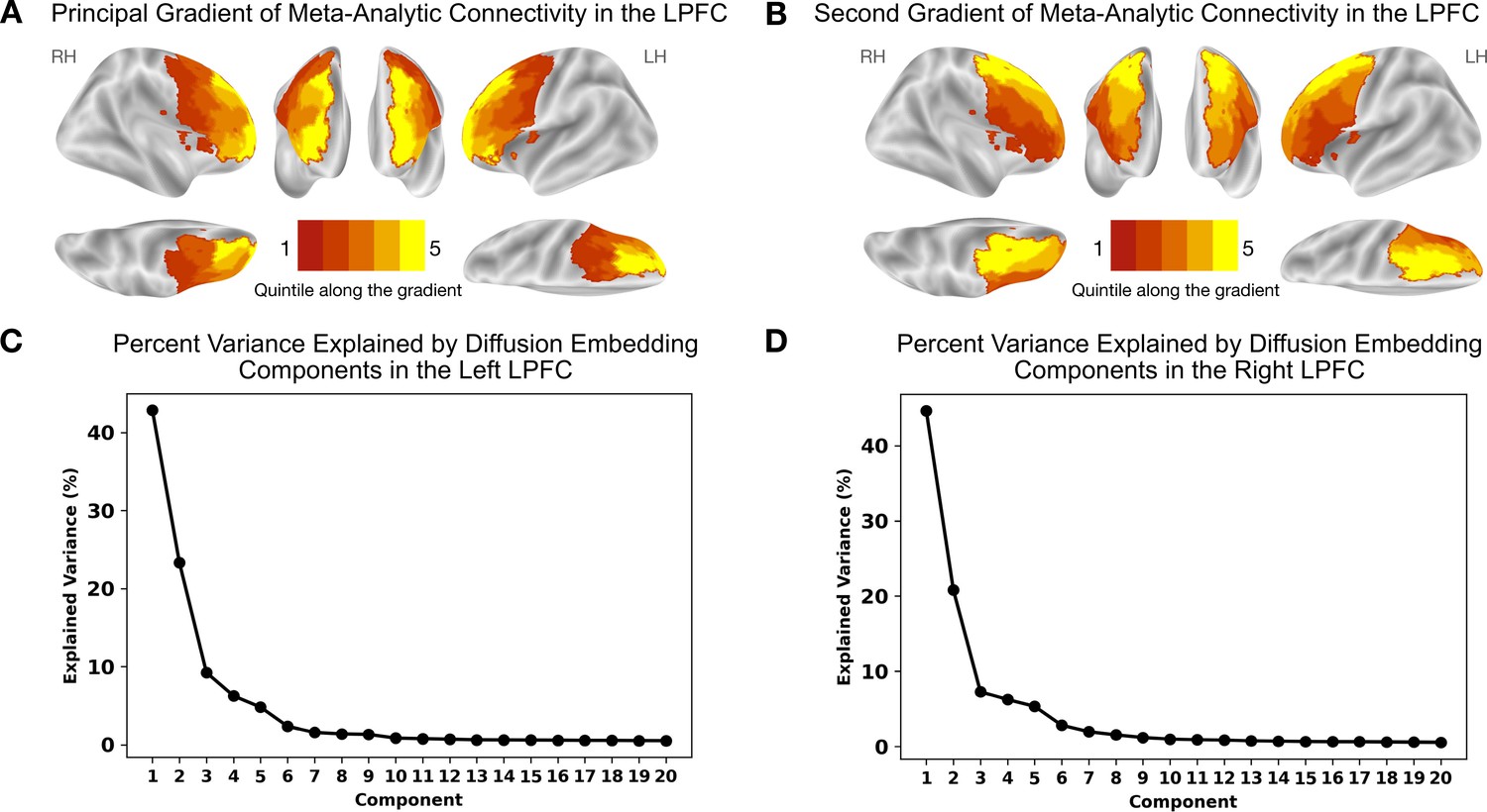
Meta-analytic connectivity gradients of the LPFC.
The rostrocaudal and dorsoventral gradients explain the greatest amount of variance in meta-analytic connectivity in the LPFC. (A) The principal gradient in both hemispheres echoes a widely proposed rostrocaudal organization in the LPFC. This gradient represents the dominant direction of variations in connectivity patterns. (B) The gradient that explains the second-most variance in meta-analytic connectivity in the LPFC echoes a dorsoventral organization extending from ventrolateral to dorsolateral PFC regions. (C) and (D) The percentage of variance explained by the first 20 diffusion embedding components in the left and right LPFC, respectively.
Figure 1—figure supplement 1
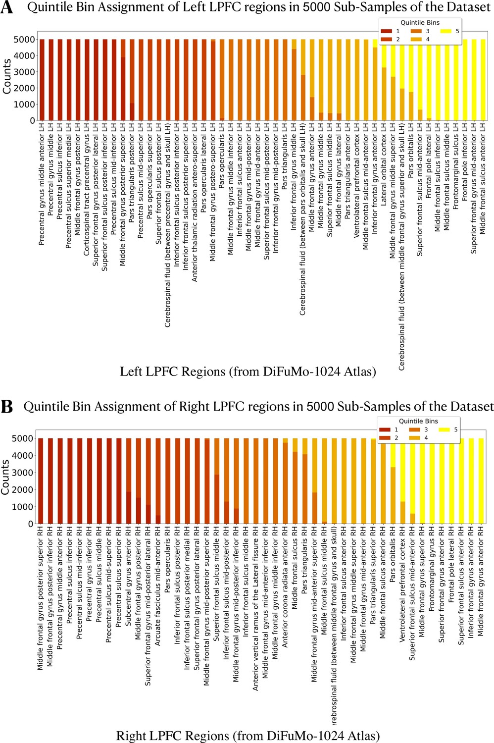
The spatial layout of the principal LPFC gradient across 5000 re-runs of the meta-analysis on random sub-samples of the Neurosynth dataset.
The spatial layout of the principal LPFC gradient across 5000 re-runs of the meta-analysis on random sub-samples of the Neurosynth dataset. We quantify the variation in the spatial layout of the principal LPFC gradient by counting the number of times each region is assigned to every quintile bin in the 5000 re-runs of the meta-analysis (each sub-sample includes 8623 studies, around 60% of the total number of studies). (A) and (B) show the number of times each region has been assigned to each of the five quintile bins in the left and right LPFC, respectively. The majority of LPFC regions in both hemispheres retain the same quintile bin assignment across the 5000 re-runs of the analysis. This result indicates that the spatial layout of the principal gradient is robust to the choice of studies provided that a large of number of studies is included.
Figure 1—figure supplement 2
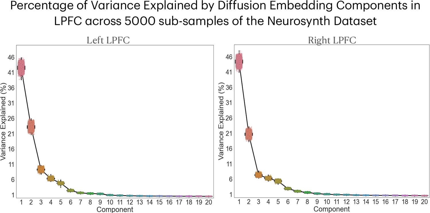
Percentage of variance explained by diffusion embedding components across 5000 re-runs of the meta-analysis.
The percentage of variance explained by diffusion embedding components in 5000 re-runs of the meta-analysis on random sub-samples of the Neurosynth dataset in the left and right LPFC. Each dot represents a run on one sub-sample. We observe that the dots cluster together for each component, indicating small variations in the percentage of variance explained across different runs of the meta-analysis.
Figure 1—figure supplement 3
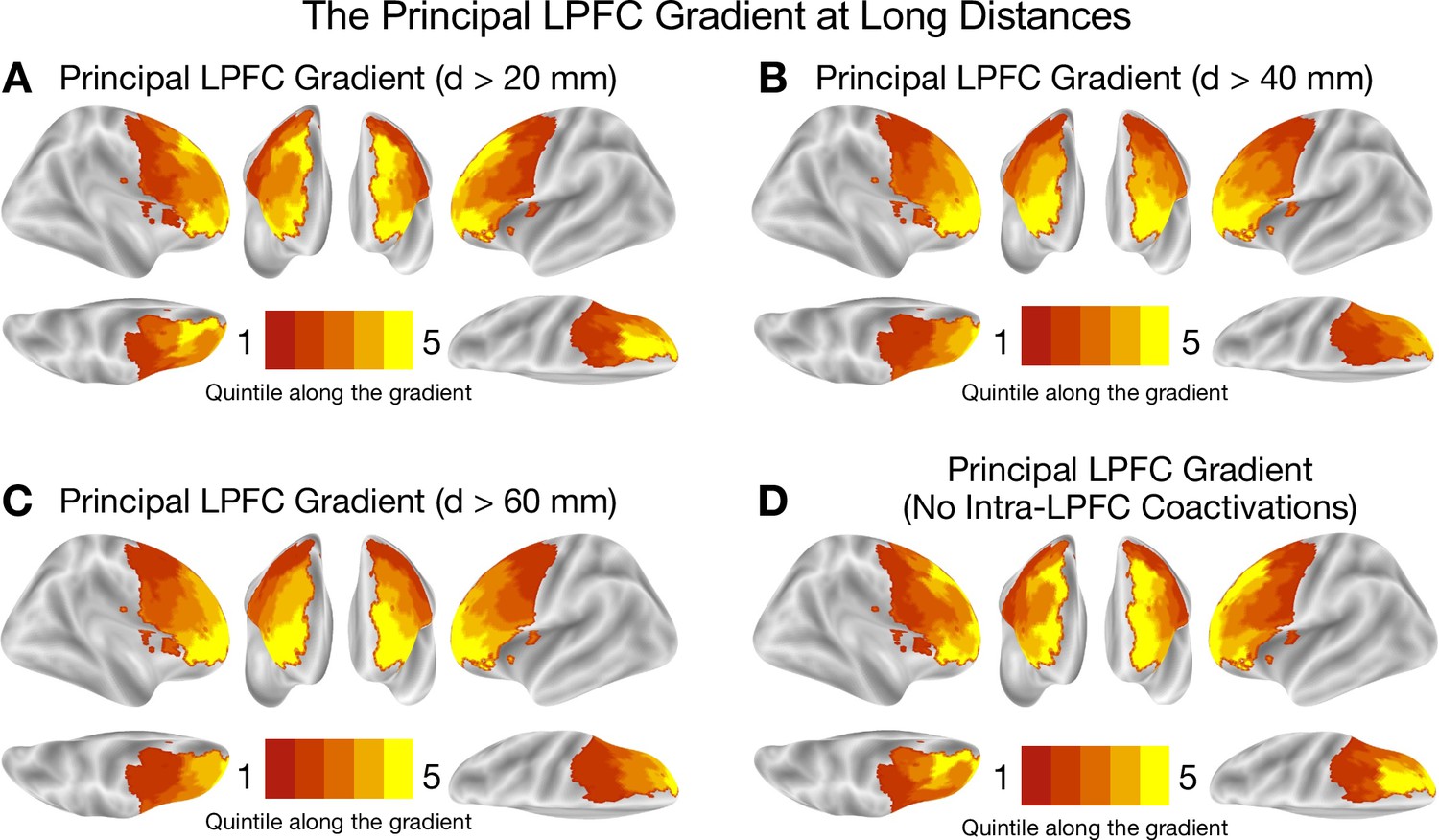
The principal LPFC gradient of long range coactivations.
The principal LPFC gradient of long range coactivations. (A), (B), (C) show the principal LPFC gradient when only taking into account regions that are at least 20 mm, 40 mm, or 60 mm apart, respectively. The coactivation distance is the Euclidean distance between the centers of mass of regions. (D) shows the principal LPFC gradient when not taking into account any intra-LPFC coactivations. The results of this analysis show that, despite changes in the shape of the gradient when varying the coactivation distance, the gross rostrocaudal spatial layout is preserved. These results suggest that the spatial layout of the principal LPFC gradient is not driven by the spatial auto-correlation among nearby LPFC regions.
Figure 1—figure supplement 4
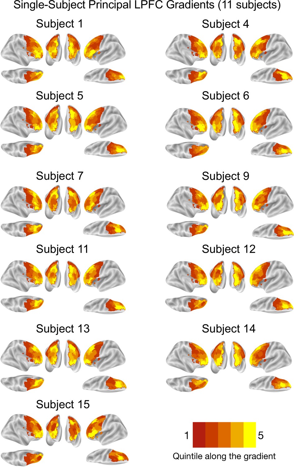
The principal LPFC gradient at the single-subject level.
The principal LPFC gradient at the single-subject level. The subject-level gradients describe changes in coactivation patterns in the LPFC of 11 healthy subjects from the Individual Brain Charting (IBC) dataset. The IBC subjects underwent an extensive set of tasks, which yielded around 750 contrasts per subject. After activation peak extraction, a meta-analysis and a gradient mapping analysis are performed to estimate the gradients. Originally, the dataset included activation maps from 12 participants, but we excluded one participant (‘Subject 8’) for insufficient data available at the time of the study. Note that the labels assigned to the subjects in the original dataset do not include ‘Subject 2’, ‘Subject 3’, and ‘Subject 10’.
Figure 1—figure supplement 5
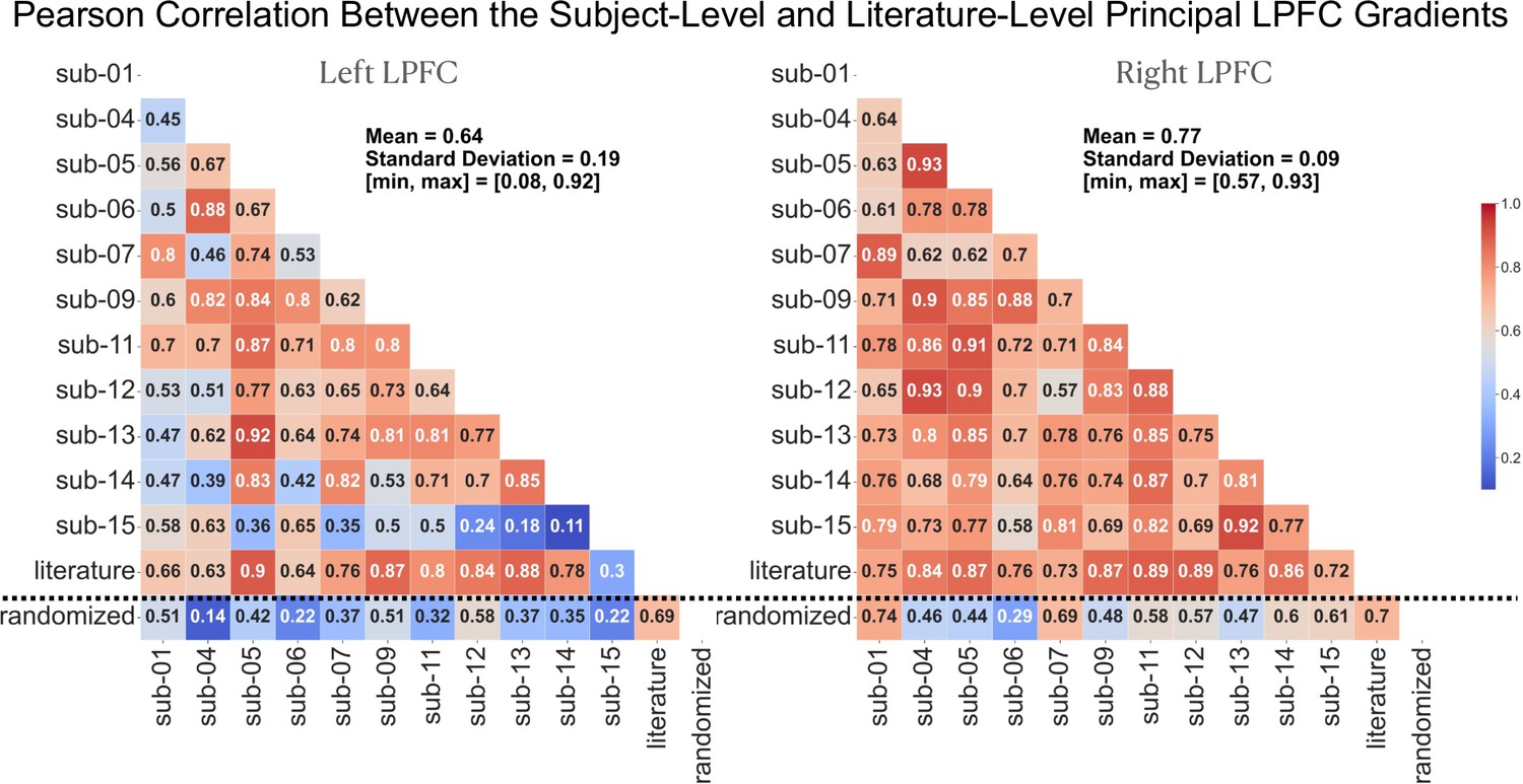
Spatial correlation between the subject-level and literature-level LPFC gradients.
Spatial correlation between the subject- and literature-level principal LPFC gradients. (left) and (right): Correlation among the principal gradients of single subjects and the literature in the left and right LPFC, respectively. The last row of each matrix, separated from other rows by a dotted line, encodes the spatial correlation of the gradients with a gradient estimated from a randomized version of the activation data. Randomization is achieved through 1000 random shuffles of the peak coordinates across the 14,371 studies. The results show strong correlation between most subjects’ principal gradients and the literature principal gradient along with a relatively weaker between-subjects correlation. The mean, standard deviation, minimum, and maximum correlation are estimated from correlation values above the dotted line. Finally, the spatial correlations with the gradient of randomized activation data are relatively weak.
Figure 2 with 1 supplement
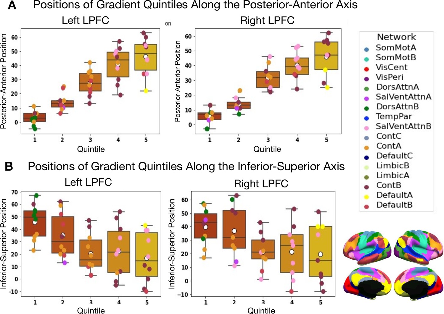
Positions along the posterior-to-anterior and inferior-to-superior axes in MNI space of quintile bins along the principal LPFC gradient.
(A) Positions of quintile bins in the left LPFC. (B) Positions of quintile bins in the right LPFC. Each colored sphere represents a brain region, with the color reflecting its network membership within the 17-Networks atlas (Yeo et al., 2011). SomMot: SomatoMotor, VisCent/Peri: Visual Central/Peripheral, SalVentAttn: Salience/Ventral Attention, DorsAttn: Dorsal Attention, TemPar: Temporo-Parietal, Cont: Executive Control, Default: Default Mode.
Figure 2—figure supplement 1
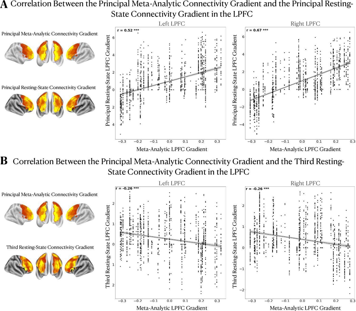
Spatial correlation between the meta-analytic and resting-state connectivity gradients in the LPFC.
Spatial correlation between the meta-analytic and resting-state connectivity gradients in the LPFC. The spatial layout of the resting-state connectivity gradients 1 and 3 from Margulies et al., 2016 is compared to that of the principal meta-analytic LPFC gradient using Pearson’s correlation. We observe (A) a moderately strong positive correlation between the principal meta-analytic and first resting-state connectivity gradient in the LPFC and (B) a weak negative correlation with the third gradient. These results suggest that the principal LPFC gradient can be understood as a local processing stream situated within a global spatial principle that explains unimodal-to-heteromodal brain activity. In the case of the LPFC, this activity organizing profile roughly extends from the caudal to rostral LPFC regions. *** p<0.0001.
Figure 3
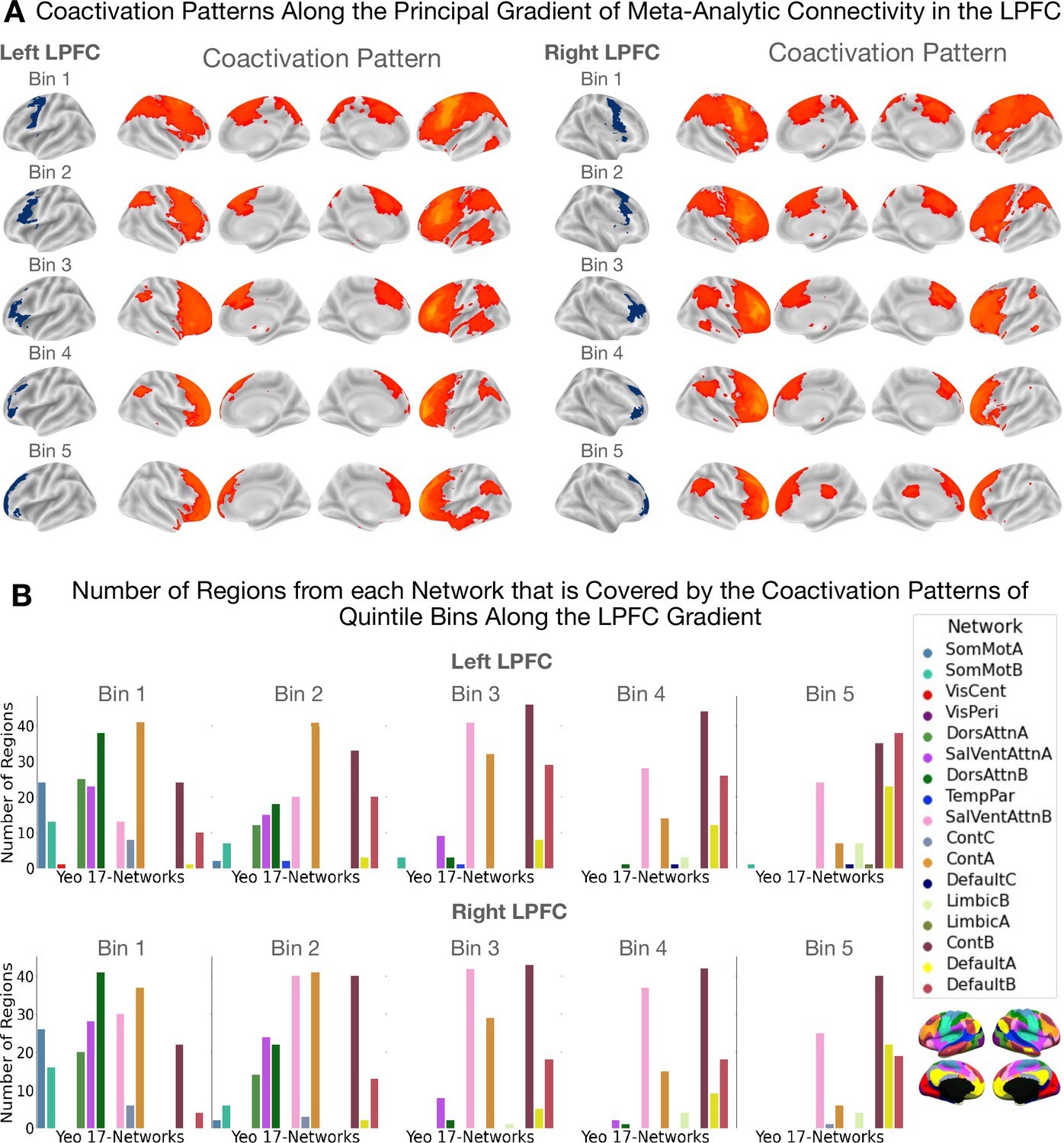
Coactivation patterns along the principal LPFC gradient.
The coactivation patterns of quintile bins along the principal gradient in the LPFC capture a unimodal-to-transmodal spatial layout in brain network connectivity. (A) Coactivation patterns along the principal gradient in the left and right LPFC. Each brain map shows the regions that have a least three times the odds of being reported active given activation in a quintile bin relative to being active when activation is not reported in the quintile bin. Note that cerebellar and sub-cortical regions, although included in the analysis, are not shown in the figures. (B) Bar plots showing the number of regions from each network that overlaps with the coactivation pattern of each quintile bin. The data shown here suggests that the dorsal attention (green) and sensorimotor networks (blue) coactivate with the caudal bins (i.e. bins 1 and 2) more than with more rostral bins. On the other hand, the default mode network coactivates more with the rostral bins (i.e. bins 4 and 5) than with caudal bins.
Figure 4 with 1 supplement

Inferring functional associations along the principal LPFC gradient using segregation queries.
Mapping functional associations using segregation queries reveals a structured ordering of topics along the principal gradient in the (A) left and (B) right LPFC. Topics are ordered by the weighted mean of their location along the principal gradient. Topics of sensorimotor processing are mostly located at the top followed by executive functions and language, and finally emotion/memory/social cognition-related topics mostly occupy the bottom. Note that although the order of topics varies between hemispheres, the general profile of topic associations is comparable. A two-headed arrow along the horizontal axis signifies a coactivation constricted in a given range of quintile bins. Log-odds ratio values are thresholded at 0 to show only positive associations. White asterisks denote the log-odds ratio values whose 95% confidence interval estimated from 5000 re-runs of the meta-analysis on random sub-samples of the Neurosynth dataset does not include the value 0.
Figure 4—figure supplement 1
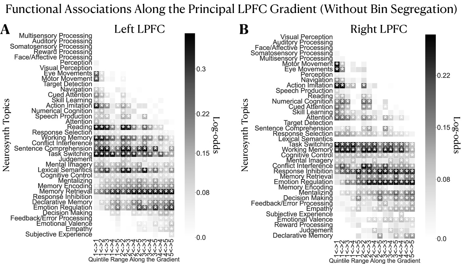
Inferring functional associations along the principal LPFC gradient without segregation queries.
Mapping functional associations without using segregation queries reveals a structured ordering of topics along the principal gradient in the (A) left and (B) right LPFC. Topics are ordered by the weighted mean of their location along the principal gradient. We observe an ordering of topics comparable to that obtained in the segregation-based inference. That is, topics of sensorimotor/attention processes are mostly located at the top followed by executive functions and language and then, emotion/memory/social cognition-related topics mostly occupy the bottom part. However, the individual associations are relatively more distributed across the gradient, indicating less specificity than the segregation based analysis. Log-odds ratio values are thresholded at 0 to show positive associations only.
Figure 5
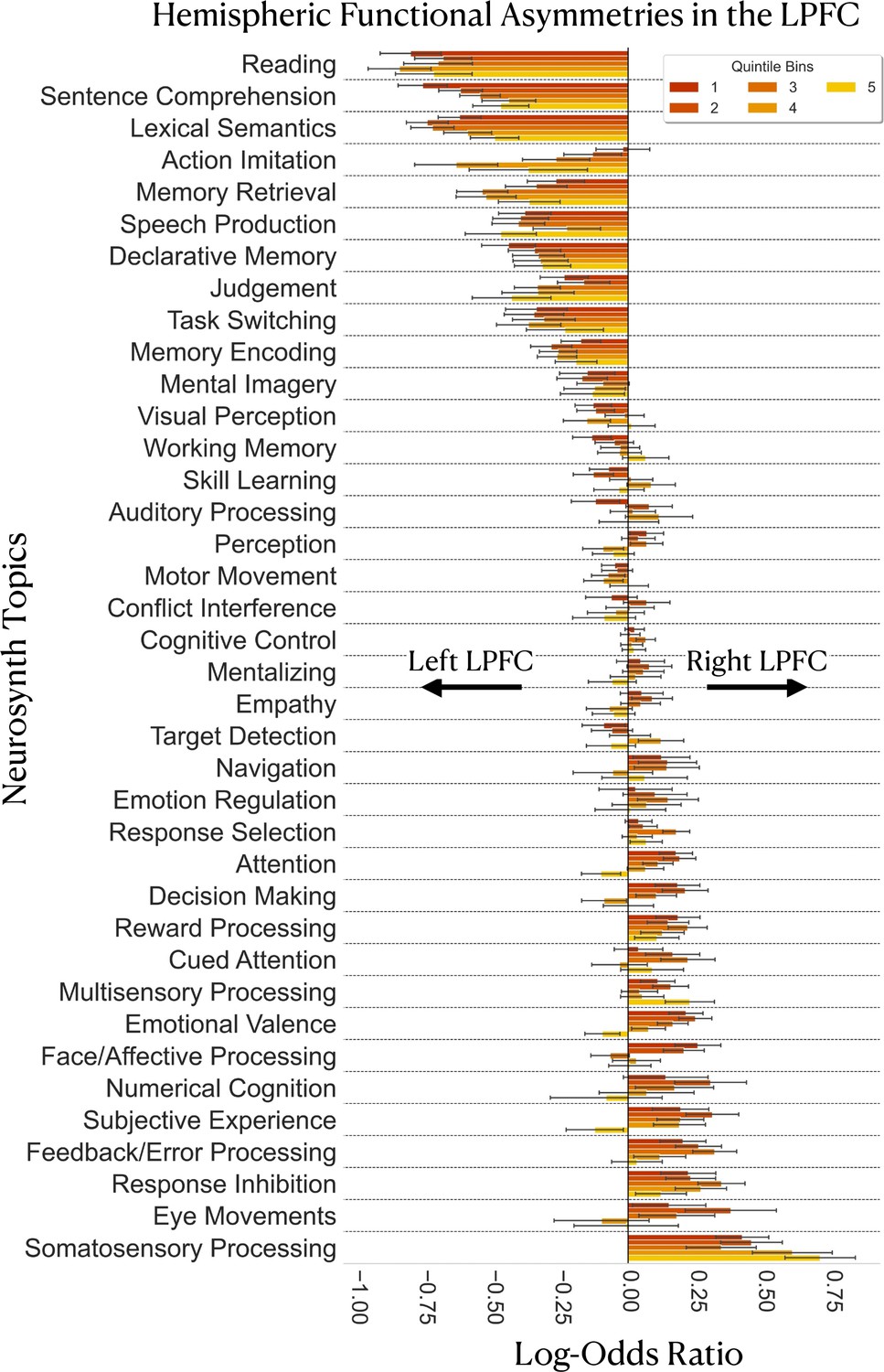
Gradient-based mapping of hemispheric asymmetries in the LPFC.
Meta-analysis of inter-hemispheric asymmetries reasserts the left-hemispheric dominance of language and memory and the right-hemispheric dominance of inhibition and sensory processing/monitoring in the LPFC. Positive log-odds ratios indicate evidence in favor of right-hemispheric preference of a topic in a given bin, whereas negative values indicate evidence in favor of left-hemispheric preference of a topic in a given bin. Error bars represent the 95% confidence intervals estimated from 5000 re-runs of the meta-analysis on random sub-samples of the Neurosynth dataset. Each random sub-sample comprises 60% of the studies of the original dataset (around 8623 studies). Topics are ordered from most-left dominant to most-right dominant based on the average of the log-odds ratio values over the five quintile bins.
Figure 6

Schematic overview of our analysis pipeline.
(A) Inputs and outputs of NeuroLang. Inputs are represented using blue arrows and include: Peak activations and topics from the Neurosynth dataset, the lateral PFC mask, and the 1024 regions from the DiFuMo atlases are represented in a unifying framework within NeuroLang. Two examples of outputs are shown here and represented using red arrows. (B) The main steps of the meta-analysis carried out in this study. (1) Spatial smoothing with 10 mm kernel around each peak. (2) The binary activation map of each study is projected onto 1024 functional regions. Varying shades of red signify that regions have different probabilities of being reported by a study depending on the location of voxels within each region. (3) The meta-analytic connectivity matrix encodes the log-odds ratios of coactivation between each region in the LPFC and every region in the brain. (4) A similarity matrix encodes the degree of correspondence between LPFC regions in their meta-analytic connectivity profiles, estimated by the eta-squared similarity metric. (5) The principal gradient of meta-analytic connectivity in each hemisphere is then derived from the similarity matrix using diffusion embedding. (6) Coactivation patterns of successive quintile gradient bins are inferred (7) Specific topic associations along the principal gradient are inferred using segregation queries. (8) Finally, a gradient-based meta-analysis of hemispheric asymmetries is performed.
Tables
Table 1
Thirty-eight topics from the Neurosynth LDA-driven 100 topics set and the top five terms loading on each topic listed in descending order of association strength.
| topics | terms |
|---|---|
| Action Imitation | mirror; observation; imitation; action; gestures |
| Attention | attention; attentional; task; visual; control |
| Auditory Processing | auditory; sounds; sound; cortex; temporal |
| Cognitive Control | task; cognitive; performance; control; executive |
| Conflict Interference | conflict; interference; control; incongruent; congruent |
| Cued Attention | spatial; cues; cue; location; attention; orienting |
| Decision Making | decision; making; risk; choice; decisions; choices |
| Declarative Memory | memory; retrieval; episodic; mtl; memories |
| Emotion Regulation | regulation; emotion; reappraisal; cognitive; amygdala |
| Emotional Valence | emotional; negative; positive; amygdala; emotion |
| Empathy | social; empathy; moral; game; people |
| Eye Movements | eye; gaze; saccade; movements; target |
| Face/Affective Processing | amygdala; emotional; faces; facial; emotion; expression |
| Feedback/Error Processing | feedback; error; learning; errors; prediction |
| Judgement | judgments; judgment; ppc; reference; drawing |
| Lexical Semantics | semantic; words; word; lexical; verbs |
| Memory Encoding | memory; encoding; hippocampus; hippocampal; retrieval |
| Memory Retrieval | memory; recognition; items; retrieval; recollection |
| Mental Imagery | imagery; mental; events; future; imagined |
| Mentalizing | reasoning; mind; mental; social; tom |
| Motor Movement | motor; movement; cortex; movements; hand |
| Multisensory Processing | visual; motion; auditory; modality; sensory integration |
| Navigation | ms; spatial; virtual; navigation; illusion |
| Numerical Cognition | number; numerical; numbers; magnitude; size |
| Perception | perceptual; perception; interaction; sensory; visual |
| Reading | reading; words; word; phonological; chinese |
| Response Inhibition | inhibition; response; control; inhibitory; task |
| Response Selection | response; stimulus; trials; trial; presented |
| Reward Processing | reward; striatum; ventral; anticipation; monetary |
| Sentence Comprehension | language; comprehension; sentences; sentence; syntax |
| Skill Learning | learning; training; sequence; performance; practice |
| Somatosensory Processing | somatosensory; stimulation; tactile; hand; cortex |
| Speech Production | speech; auditory; production; perception; temporal |
| Subjective Experience | pictures; images; aversive; neutral; unpleasant |
| Target Detection | target; detection; targets; awareness; presented |
| Task Switching | task; switching; rule; set; switch |
| Visual Perception | object; objects; visual; category; cortex |
| Working Memory | memory; working; wm; task; load; verbal |
Additional files
Download links
A two-part list of links to download the article, or parts of the article, in various formats.
Downloads (link to download the article as PDF)
Open citations (links to open the citations from this article in various online reference manager services)
Cite this article (links to download the citations from this article in formats compatible with various reference manager tools)
Functional gradients in the human lateral prefrontal cortex revealed by a comprehensive coordinate-based meta-analysis
eLife 11:e76926.
https://doi.org/10.7554/eLife.76926


