Retinoic acid-gated BDNF synthesis in neuronal dendrites drives presynaptic homeostatic plasticity
- Departments of Neurosurgery, Neuropsychiatry and Behavioral Sciences, Stanford University School of Medicine, United States
Figures
Figure 1 with 1 supplement
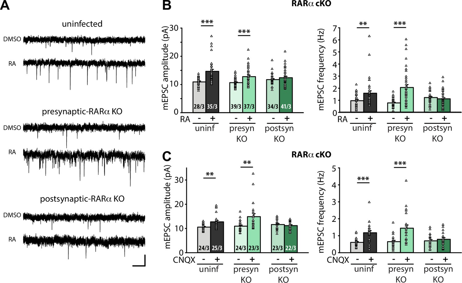
Postsynaptic RARα expression is required for presynaptic homeostatic plasticity.
(A) Example traces of mEPSCs recorded from hippocampal pyramidal neurons in organotypic slices from WT (uninfected), presynaptic RARα KO (Cre expression in CA3) and postsynaptic RARα KO (Cre expression in CA1) groups treated with DMSO or RA (10 μM, 4 hr). Scale bars, 10 pA, 0.5 sec. (B) Quantification of mEPSC amplitudes and frequencies recorded from WT, presynaptic and postsynaptic RARα KO neurons treated with DMSO or RA. **, p < 0.01; ***, p < 0.001; two-way ANOVA followed by Mann Whitney test. Amp: F(2,208) = 5.413, p < 0.01; freq: F(2,208) = 11.81, p < 0.0001. (C) Quantification of mEPSC amplitudes and frequencies recorded from WT, presynaptic and postsynaptic RARα KO neurons treated with DMSO or CNQX (36 hours). **, p < 0.01; ***, p < 0.001; two-way ANOVA followed by Mann Whitney test. Amp: F(2,135) = 6.004, p < 0.01; freq: F(2,135) = 5.23, p < 0.01. n/N represent number of neurons/number of independent experiments (pups). All graphs represent mean ± SEM.
-
Figure 1—source data 1
Individual data spreadsheet in Figure 1B and C.
- https://cdn.elifesciences.org/articles/79863/elife-79863-fig1-data1-v3.xlsx
Figure 1—figure supplement 1
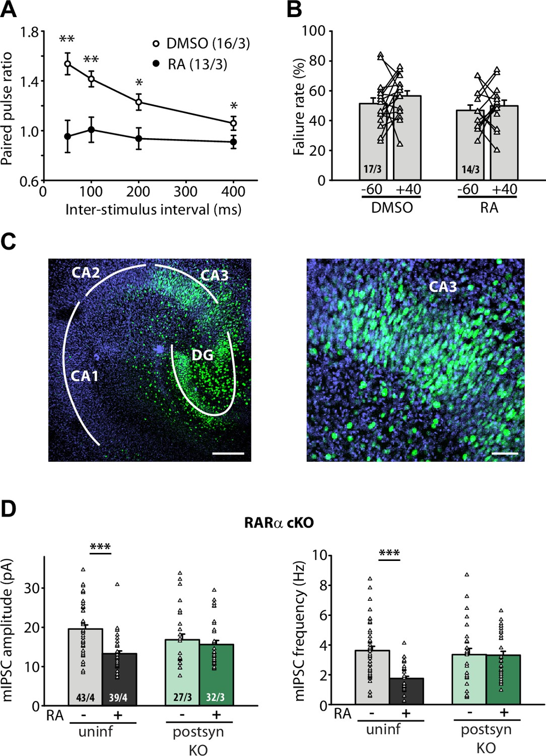
Additional data related to Figure 1: paired-pulse ratio and failure rate of evoked excitatory postsynaptic currents (eEPSCs) at CA3-CA1 synapses in retinoic acid (RA)-treated slices; viral expression efficacy in CA3; homeostatic plasticity of mIPSCs in older neurons.
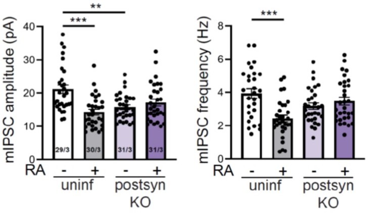
(A) Paired-pulse ratio measured at the CA3-CA1 synapses in DMSO- and RA-treated 21–25 days in vitro (DIV) organotypic hippocampal slices. *, p<0.05; **, p<0.01; two-way ANOVA with repeated measure followed by paired-wise comparison at each ISI. (B) Failure rate of CA3-CA1 eEPSCs at –60 and +40 mV with minimal stimulation in DMSO- and RA-treated organotypic hippocampal slices. p>0.25; paired t-test. (C) Left: a representative image of a 21 DIV organotypic hippocampal slice infected with Cre-GFP-expressing AAVs in the CA3 and part of the DG region. Scale bar: 200 µm. Right: a zoomed in image of CA3 showing AAV-infected neurons (green) represent more than 90% of the neuronal population in the CA3. Scale bar: 500 µm. (D) Quantification of mIPSC amplitudes and frequencies recorded from WT and postsynaptic RARα knockout (KO) neurons treated with DMSO or RA. ***, p<0.001; two-way ANOVA followed by Tukey’s test. Amp: F(1,137) = 5.78, p<0.001; freq: F(1,137) = 10.76, p=0.001. n/N represents number of neurons/number of independent experiments. All graphs represent mean ± SEM.
-
Figure 1—figure supplement 1—source data 1
Individual data spreadsheet in Fig.S1A, S1B and S1D.
- https://cdn.elifesciences.org/articles/79863/elife-79863-fig1-figsupp1-data1-v3.xlsx
Figure 2 with 1 supplement
RARα binds specific brain-derived neurotrophic factor (Bdnf) transcript isoforms.
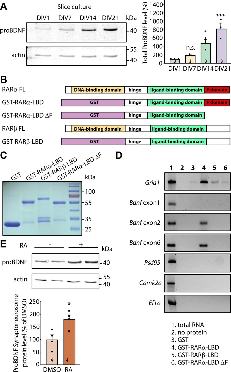
(A) Representative immunoblots (left) and quantification (right) depicting proBDNF expression profiles in cultured hippocampal slices collected at 1, 7, 14, and 21 days in culture. Actin was used as a loading control and all expression levels were normalized to that of 1 day in vitro (DIV) (one-way ANOVA with Dunnett’s multiple comparison test, ***, p<0.001; *, p<0.05). (B)Schematic diagram of recombinant GST fusion proteins of RARα LBD, RARα LBD ΔF, and RARβ LBD used in RNA-binding assays. Full-length RARα and RARβ protein structures are shown as references. (C) Representative imaging of Coomassie brilliant blue-stained SDS-polyacrylamide gel showing the expression of purified recombinant GST and GST fused RARα and RARβ LBD proteins (n=3). (D) Representative images for semi-quantitative RT-PCR of specific Bdnf transcripts pulled down from total hippocampal RNAs in in vitro selection with purified GST fusion proteins. Gria1 mRNA was used as a positive control. Psd95, Camk2a, and Ef1a mRNAs served as negative controls. The representative image shown here is from one of the three experiments with similar results. (E) Representative immunoblot (left) and quantification (right) showing induced proBDNF synthesis in synaptoneurosomal fraction following 30 min of retinoic acid (RA) treatment. Actin was used as a loading control (two- tailed unpaired t-test, *, p<0.05). N represents number of independent experiments. All graphs represent mean ± SEM.
-
Figure 2—source data 1
Individual data spreadsheet in Figure 2A and E.
- https://cdn.elifesciences.org/articles/79863/elife-79863-fig2-data1-v3.xlsx
-
Figure 2—source data 2
Figure 2A ProBDNF and FigureS2A ProBDNF: Immunoblots depicting proBDNF expression profile in cultured hippocampal slices.
Figure 2A Actin and FigureS2A Actin: Immunoblots depicting actin expression profile in cultured hippocampal slices.
Figure 2C Coommassie: Coomassie brilliant blue-stained SDS-polyacrylamide gel showing the expression of purified recombinant proteins.
Figure 2D GluR1: Representative image for semi-quantitative RT-PCR of GluA1 in in vitro selection assay.
Figure 2D BDNF Exon 1: Representative image for semi-quantitative RT-PCR of Bdnf exon 1 in in vitro selection assay.
Figure 2D BDNF Exon 2: Representative image for semi-quantitative RT-PCR of Bdnf exon 2 in in vitro selection assay.
Figure 2D BDNF Exon 6: Representative image for semi-quantitative RT-PCR of Bdnf exon 6 in in vitro selection assay.
Figure 2D CamKII PSD95: Representative image for semi-quantitative RT-PCR of Psd95 and Camkii in in vitro selection assay.
Figure 2D EF1a: Representative image for semi-quantitative RT-PCR of Ef1a in in vitro selection assay.
Figure 2E ProBDNF: Immunoblot showing proBDNF synthesis in synaptoneurosomal fraction following retinoic acid (RA) treatment.
Figure 2E Actin: Immunoblot showing actin levels in synaptoneurosomal fraction following RA treatment.
- https://cdn.elifesciences.org/articles/79863/elife-79863-fig2-data2-v3.zip
Figure 2—figure supplement 1
Additional data related to Figure 2: Mouse brain-derived neurotrophic factor (Bdnf) gene structure and expression; local translation of specific proteins induced by retinoic acid (RA).
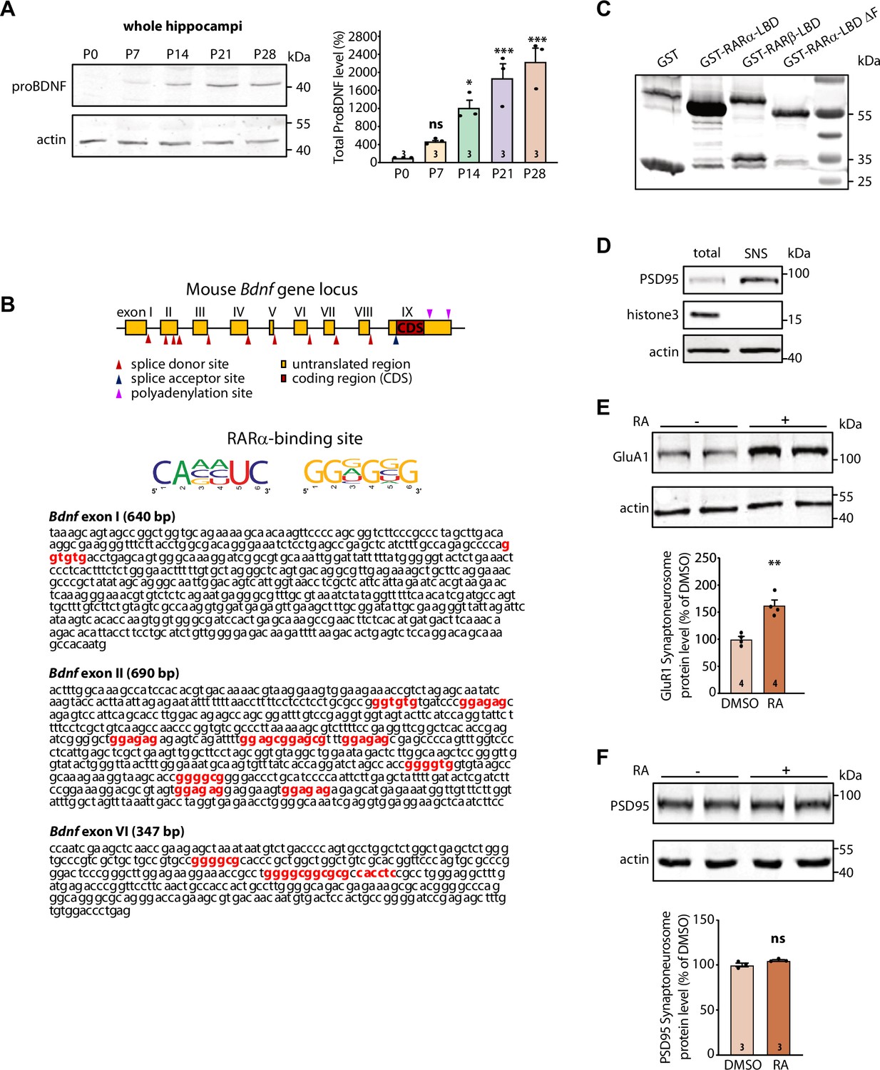
(A) Representative immunoblots (left) and quantification (right) depicting proBDNF expression profile in whole hippocampi collected from 1-, 7-, 14-, 21-, and 28-day-old mouse pups. Actin was used as a loading control. All expression levels were normalized to that of P0 (one-way ANOVA with Dunnett’s multiple comparison test, ***, p<0.001; *, p<0.05). Graph represents mean ± SEM. (B) Schematic diagram of mouse Bdnf gene structure (top). Exons are indicated by boxes. Maroon box inside exon IX represents the coding region of the Bdnf gene. Red arrow heads indicate the alternative splice donor sites giving rise to Bdnf transcripts with different exon choices. The middle panel shows the specific mRNA sequence consensus recognized by RARα. The bottom panel indicates the presence of RARα-binding motifs (in red) in the 5’UTRs of three Bdnf transcripts with exons I, II, and VI. (C) Representative GST-immunoblot showing the expression levels of purified recombinant GST and GST-tagged RARα and RARβ LBD proteins (n=2). (D) Representative immunoblots to confirm the purity of synaptoneurosome preparation. PSD95 was enriched in, and lysate.histone H3 was selectively absent from, the hippocampal synaptoneurosome fraction relative to the whole cell. (E) Representative immunoblot (top) and quantification (bottom) showing induced GluA1 synthesis in synaptoneurosomal fraction following 30 min of retinoic acid (RA) (1 μM) treatment. Actin was used as a loading control (two-tailed unpaired t-test, **, p<0.005). (F) Representative immunoblot (top) and quantification (bottom) showing no induction of PSD95 synthesis in synaptoneurosomes following 30 min of RA treatment. Actin was used as a loading control. N represents number of independent experiments. All graphs represent mean ± SEM.
-
Figure 2—figure supplement 1—source data 1
Individual data spreadsheet in Fig.S2A, S2E and S2F.
- https://cdn.elifesciences.org/articles/79863/elife-79863-fig2-figsupp1-data1-v3.xlsx
-
Figure 2—figure supplement 1—source data 2
Figure 2A ProBDNF and FigureS2A ProBDNF: Immunoblots depicting proBDNF expression profile in whole hippocampi collected from mouse pups.
Figure 2A Actin and Figure S2A Actin: Immunoblots depicting actin expression profile in whole hippocampi collected from mouse pups. Figure 2: GST-immunoblot showing the expression levels of purified recombinant proteins. FigureS2D Histone H3: Immunoblot to confirm histone H3 was selectively absent from the hippocampal synaptoneurosome fraction relative to the whole-cell lysate. Figure 2 PSD95: Immunoblot to confirm PSD95 was enriched in the hippocampal synaptoneurosome fraction relative to the whole-cell lysate. FigureS2D Actin: Immunoblot depicting actin levels in hippocampal synaptoneurosome fraction and whole-cell lysate. FigureS2E GluA1 and actin: Immunoblot showing GluA1 synthesis in synaptoneurosomal fraction following retinoic acid (RA) treatment. Actin is shown as a loading control. FigureS2F PSD95 and actin: Immunoblot showing no PSD95 synthesis in synaptoneurosomal fraction following RA treatment. Actin is shown as loading control.
- https://cdn.elifesciences.org/articles/79863/elife-79863-fig2-figsupp1-data2-v3.zip
Figure 3 with 1 supplement
Retrograde BDNF signalling is required for RARα-mediated regulation of presynaptic homeostatic scaling.
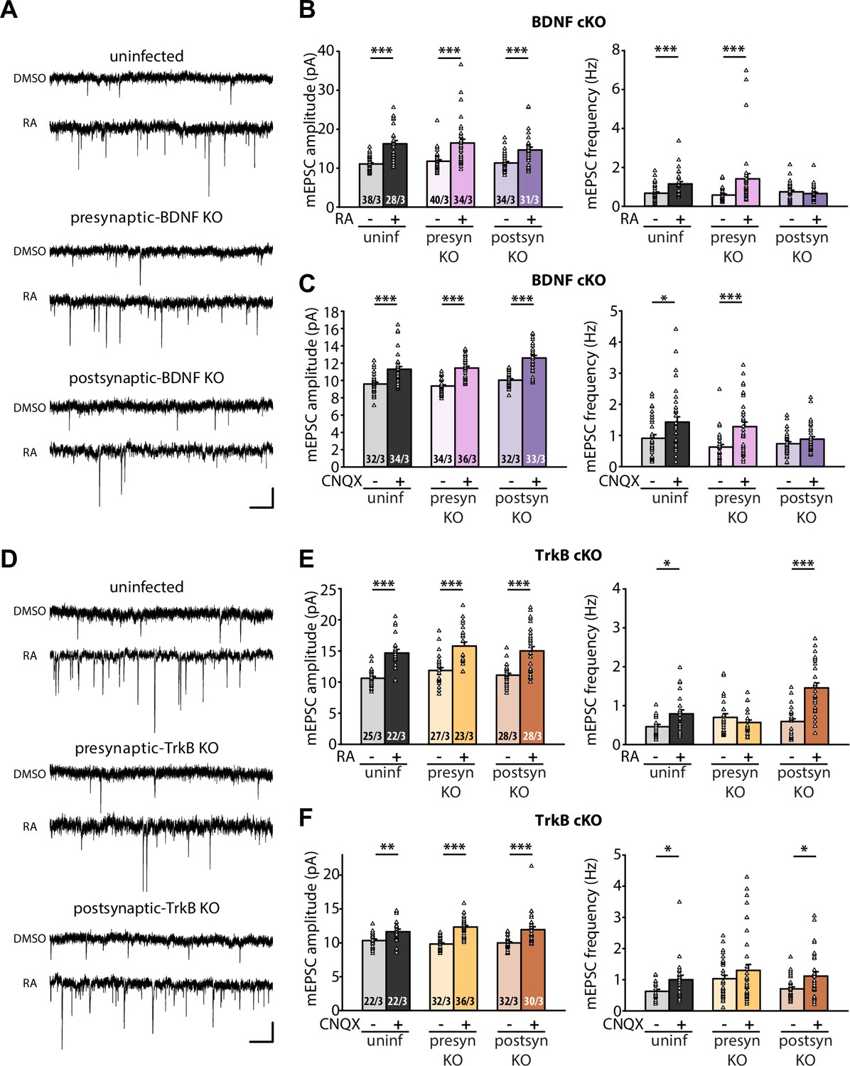
(A) Example traces of mEPSCs recorded from hippocampal pyramidal neurons in organotypic slices from WT (uninfected), presynaptic BDNF KO (Cre expression in CA3) and postsynaptic BDNF KO (Cre expression in CA1) groups treated with DMSO or RA (10 μM, 4 hr). Scale bars, 10 pA, 0.5 sec. (B) Quantification of mEPSC amplitudes and frequencies recorded from WT, presynaptic and postsynaptic BDNF KO neurons treated with DMSO or RA. ***, p < 0.001; two-way ANOVA followed by Mann Whitney test. Amp: F(2,199) = 1.062, p > 0.3; freq: F(2,199) = 6.244, p < 0.01. (C) Quantification of mEPSC amplitudes and frequencies recorded from WT, presynaptic and postsynaptic BDNF KO neurons treated with DMSO or CNQX (36 hours). *, p<0.05, **, p < 0.01; ***, p < 0.001; two-way ANOVA followed by Mann Whitney test. Amp: F(2,195) = 1.77, p > 0.15; freq: F(2,195) = 2.53, p >0.05. (D) Example traces of mEPSCs recorded from hippocampal pyramidal neurons in organotypic slices from WT (uninfected), presynaptic TrkB KO (Cre expression in CA3) and postsynaptic TrkB KO (Cre expression in CA1) groups treated with DMSO or RA (10 μM, 4 hr). Scale bars, 10 pA, 0.5 sec. (E) Quantification of mEPSC amplitudes and frequencies recorded from WT, presynaptic and postsynaptic TrkB KO neurons treated with DMSO or RA. **, p < 0.01; ***, p < 0.001; two-way ANOVA followed by Mann Whitney test. Amp: F(2,147) = 0.01, p > 0.9; freq: F(2,147) = 14.87, p < 0.0001. (F) Quantification of mEPSC amplitudes and frequencies recorded from WT, presynaptic and postsynaptic TrkB KO neurons treated with DMSO or CNQX (36 hours). *, p<0.05,**, p < 0.01; ***, p < 0.001; two-way ANOVA followed by Mann Whitney test. Amp: F(2,168) = 2.33, p > 0.1; freq: F(2,168) = 0.17, p> 0.8.n/N represent number of neurons/number of independent experiments. All graphs represent mean ± SEM.
-
Figure 3—source data 1
Individual data spreadsheet in Figure 3B, C, E and F.
- https://cdn.elifesciences.org/articles/79863/elife-79863-fig3-data1-v3.xlsx
Figure 3—figure supplement 1
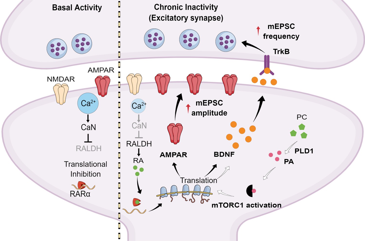
A working model for retinoic acid (RA)-mediated regulation of presynaptic and postsynaptic homeostatic plasticity.
When synaptic transmission is normal, basal dendritic calcium levels inhibit RA synthesis through a calcineurin-dependent pathway. This allows RARα to inhibit the translation of specific dendritic mRNAs. During chronic synaptic inactivity, decreased calcium levels in dendrites remove the inhibition on RA synthesis. Newly synthesized RA binds to RARα and de-represses local translation of specific dendritic mRNAs, including those for GluA1 and brain-derived neurotrophic factor (BDNF). Newly synthesized α-amino-3-hydroxy-5-methyl-4-isoxazolepropionic acid (AMPA) receptors supports enhanced excitatory postsynaptic strength, while newly synthesized BDNF acts retrogradely on presynaptic tropomyosin receptor kinase B (TrkB) receptors to augment presynaptic functions. Previous studies have also identified mTORC1 activation as a key regulatory step in de novo BDNF synthesis, and showed that AMPA receptor blockade results in enhanced phospholipase D (PLD1) activity that converts phosphatidylcholine (PC) to phosphatidic acid (PA), which in turn engages mTORC1 signaling pathway. Thus, multiple signaling pathways may work in concert to regulate postsynaptic protein synthesis in the context of homeostatic plasticity.
Author response image 1
Effect of RA and postsynaptic BDNF deletion on mIPSCs.
Quantification of mEPSC amplitudes (left) and frequencies (right) recorded from WT and postsynaptic BDNF KO neurons treated with DMSO or RA. **, p < 0.01; ***, p < 0.001, two-way ANOVA followed by Tukey’s test. Amp: F(1,117) = 17.55, p < 0.0001. Freq: F(1,117) = 15.59, p = 0.001. n/N = # of neurons/# of mice. Data shown as mean ± SEM.
Tables
Key resources table
| Reagent type (species) or resource | Designation | Source or reference | Identifiers | Additional information |
|---|---|---|---|---|
| Strain, strain background (Mus musculus) | Rarafl/fl | Chapellier et al., 2002 | N/A | |
| Strain, strain background (Mus musculus) | Bdnffl/fl | Rios et al., 2001 (The Jackson Lab) | JAX#004339 RRID: IMSR_JAX:004339 | |
| Strain, strain background (Mus musculus) | Ntrk2fl/fl | Luikart, 2005 | N/A | |
| Strain, strain background (Mus musculus) | CD-1 IGS | Charles River Laboratories | Strain code: 022 | |
| Antibody | Anti-Glutathione-S-transferase (GST) (Mouse monoclonal) | Sigma | SAB4200237-200UL; Clone 2H3-D10 | WB (1:1000) |
| Antibody | Anti-BDNF (Rabbit monoclonal) | Abcam | ab108319 | WB (1:1000) |
| Antibody | Anti-Glur1-NT (N-terminus) (Mouse monoclonal) | Millipore | MAB2263; Clone RH95 | WB (1:2000) |
| Antibody | Anti-PSD95 (Mouse monoclonal) | Invitrogen | MA1-046 | WB (1:1000) |
| Antibody | Anti-Histone H3 (Rabbit polyclonal) | Millipore | 07-690 | WB (1:5000) |
| Antibody | Anti-Actin (Mouse monoclonal) | Millipore | MAB1501; Clone C4 | WB (1:5000) |
| Antibody | IRDye 800CW IgG Secondary Antibody (Donkey anti-Rabbit) | Li-cor | P/N: 926-32213 | WB (1:10,000) |
| Antibody | IRDye 680RD IgG Secondary Antibody (Donkey anti-Mouse) | Li-cor | P/N: 926–68072 | WB (1:10,000) |
| Recombinant DNA reagent | pGEX-KG | Expression vector with GST tag | ||
| Recombinant DNA reagent | pGEX-KG-RARα LBD | This paper | Nucleotides 460 to end | |
| Recombinant DNA reagent | pGEX-KG-RARα LBD ΔF | This paper | Nucleotides 460–1,251 | |
| Recombinant DNA reagent | pGEX-KG- RARβ LBD | This paper | 496 to end | |
| Sequence-based reagent | Gria1_F | This paper | PCR primers | caatcacaggaacatgcggc |
| Sequence-based reagent | Gria1_R | This paper | PCR primers | cctgccagttcttctcggcggc |
| Sequence-based reagent | Exon 1 Bdnf _F | This paper | PCR primers | ctccctcactttctctggg |
| Sequence-based reagent | Exon 1 Bdnf _R | This paper | PCR primers | ctgagagacacgtttccc |
| Sequence-based reagent | Exon 2 Bdnf _F | This paper | PCR primers | cgagccccagtttggtcccc |
| Sequence-based reagent | Exon 2 Bdnf _R | This paper | PCR primers | ggtggctagatcctggtg |
| Sequence-based reagent | Exon 6 Bdnf _F | This paper | PCR primers | gacccggttccttcaactgcc |
| Sequence-based reagent | Exon 6 Bdnf _R | This paper | PCR primers | ctcagggtccacacaaagctctcgg |
| Sequence-based reagent | Dlg4 (Psd95)_F | This paper | PCR primers | catcgaaggaggcgctgccc |
| Sequence-based reagent | Dlg4 (Psd95)_R | This paper | PCR primers | cattgtccaggtgctgagaata |
| Sequence-based reagent | Camk2a_F | This paper | PCR primers | cattgtggcccgggagtatt |
| Sequence-based reagent | Camk2a_R | This paper | PCR primers | ggtgatgggaaatcataggcacc |
| Sequence-based reagent | Ef1a_F | This paper | PCR primers | cgagaccagcaaatactatgtgacc |
| Peptide, recombinant protein | Ef1a_R | This paper | PCR primers | ggcatattagcacttggctcc |
| Commercial assay or kit | PrimeScript RT Reagent Kit with gDNA Eraser (Perfect Real Time) | Takara | RR047B | |
| Chemical compound, drug | Retinoic acid (RA) | Sigma | R2625-50MG | |
| Chemical compound, drug | CNQX | Tocris | 0190 | |
| Chemical compound, drug | cOmplete, EDTA-free Protease Inhibitor Cocktail | Sigma | 4693132001 | |
| Chemical compound, drug | RNasin Ribonuclease Inhibitors | Promega | N2115 | |
| Chemical compound, drug | TRIzol Reagent | Thermo Fisher Scientific | 15596026 | |
| Software, algorithm | Image Studio 5.2.5 | LI-COR Bioscience | https://www.licor.com/bio/pr oducts/software/image_studi o_lite/ | |
| Software, algorithm | Prism 9 | GraphPad | https://www.graphpad.com/s cientific-software/prism/ | |
| Software, algorithm | ImageJ | NIH | https://imagej.nih.gov/ij/ | |
| Software, algorithm | Mind the graph | https://mindthegraph.com/ | ||
| Other | Glutathione Sepharose 4B | Sigma Millipore | GE17-0756-05 | In vitro RNA-binding assay |
Additional files
Download links
A two-part list of links to download the article, or parts of the article, in various formats.
Downloads (link to download the article as PDF)
Open citations (links to open the citations from this article in various online reference manager services)
Cite this article (links to download the citations from this article in formats compatible with various reference manager tools)
Retinoic acid-gated BDNF synthesis in neuronal dendrites drives presynaptic homeostatic plasticity
eLife 11:e79863.
https://doi.org/10.7554/eLife.79863


