Insight into the evolutionary assemblage of cranial kinesis from a Cretaceous bird
- Key Laboratory of Vertebrate Evolution and Human Origins, Institute of Vertebrate Paleontology and Paleoanthropology, Chinese Academy of Sciences, China
- Center for Excellence in Life and Paleoenvironment, Chinese Academy of Sciences, China
- University of Chinese Academy of Sciences, China
- Field Museum of Natural History, United States
Figures
Figure 1 with 2 supplements
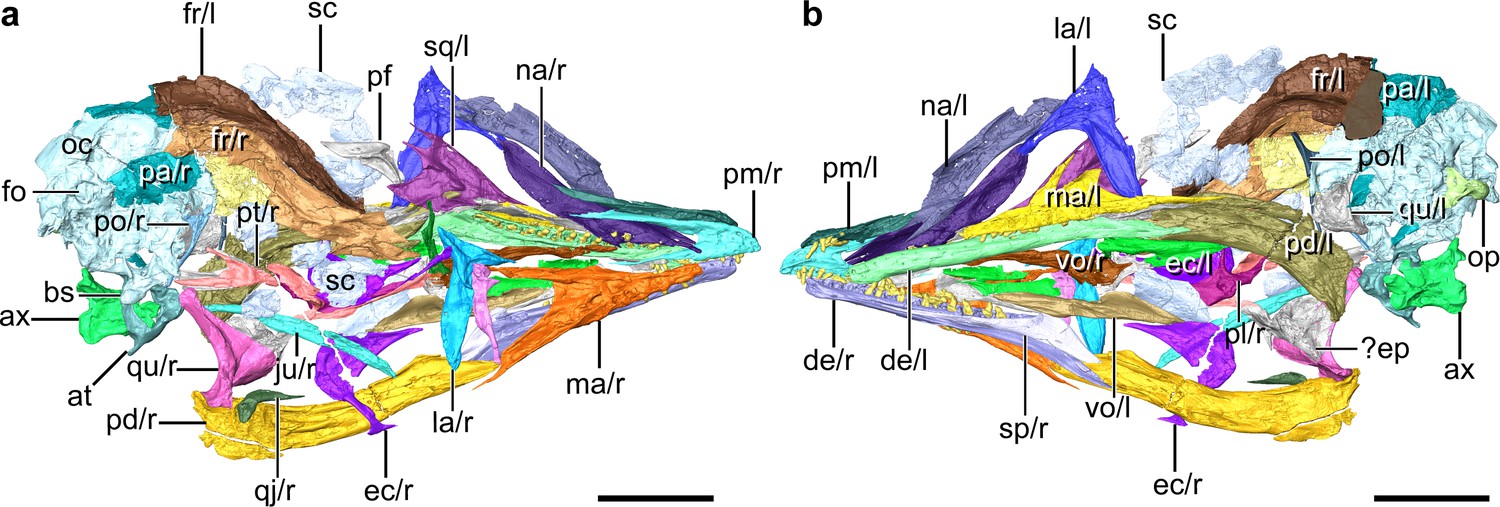
Digital reconstruction of the skull of Yuanchuavis, IVPP V27883.
(a and b) Skull in right (a) and left (b) view, respectively. ax, axis; at, atlas; bs, basipterygoid process; de, dentary; ec, ectopterygoid; ep, epipterygoids; fo, foramen magnum; fr, frontal; ju, jugal; la, lacrimal; ma, maxilla; na, nasal; oc, occipital region; op, occipital condyle; pa, parietal; pd, post-dentary mandible; pl, palatine; pm, premaxilla; po, postorbital; pt, pterygoid; qj, quadratojugal; qu, quadrate; sc, scleral ossicles; sp, splenial; sq, squamosal; vo, vomer; and r/l, right/left side. Scale bars, 10 mm.
Figure 1—figure supplement 1
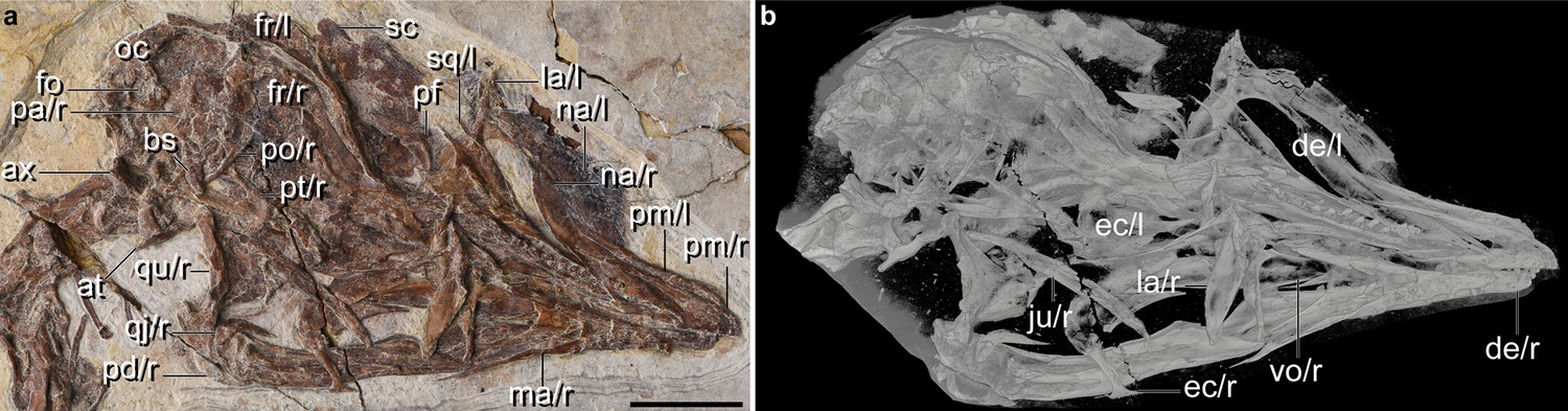
Cranial anatomy of Yuanchuavis, IVPP27883.
(a) Photograph. (b) CT imaging. ax, axis; at, atlas; bs, basipterygoid process; de, dentary; ec, ectopterygoid; fo, foramen magnum; fr, frontal; ju, jugal; la, lacrimal; ma, maxilla; na, nasal; oc, occipital region; pa, parietal; pf, prefrontal; pd, post-dentary mandible; pm, premaxilla; po, postorbital; pt, pterygoid; qj, quadratojugal; qu, quadrate; sc, sclerotic bone; sp, splenial; sq, squamosal; vo, vomer; r/l, right/left side. Scale bar, 10 mm.
Figure 1—figure supplement 2
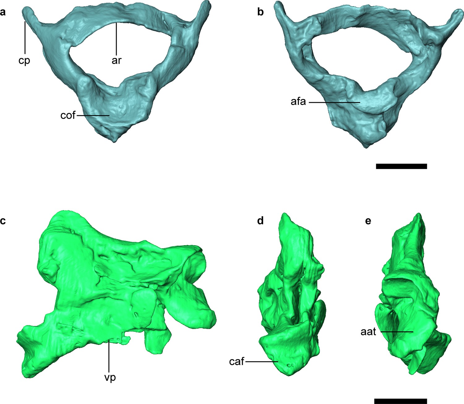
Digital reconstruction of atlas and axis of Yuanchuavis.
(a and b) Atlas in cranial (a) and caudal view (b). (c–e) Axis in lateral (c), caudal (d), and cranial view (e). aat, articular facet for atlas; afa, articular facet for axis; ar, arcus atlantis; caf, caudal articular facet; cof, condyloid fossa; cp, costal process; tv, transverse process; Scale bars, 2 mm.
Figure 2 with 1 supplement
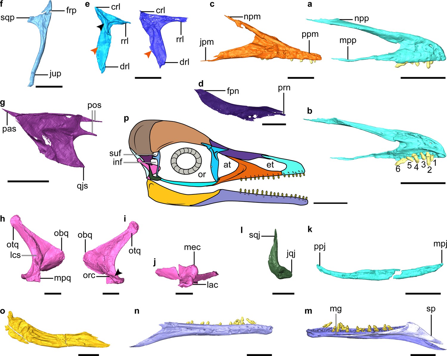
Digital reconstruction of facial and jaw bones of Yuanchuavis.
(a and b) Right premaxilla in lateral and medial view, respectively (numbers 1–6 denote premaxillary teeth; (b) is mirrored). (c) Right maxilla. (d) Right nasal. (e) Right (right column) and left lacrimal in lateral and medial view, respectively (black and red arrowhead denote the lateral flange and foramen, respectively). (f) Right postorbital. (g) Left squamosal. (h–j) Right quadrate (i) arrowhead denotes the absence of a pterygoid condyle. (k) Right jugal. (l) Right quadratojugal. (m and n) Right dentary. (o) Right post-dentary mandible. (p) Reconstruction of Yuanchuavis skull in lateral aspect. (a, c–f, h, k–m, and o) Lateral view; (b, g, i, and n) medial view; (j) ventral view. at, antorbital fenestra; crl, caudal ramus of lacrimal; drl, descending ramus of lacrimal; et, external naris; frp, frontal process of postorbital; inf, infratemporal fenestra; jpm, jugal process of maxilla; jqj, jugal process of quadratojugal; jup, jugal process of postorbital; lac, lateral condyle; lsc, lateral crest; mec, medial condyle; mg, Meckel’s groove; mpj, maxillary process of jugal; mpp, maxillary process of premaxilla; mpq, mandiblar process of quadrate; npm, nasal process of maxilla; npp, nasal process of premaxilla; obq, orbital process of quadrate; or, orbit; orc, orbitocondylar crest; otq, otic process of quadrate; pas, paroccipital process of squamosal; pf, prefrontal; pos, postorbital process of squamosal; ppj, postorbital process of jugal; ppm, premaxillary process of maxilla; qjs, quadratojugal process of squamosal; rrl, rostral ramus of lacrimal; sp, splenial; sqj, squamosal process of quadratojugal; sqp, squamosal process of postorbital; suf, supratemporal fenestra. Scale bars, 5 mm (a–e, g–k, and m–o), 2.5 mm (f and l), and 10 mm (p).
Figure 2—figure supplement 1
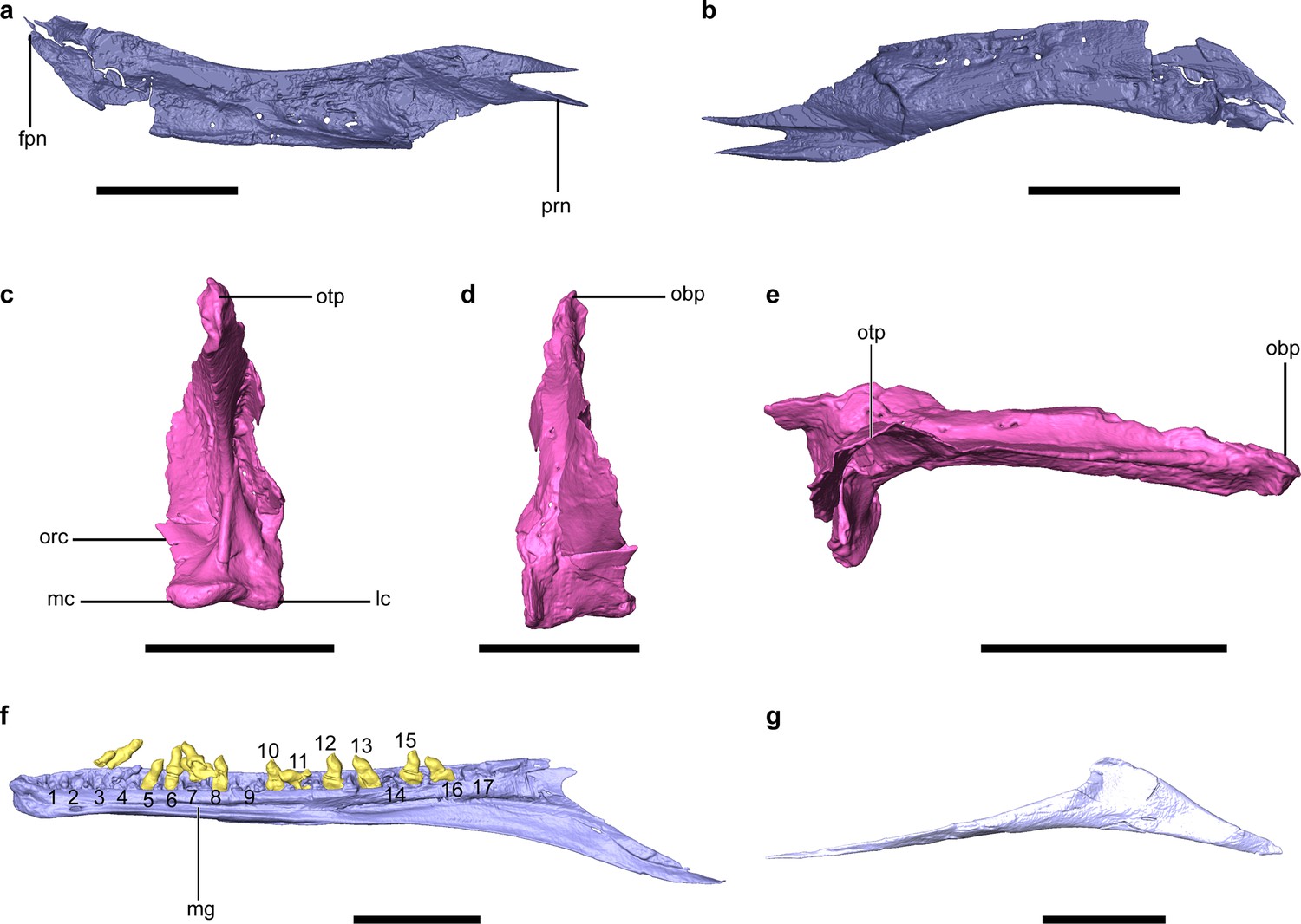
Additional cranial anatomy of Yuanchuavis.
(a–g) Digital reconstruction of the left nasal in medial (a) and lateral (b) view, right quadrate in caudal (c), rostral (d), and dorsal (e) view, right dentary in medial view (f), and right splenial in medial view (g). fpn, frontal process of nasal; lc, lateral condyle; mc, medial condyle; mg, Meckel’s groove; obp, orbital process; orc, orbitocondylar crest; otp, otic process; prn, premaxillary process of nasal; 1–17, dentary tooth count. Scale bars, 10 mm (a, b, f, and g), and 5 mm (c–e).
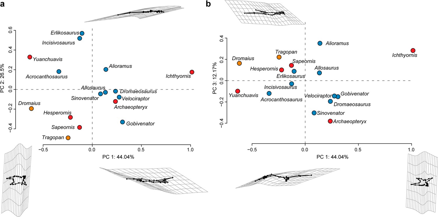
Figure 3 with 5 supplements
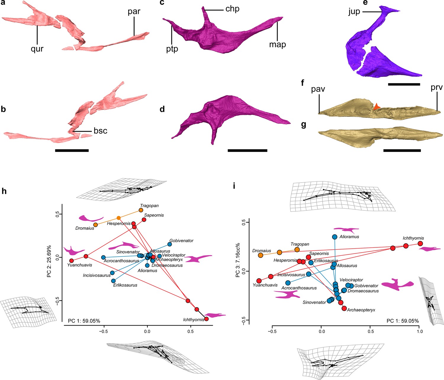
Palate anatomy of Yuanchuavis.
(a–f) Digital reconstruction of the right pterygoid in lateral (a) and ventral (b) view; right palatine in dorsal (c) and ventral (d) view; right ectopterygoid in dorsal view (e); and left vomer in dorsal (f) and ventral (g) view (arrowhead denotes the dorsal transverse ridge). (h and i) Phylomorphospace showing the diversity of palatine shape in early-diverging avialans and their close non-avialan theropod relatives based on the first three principal components (PC1–PC3), with deformation grids and wireframes from average to extreme; line drawings of the palatine in dorsal/ventral view are placed nearby corresponding taxa (blue circles: non-avialan theropods; red circles: Mesozoic avialans; orange circles: crown birds). bsc, basipterygoid process cotyle; chp, choanal process; jup, jugal process; map, maxillary process; par, palatine ramus; pav, palatine ramus of vomer; prv, premaxillary ramus of vomer; ptp, pterygoid process; qur, quadrate ramus. Scale bars, 10 mm (a–d, f, and g), 2 mm (e).
Figure 3—figure supplement 1

Comparison of pterygoid morphology.
(a and b) Digital reconstruction of the pterygoid of Yuanchuavis (a) right element in lateral view, and enantiornithine IVPP V12707 (b), left element in medial view. (c and d) Line drawing of the pterygoid of Sinornithosaurus millenii (Theropod: Dromaeosauridae; left in medial view; c), and Sinovenator changii (Theropod: Troodontidae; left in medial view; d). (e and f) Digital reconstruction of the pterygoid of Tragopan caboti (e) left element in ventrolateral view, and Dromaius novaehollandiae (f), left element in lateral view. afq, articular facet for quadrate; afp, articular facet for parasphenoid rostrum; bsc, basipterygoid cotyla; par, palatine ramus; qur, quadrate ramus. The blue arrowhead in (a) denotes the caudal extension that is absent in other enantiornithines and non-avialan theropods (b–d). The green arrowhead in (f) indicates the potential homologous but reduced quadrate ramus present in some paleognaths. All figures are not scaled.
Figure 3—figure supplement 2
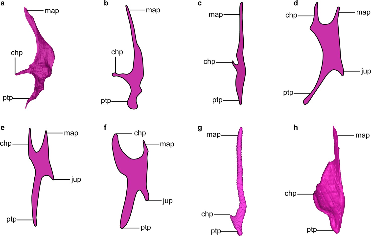
Comparison of palatine morphology.
(a, g, and h) Digital reconstruction of the palatine of Yuanchuavis (a) right element in dorsal view, Tragopan caboti (g) left element in ventral view, and Dromaius novaehollandiae (h) left element in ventral view. (b–f) Interpretative line drawing of the palatine of selected early-diverging avialans and non-avialan theropods (converted to right element in ventral view): Sapeornis (b), Hesperornis (c), Archaeopteryx (d; modified from Bhullar et al., 2016), Velociraptor (e; modified from Barsbold and Osmólska, 1999), and Dromaeosaurus (f; modified from Currie, 1995). chp, choanal process; jup, jugal process; map, maxillary process; ptp, pterygoid process. All figures are not scaled.
Figure 3—figure supplement 3
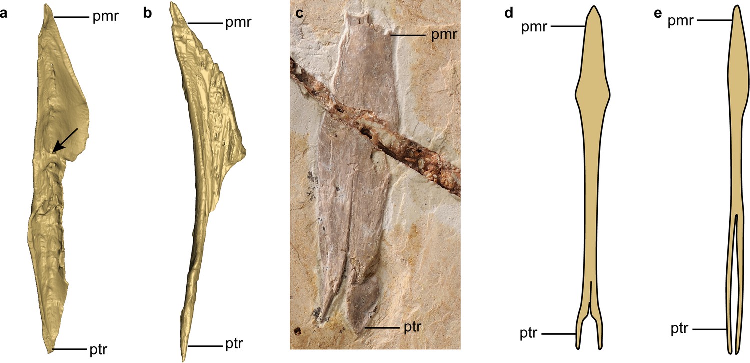
Comparison of vomer morphology.
(a and b) Digital reconstruction of the right vomer of Yuanchuavis (a) and left vomer of enantiornithine IVPP V127071 (b) in dorsal aspect (the left vomer of IVPP V127071 has been mirrored). (c) Photograph of Sapeornis (IVPP V19058). (d and e) Line drawing of the vomer of Dromaeosaurus (d) and Allosaurus (e) in dorsal view (modified from Currie, 1995; Barsbold and Osmólska, 1999). The rostral ends of the vomers are fused in (c–e), which are not in the two enantiornithines (a and b). pmr, premaxillary ramus; ptr, pterygoid ramus. The arrowhead in (a) denotes the transverse ridge that is absent in other taxa. All figures are not scaled.
Figure 3—figure supplement 4
Diversity of palatine shape in early-diverging avialans and non-avialan theropods.
Morphospace based on the first three principal components (PC1–PC3), with deformation grids and wireframes from average to extreme (blue circles: non-avialan theropods; red circles: Mesozoic avialans; orange circles: crown birds).
Figure 3—figure supplement 5
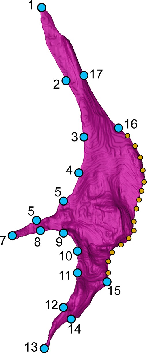
Landmark scheme.
17 landmarks (blue dots) and 15 semi-landmarks (orange dots) were digitized on the CT reconstructed right palatine of Yuanchuavis in dorsal view. See Appendix 1 for anatomical description of landmarks and semi-landmark placements.
Figure 4
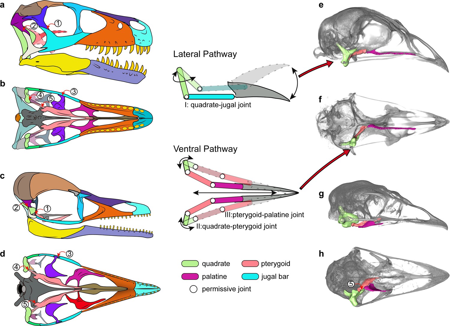
Cranial kinesis in early bird evolution.
The typical avian cranial kinesis is realized through two pathways (schematic drawing in the middle): the quadrate-jugal bar-rostrum on the lateral side and quadrate-pterygoid-palatine on the ventral side, highlighted in the galliform Tragopan caboti (e and f). These two pathways are restricted by the postorbital bar (1), squamosal-quadratojugal contact (2), ectopterygoid (3), the scarf joint between quadrate-pterygoid (4), and the prominent pointed basipterygoid process (5) in non-avialan theropods (a and b) and enantiornithines (c and d), indicating an akinetic skull. The ventral pathway is also absent in most paleognaths (h) at least restricted by non-permissive contact (5). (a–d) Skull reconstructions of Dromaeosaurus (Theropod: Dromaeosauridae) in lateral (a) and ventral (b) view (modified from Currie, 1995), Yuanchuavis (Avialae: Enantiornithes) in lateral (c) and ventral (d) view. (e–g) Digital renderings of skulls of Tragopan caboti (Galloanserae: Galliformes) in lateral (e) and ventral (f) view, and Dromaius novaehollandiae (Palaeognathae: Casuariiformes) in lateral (g) and ventral (h) view.
Videos
Video 1
Three-dimensional digital model of Yuanchuavis kompsosoura with rotation around vertical axis.
Video 2
Three-dimensional digital model of Yuanchuavis kompsosoura with rotation around horizontal axis.
Additional files
-
MDAR checklist
- https://cdn.elifesciences.org/articles/81337/elife-81337-mdarchecklist1-v1.docx
-
Supplementary file 1
Taxa used in geometric morphometric analysis of palatine shape.
- https://cdn.elifesciences.org/articles/81337/elife-81337-supp1-v1.docx
Download links
A two-part list of links to download the article, or parts of the article, in various formats.
Downloads (link to download the article as PDF)
Open citations (links to open the citations from this article in various online reference manager services)
Cite this article (links to download the citations from this article in formats compatible with various reference manager tools)
Insight into the evolutionary assemblage of cranial kinesis from a Cretaceous bird
eLife 11:e81337.
https://doi.org/10.7554/eLife.81337


