Cholecystokinin facilitates motor skill learning by modulating neuroplasticity in the motor cortex
- Departments of Neuroscience and Biomedical Sciences, City University of Hong Kong, China
- Centre for Regenerative Medicine and Health, Hong Kong Institute of Science & Innovation, Chinese Academy of Sciences, China
- The Brain Cognition and Brain Disease Institute (BCBDI), Shenzhen Institutes of Advanced Technology, Chinese Academy of Sciences, China
- Department of Neuroscience, Del Monte Institute for Neuroscience, University of Rochester Medical Center, United States
Figures
Figure 1 with 1 supplement

Single pellet reaching task for Cck−/− and WT mice.
(A) Task schematic. A mouse reaches for the food pellet through the slit. (B) Procedure. Three days before training, the mouse was placed in the chamber and allowed to acclimate to the environment and determine the dominant hand. Throughout the procedure, the mouse was food restricted, keeping the body weight at approximately 90% of the original weight. (C) Success rate of wildtype (WT, C57BL/6) (N = 10) and Cck−/− (N = 8) mice performing the single pellet reaching task. *p < 0.05, **p < 0.01. Two-way mixed analysis of variance (ANOVA), post hoc comparison between two groups. (D) Representative trajectories of WT and Cck−/− mice at Days 1, 3, and 6. (E) The pairwise Hausdorff distances of the trajectories were calculated to compare the variation in the trajectories of WT (N = 10) and Cck−/− mice (N = 8). Left, blue and red solid square represent for average of the Hausdorff distance of WT and Cck−/− mice, respectively. **p < 0.01, N.S. means not significant. Paired t-test. Right, Hausdorff distance changes with 3-day training of WT and Cck−/− mice. ***p < 0.001, N.S. means not significant. t-test. (F) Diagram shows the task phases (reach, grasp, and retrieval) and different reaching results (miss, no-grasp, drop, and success). (G) Detailed reaching results for WT and Cck−/− mice on experimental Days 1 and 6. *p < 0.05; paired t-test and t-test. (H) Normalized field excitatory postsynaptic potential (EPSP) amplitude before and after high-frequency stimulation (HFS) for both WT (N = 6, n = 21) and Cck−/− mice (N = 3, n = 7). (I) The average normalized field excitatory postsynaptic potential (fEPSP) amplitude 10 min before HFS (−10 to 0 min, before) and 10 min after HFS (50 to 60 min, after) in the two groups of mice. ***p < 0.001, N.S. means not significant. Two-way mixed ANOVA, pairwise comparison.
Figure 1—figure supplement 1
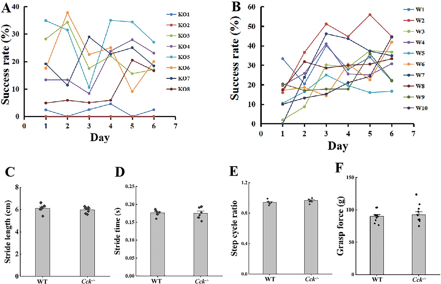
Learning curve of single mouse of Cck−/− (A) and wildtype (B) group and basic movement ability of WT and Cck−/−, including stride length (C, t-test, p = 0.405), stride time (D, t-test, p = 0.973), step cycle ratio (E, t-test, p = 0.093), and grasp force (F, t-test, p = 0.543).
Figure 2 with 1 supplement
Effect of local injection of CCK-B receptor (CCKBR) antagonist on motor learning.
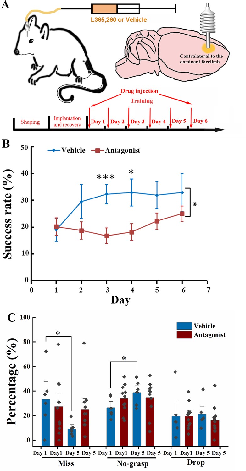
(A) A cannula was implanted into the motor cortex contralateral to the dominant hand. One microliter of L365.260 or vehicle was injected into the motor cortex through the cannula every day before training. (B) Success rate of the mice injected with CCKBR antagonist (N = 11) and vehicle (N = 6). *p < 0.05, ***p < 0.001. Two-way mixed analysis of variance (ANOVA), post hoc comparison between two groups. (C) Detailed reaching results, in terms of miss, no-grasp, drop, on Days 1 and 5 for mice injected with CCKBR antagonist and vehicle. *p < 0.05, paired t-test.
Figure 2—figure supplement 1
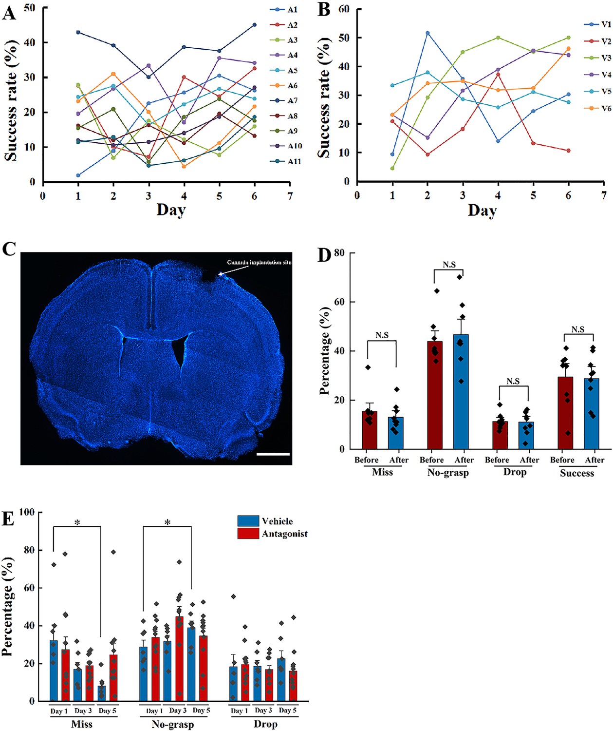
Learning curve of every single mouse administrated with CCK-B receptor (CCKBR) Antagonist (A) and Vehicle (B) and example of drug cannula implantation site in the motor cortex (MC) (C) and detailed learning results of the learned mice before and after injected with antagonist (D), paired t-test, Before vs. after, Miss, p = 0.099, No-grasp, p = 0.506, Drop, p = 0.853, success, p = 0.854.
Detailed reaching result including Days 1, 3, and 5 (E), paired t-test, Vehicle, Day 1 vs. Day 3, Miss, p = 0.258, No-grasp, p = 0.082, Drop, p = 0.512, Antagonist, Day 1 vs. Day 3, Miss, p = 0.133, No-grasp, p = 0.645, Drop, p = 0.941. N.S means not significant. *p<0.05. Scale bar represents 1000 µm.
Figure 3 with 2 supplements

Calcium imaging of the MC during motor skill learning.
(A) Experiment setup. C57BL/6, Cck−/−, and C57BL/6 mice injected with CCK-B receptor (CCKBR) antagonist were applied for single pellet reaching task training and calcium imaging. (B) Schematic diagram of calcium imaging. A wide-tip glass pipette tightly touched the brain by being lowered to a depth of 400–500 µm, and strong GCaMP6s virus expression was observed in the superficial layer of the motor cortex with a high contrast compared with the deep layers after >14 days of expression. A baseplate was implanted on the skull, which was connected to the miniscope for calcium imaging during motor skills training (right panel). (C) Representative traces of extracted neurons from miniscope using the CNMF-E algorithm. The scale bar represents 5 units of the scaled ΔF/F. (D) Neuronal activity pattern of C57BL/6 (N = 10), Cck−/− (N = 7), and C57BL/6 mice injected with L365.260 (N = 7). Upper line is from training Day 1 and the bottom is from training Day 6. (E) Neuronal population activity from C57BL/6, Cck−/−, and C57BL/6 mice injected with L365.260. (F) Activated population activity (peak activity minus baseline activity) was calculated for C57BL/6, Cck−/−, and C57BL/6 mice injected with L365.260 at Days 1 and 6. *p < 0.05, N.S. not significant. Paired t-test. (G) Trial-to-trial population activity correlation at Days 1 and 6 for C57BL/6, Cck−/−, and C57BL/6 injected with L365.260. (H) The pairwise Hausdorff distances of the trajectories for C57BL/6, Cck−/−, and C57BL/6 injected with L365.260 at Days 1 and 6. *p < 0.05, **p<0.01 N.S., not significant. One-way RM analysis of variance (ANOVA).
Figure 3—figure supplement 1
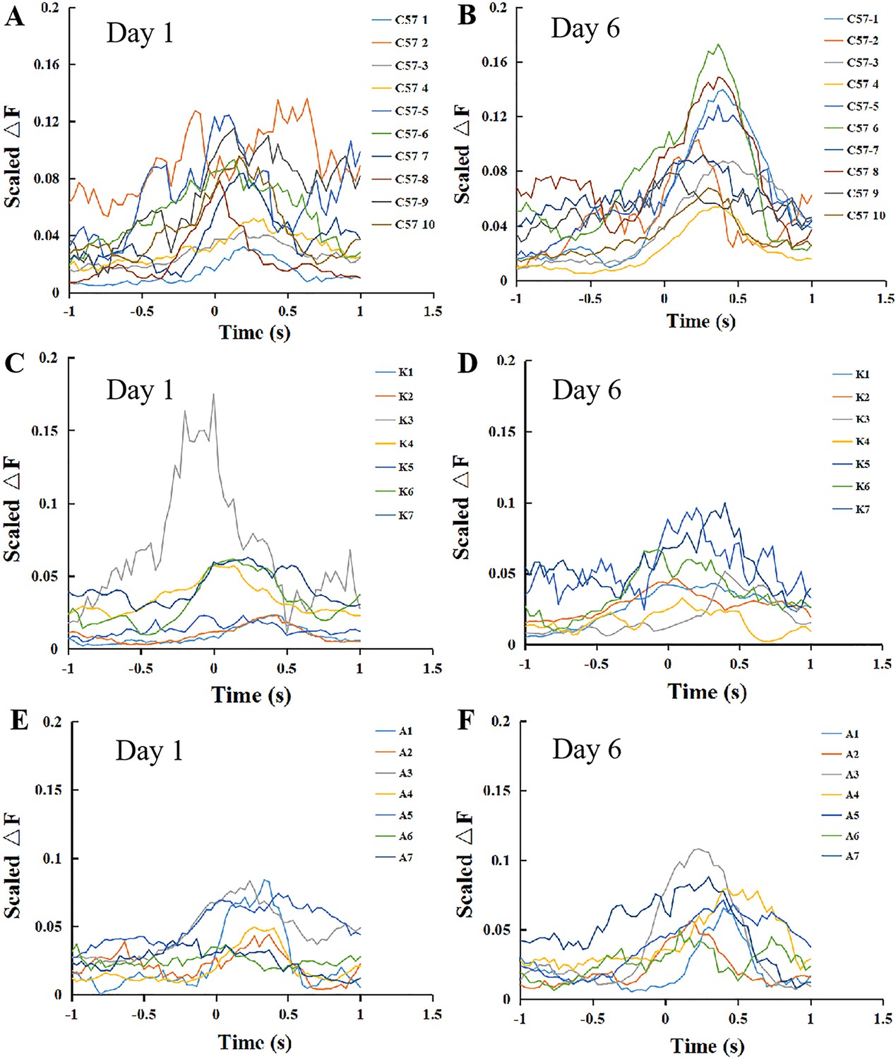
Neuronal activity relative to the movement of different groups, including C57BL/6 (A, B), Cck−/− (C, D), and L365,260 injection (E, F) mice at Days 1 and 6.
Figure 3—figure supplement 2
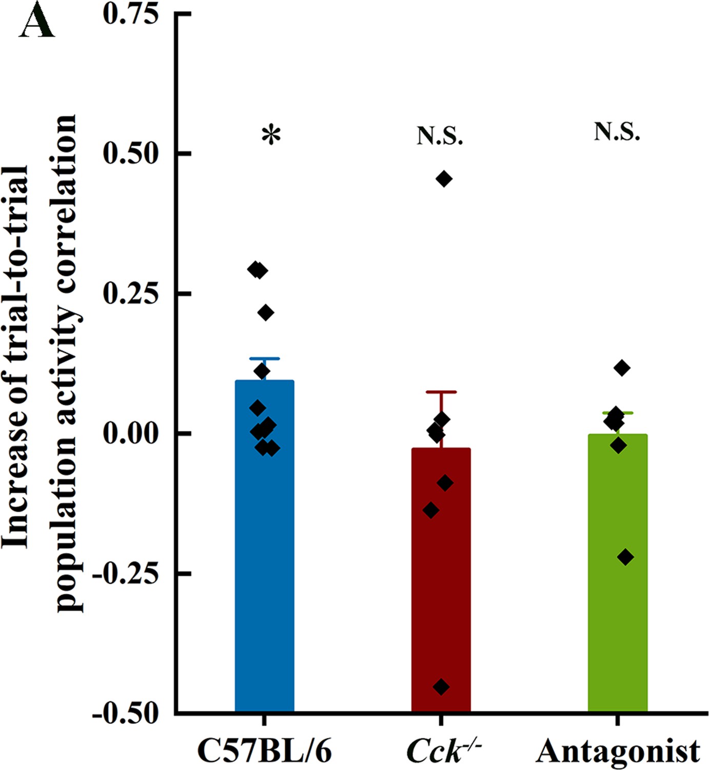
Increase of trial-to-trial population activity correlation (C57BL/6, p = 0.01; Cck−/−, p = 0.61; Antagonist, p = 0.53).
*p<0.05.
Figure 4 with 1 supplement
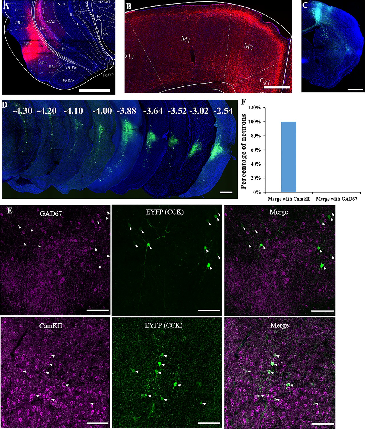
Labeling of cholecystokinin (CCK) neuron projections from the rhinal cortex (RC) to the MC.
(A) Coronal section showing the virus injection site. The Cre-dependent AAV-hsyn-DIO-mCherry virus was injected into Cck-Cre mice. (B) Effective labeling of CCK neuron fibers in the MC. (C) Cre-dependent retrograde AAV virus injection site in the MC of the Cck-Cre mouse. (D) Continuous coronal brain sections showing EYFP in the lateral EC. The numbers (mm) indicate the position of the sections relative to the bregma. (E) GAD67 staining did not merge with the retrograde tracking CCK-positive neurons in the EC and CaMKII staining merged with the signal of retrograde tracking CCK neurons EC projecting. Arrowhead indicate the positions of CCK neurons. (F) Percentage of retrogradely labeled neurons merged with CamKII and GAD67 (N = 4, a total of 140 neurons for CamKII and 136 for GAD67). Scale bars represent 1000 µm in (A), (B), (C), and (D) and 100 µm in (E).
Figure 4—figure supplement 1
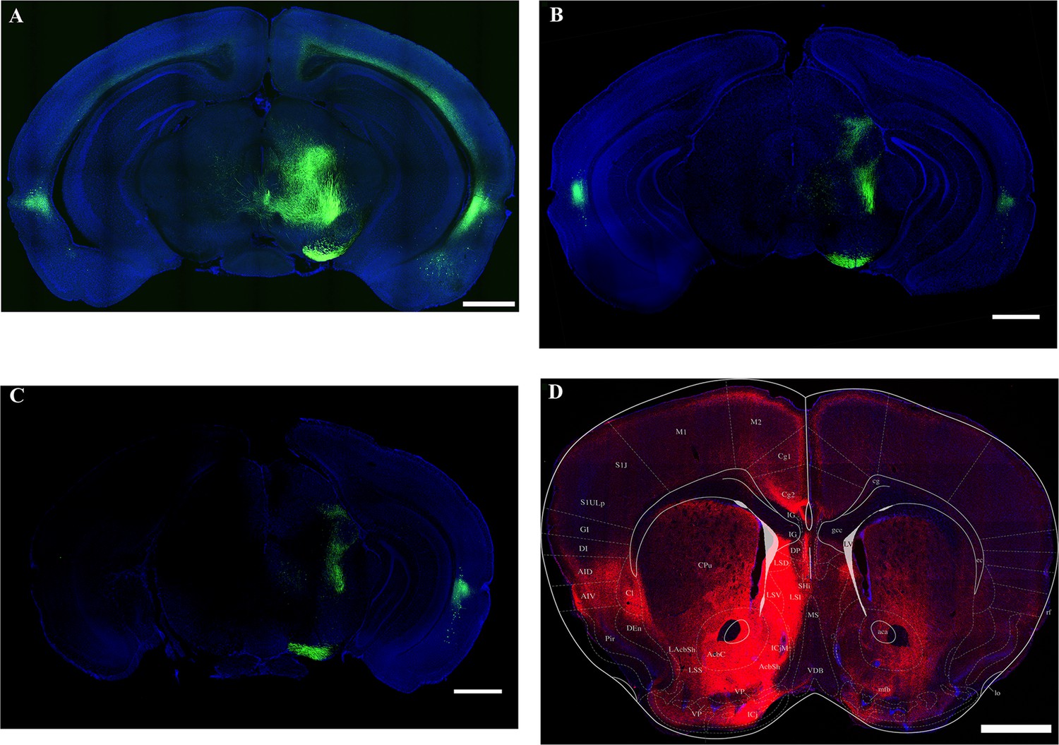
Three examples of CCK+ neurons in the RC of the Cck-Cre mouse injected with AAV(retro)-EF1a-DIO-EGFP in the MC (A–C).
An example of the cholecystokinin (CCK) projections from the rhinal cortex (D). Scale bar is 1000 µm.
Figure 5 with 1 supplement

Effect of inhibition of the RC cholecystokinin (CCK) neurons on motor learning.
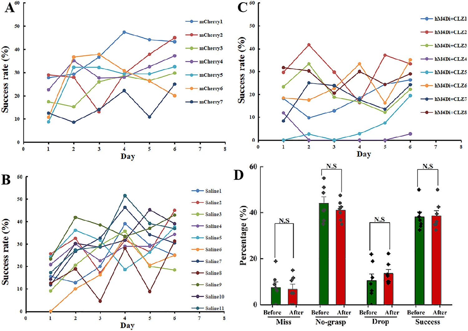
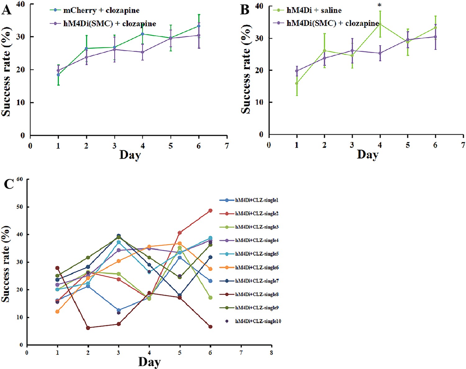
(A) Experimental paradigm for the chemogenetic experiment. Cre-dependent AAV-DIO-hM4Di-mCherry or AAV-DIO-mCherry was infused into the rhinal cortex of Cck-Cre mice. After 4 weeks, clozapine or saline was intraperitoneally injected 30 min before training. (B) Success rate of Cck-Cre mice injected with hM4Di containing virus together with clozapine (hM4Di + clozapine) (N = 10) and control virus with clozapine (mCherry + clozapine) (N = 8). (C) Success rate of Cck-Cre mice injected with hM4Di containing virus plus clozapine (hM4di + clozapine, shared with (B)) and hM4Di plus saline (hM4Di + saline) (N = 11). The hM4Di + clozapine curve in C shared that in (B). *p < 0.05, **p < 0.01. Two-way mixed analysis of variance (ANOVA), post hoc comparison between two groups on different days.
Figure 5—figure supplement 1
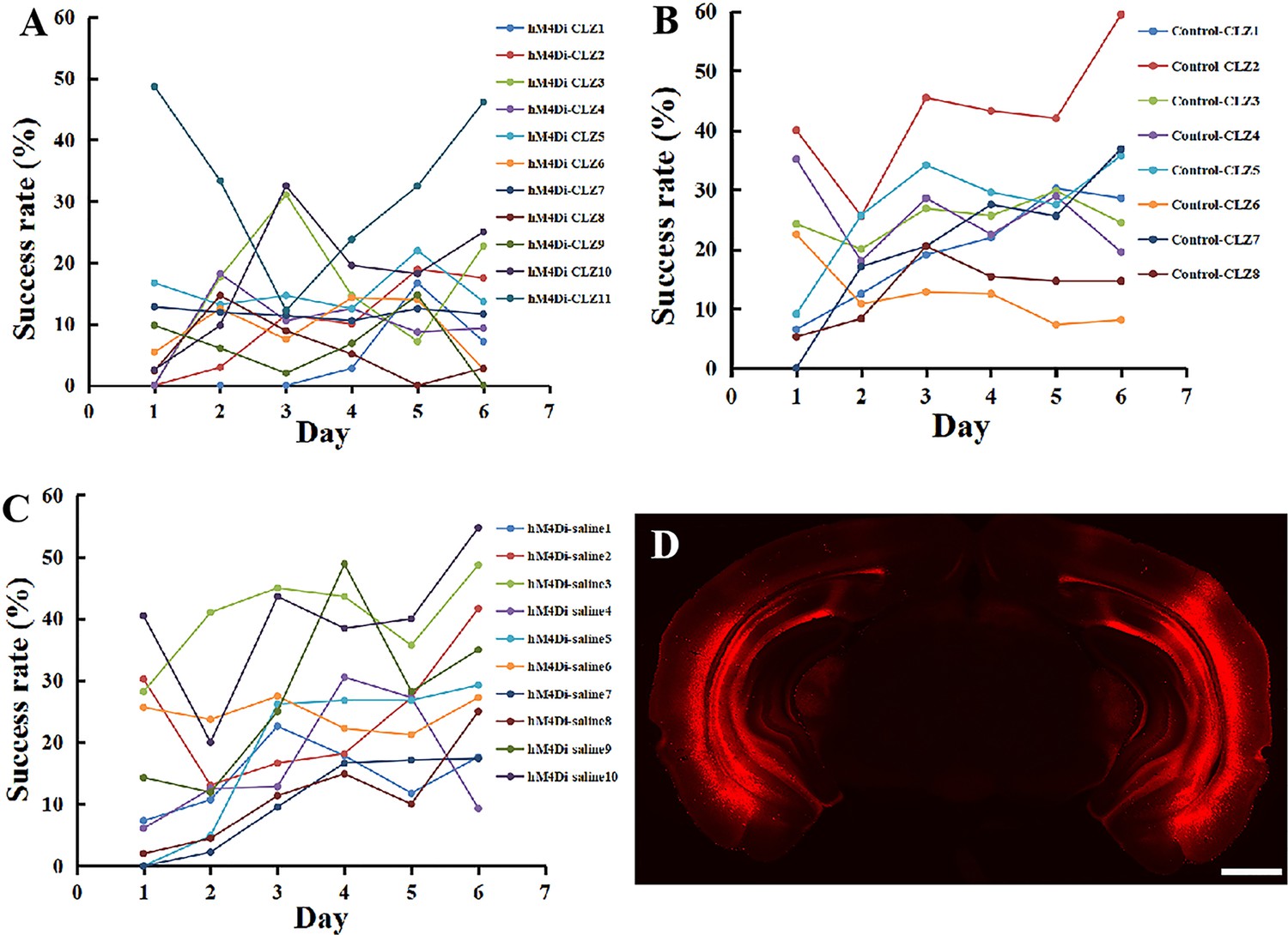
Learning curve of single Cck-Cre mouse injected with hM4Di-clozapine (A), Control-clozapine (B), and hM4Di-saline (C), and example of the expression of hM4Di virus in the rhinal cortex (D).
Scale bar represents 1000 µm.
Figure 6 with 5 supplements

Effect of inhibition of neurons projecting from the RC to the MC on motor learning.
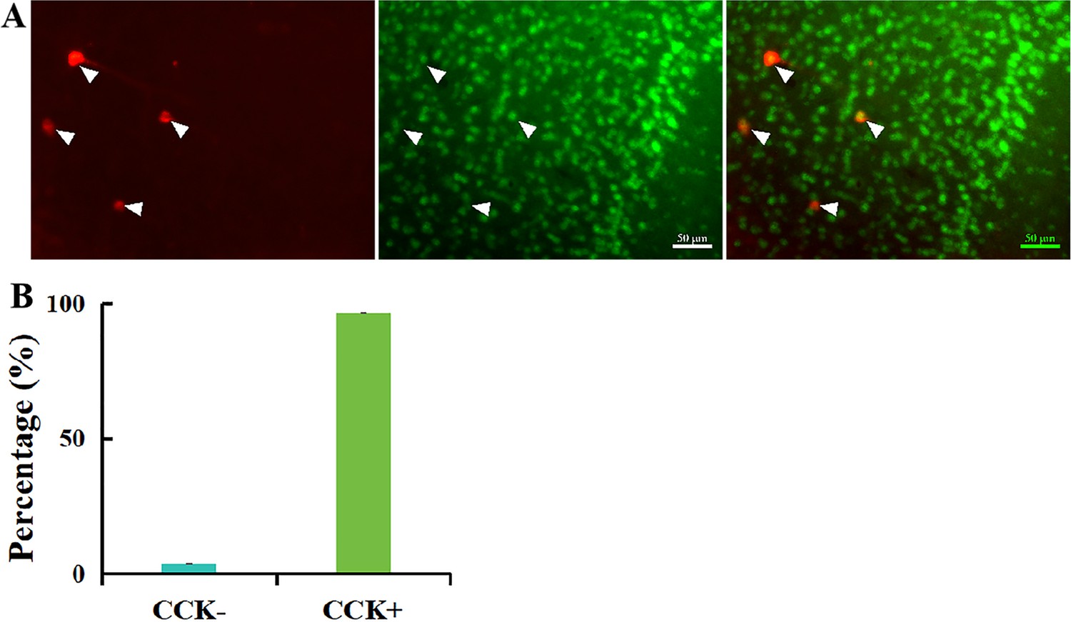
(A) Retrograde neurons in the EC of Cck-Cre mice injected with AAV(retro)-EF1a-DO-mCherry-DIO-EGFP in the MC. (B) Percentage of EGFP (CCK+) and mCherry (CCK−) in the EC. N = 3, n = 10, a total of 111 neurons. (C) Experimental paradigm for the chemogenetic experiment. A retro-Cre virus was injected in the MC of two hemispheres and a Cre-dependent hM4Di (or not, as a negetive control) virus was injected in the EC. After 4 weeks of expression, mice were trained to learn the single pellet reaching task. Thirty minutes before training, mice were injected with clozapine (i.p.) every day. (D) Success rate of mice injected with hM4Di containing virus together with clozapine (hM4Di + clozapine) (N = 7) and control virus with clozapine (mCherry + clozapine) (N = 8). (E) Success rate of mice injected with hM4Di containing virus plus clozapine (hM4di + clozapine, shared with (D)) and hM4Di combined with saline (hM4Di + saline) (N = 11). The hM4Di + clozapine curve in Figure 5C shared that in (D). (F) Detailed reaching results for the three different treatments on Days 1 and 4. *p < 0.05, **p < 0.01. Two-way mixed analysis of variance (ANOVA), post hoc comparison between two groups on different days.
Figure 6—figure supplement 1
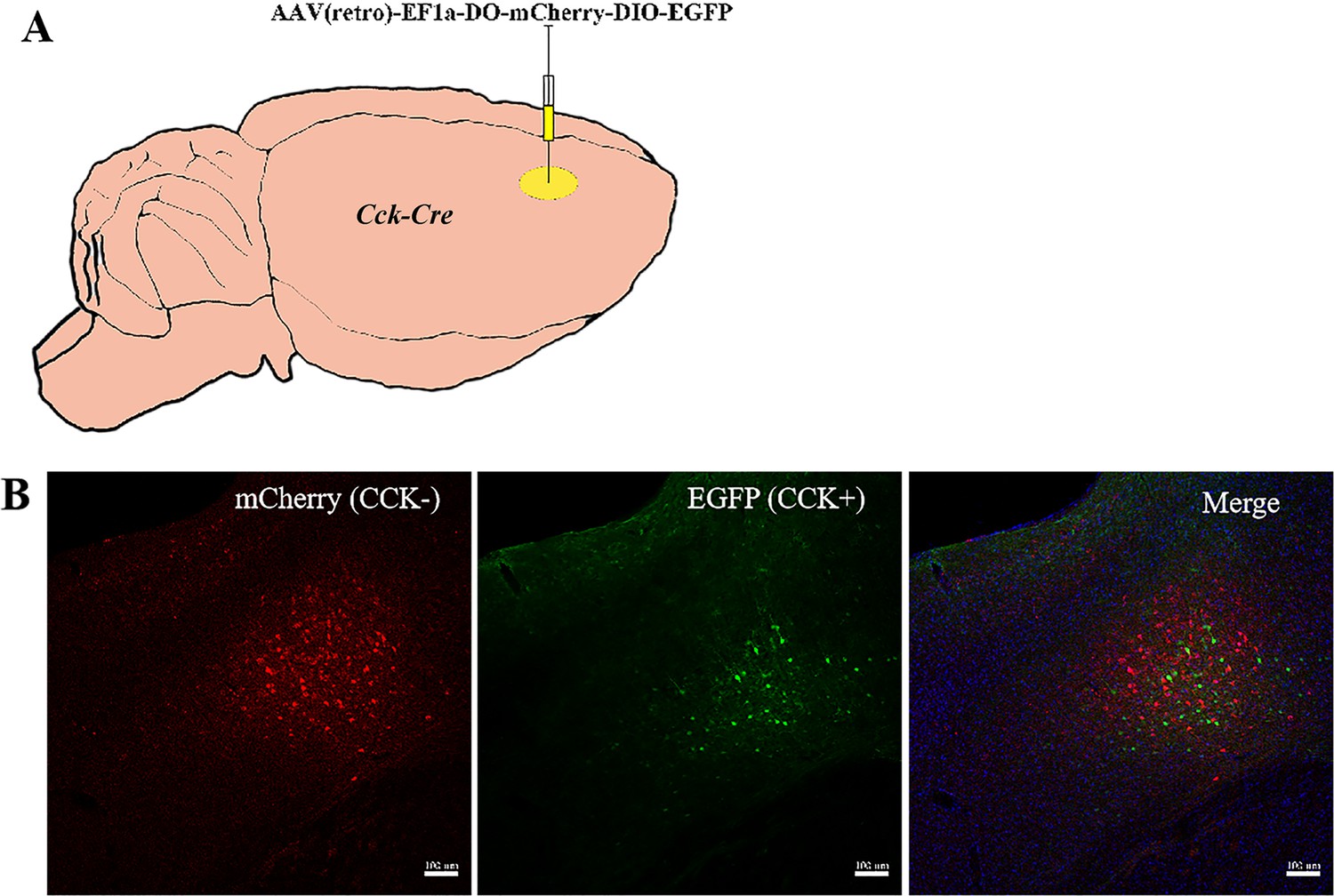
Experimental paradigm of AAV(retro)-EF1a-DO-mCherry-DIO-EGFP injected in the MC of the Cck-Cre mice (A).
Labbeled neurons in the MC (B). Scale bar represents 100 µm.
Figure 6—figure supplement 2
AAV(retro)-hSyn-mCherry injected in the MC of Cck-Cre mice.
Retrograde neurons (mCherry) and the stained Cre (green) in the RC (A), and the percentage of CCK+ and CCK− neurons from the RC projecting to the MC (B). N = 2, a total of 56 retrograde neurons were involved. p < 0.001.
Figure 6—figure supplement 3
Learning curve of single C57BL/6 mouse injected with Cre-dependent ± hM4Di (EC) and retro-Cre (MC, two sides).
Cre-dependent control virus together with clozapine injection (A), Cre-dependent hM4Di together with saline injection (B), Cre-dependent hM4Di and retro-Cre in MC (two sides) together with clozapine injection (C); and detailed learning results of the learned mice before and after injected with antagonist (D, paired t-test, Before vs. afrer, Miss, p = 0.769, No-grasp, p = 0.379, Drop, p = 0.402, Success, p = 0.596).
Figure 6—figure supplement 4
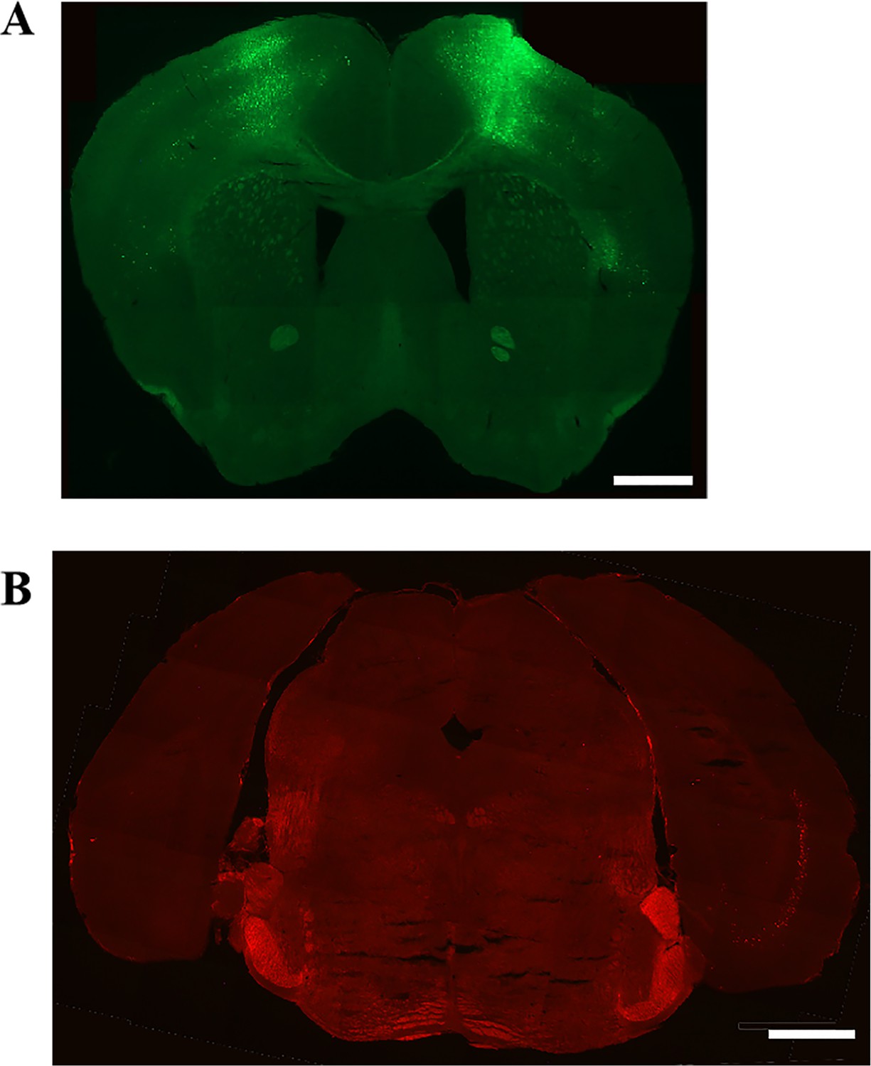
Expression of retro-Cre in the single side of the MC (A) and Cre-dependent hM4Di-mCherry in th RC (B).
Scale bar represents 1000 µm.
Figure 6—figure supplement 5
Learning curve of mice injected with hM4Di (SMC, retro-Cre in the single motor cortex contralateral to the dominant hand and hM4Di in the entorhinal cortex) and clozapine compared with mice injected with control virus and clozapine (A, shared with Figure 6B), hM4Di and saline (B, shared with Figure 6C), and learning curve of single mouse (C).
*p < 0.05.
Figure 7 with 1 supplement
Rescuing the motor learning ability of Cck−/− mice by CCK4.
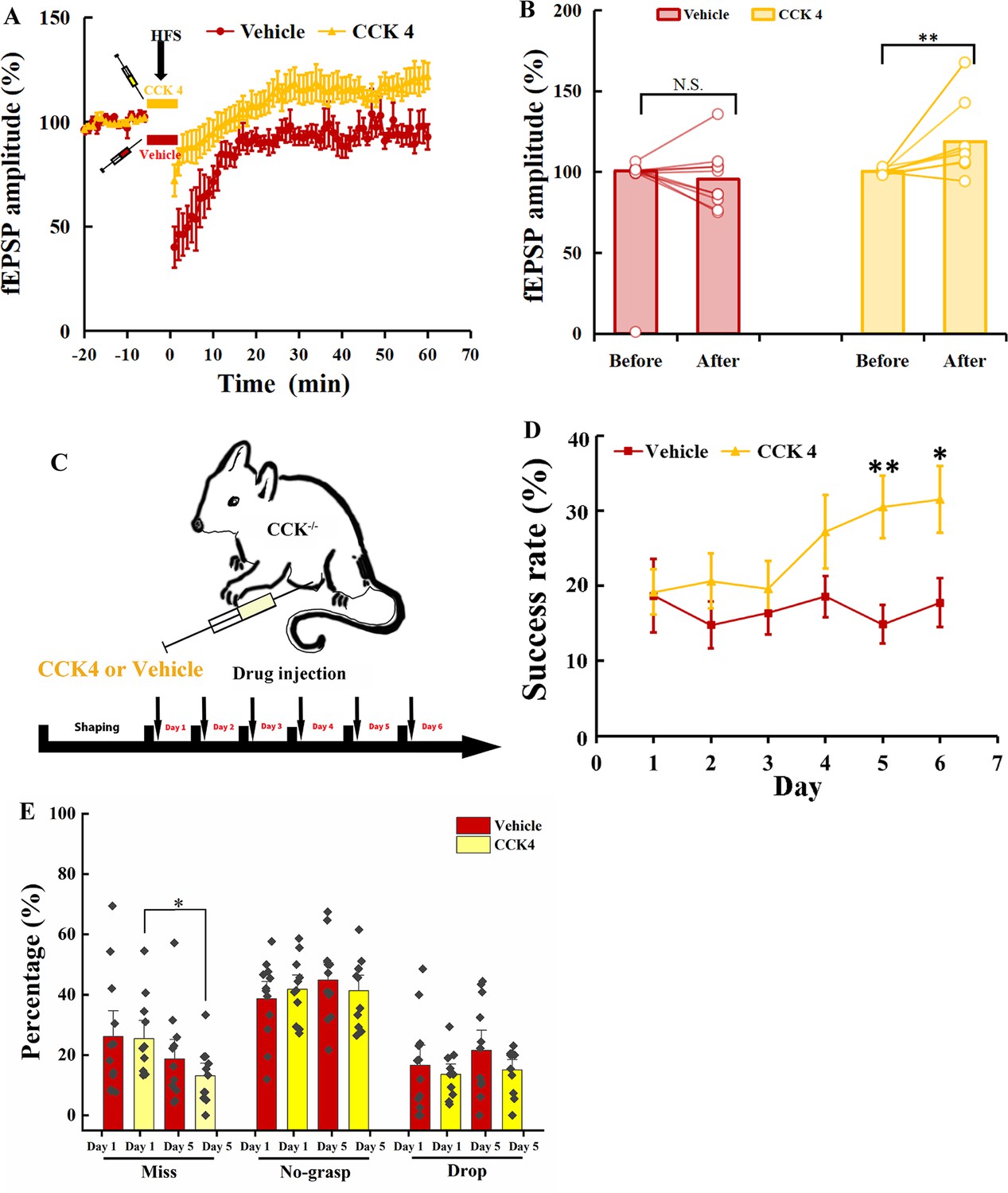
(A) Normalized field excitatory postsynaptic potential (fEPSP) amplitude before and after high-frequency stimulation (HFS) of the MC of Cck-−/− mice applied with CCK4 (N = 6, n = 14) or vehicle (N = 6, n = 11). (B) The average normalized fEPSP amplitude 10 min before HFS (−10 to 0 min, before) and 10 min after HFS (50 to 60 min, after) in the MC of Cck−/− mice injected with CCK4 or vehicle. *p < 0.05, **p < 0.01. Two-way mixed analysis of variance (ANOVA) with Bonferroni pairwise comparison. (C) Experimental paradigm for cholecystokinin (CCK) rescuing experiment. CCK4 or vehicle was injected (i.p.) every day before training. (D) Success rate of Cck−/− mice injected with CCK4 (N = 11) or vehicle (N = 10). *p < 0.05, **p < 0.01. Two-way mixed ANOVA, post hoc comparison between two groups on Days 5 and 6. (E) Detailed reaching results or Cck−/− mice injected (i.p.) with vehicle and CCK4 on Days 1 and 5. *p < 005, N.S., not significant. Paired t-test.
Figure 7—figure supplement 1
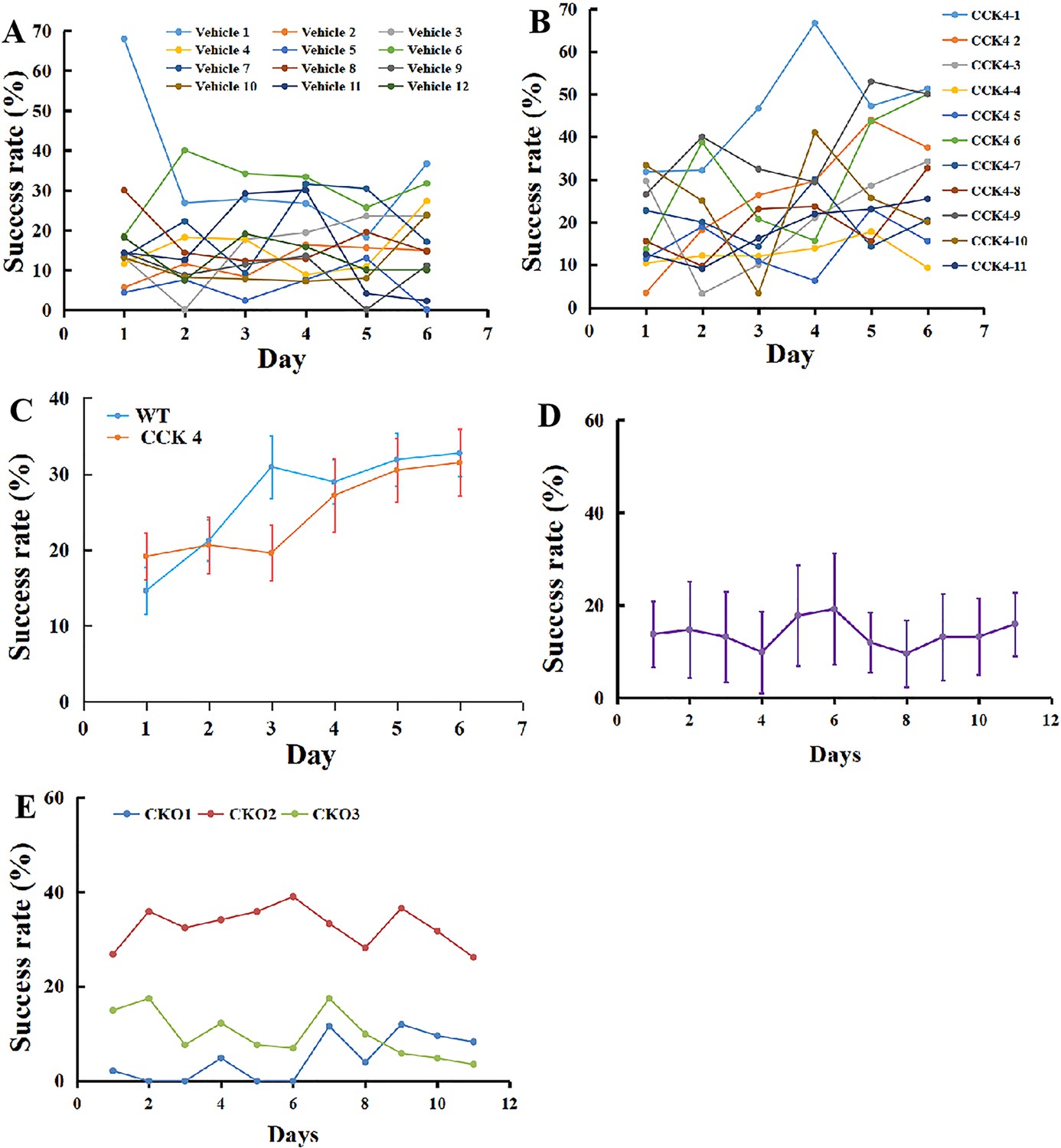
Learning curve of single Cck−/− mouse administrated with Vehicle (A) and CCK4 (B), and comparison between Cck−/− mice injected with CCK4 and WT group (C) and learning curve of Cck−/− mice injected with CCK8 in the motor cortex, for the average (D) and every single mouse (E).
Videos
Video 1
Example of ‘miss’ performance of a mouse.
Video 2
Example of ‘no-grasp’ performance of a mouse.
Video 3
Example of ‘drop’ performance of a mouse.
Video 4
Example of ‘success’ performance of a mouse.
Video 5
Example of calcium signals of neurons in the motor cortex under the miniscope.
Tables
Appendix 1—key resources table
| Reagent type (species) or resource | Designation | Source or reference | Identifiers | Additional information |
|---|---|---|---|---|
| Antibody | Anti-CamKIIa (Mouse monoclonal) | Abcam | Cat# Ab22609 | 1:500 |
| Antibody | Anti-GAD67 (Mouse monoclonal) | Millipore | Cat# MAB5406 | 1:1000 |
| Antibody | Anti-mCherry (Rabbit monoclonal) | Invitrogen | Cat# M11217 | 1:1000 |
| Antibody | Anti-mouse IgG Alexa 594 (Goat polyclonal) | Jackson ImmunoResearch | Cat# 115-585-003 | 1:500 |
| Antibody | Anti-rabbit IgG Alexa 594 (Goat polyclonal) | Jackson ImmunoResearch | Cat# 111-585-003 | 1:500 |
| Recombinant DNA reagent | AAV-hSyn-DIO-mCherry | Addgene | RRID: Addgene_50459 | |
| Recombinant DNA reagent | AAV(retro)-EF1a-Cre-EGFP | WZ Biosciences lnc | NA | |
| Recombinant DNA reagent | AAV-hSyn-DIO-hM4Di-mCherry | WZ Biosciences lnc | NA | |
| Recombinant DNA reagent | AAV-EF1a-DO-mCherry-DIO-EGFP | Braincase | Cat# BC0658 | |
| Recombinant DNA reagent | AAV-hSyn-DIO-hM4Di-mCherry | Addgene | RRID: Addgene_44362 | |
| Recombinant DNA reagent | retroAAV-EF1a-DIO-EYFP | Addgene | RRID: Addgene_27056 | |
| Recombinant DNA reagent | AAV-CamKIIa-GCaMP6s-WPRE-SV40 | Addgene | RRID: Addgene_107790 | |
| Chemical compound, drug | Pentobarbital | Alfasan International B.V. | NA | |
| Chemical compound, drug | Carprofen | Sigma-Aldrich | Cat# PHR1452 | |
| Chemical compound, drug | CCK-4 | Abcam, Cambridge, UK | Cat# ab141328 | |
| Chemical compound, drug | Dexamethasone | Sigma-Aldrich | Cat# D4902 | |
| Chemical compound, drug | Clozapine | Sigma-Aldrich | Cat# C6305 | |
| Chemical compound, drug | DAPI | Santa Cruz Biotechnology | Cat# sc-3598 | |
| Chemical compound, drug | Food pellet | TestDiet | Cat# 1811223 | |
| Genetic reagent (Mus musculus) | Mouse: C57BL/6 | The Laboratory Animal Services Centre, Chinese University of Hong Kong | NA | |
| Genetic reagent (Mus musculus) | Mouse: C57BL/6 | Laboratory Animal Research Unit, City University of Hong Kong | NA | |
| Genetic reagent (Mus musculus) | Mouse: Cck-ires-Cre | Jackson Laboratories | Stock# 012706 | |
| Genetic reagent (Mus musculus) | Mouse: Cck-CreER | Jackson Laboratories | Stock# 012710 | |
| Software, algorithm | Excel | Microsoft | https://www.microsoft.com/en-us/microsoft365/excel | |
| Software, algorithm | Matlab R2020a | Mathworks | https://www.mathworks.com/products/new_products/release2020a.html | |
| Software, algorithm | Fiji | Schindelin et al., 2012 | https://imagej.net/Fiji | |
| Software, algorithm | Photoshop | Adobe | https://www.adobe.com/products/photoshop.html | |
| Software, algorithm | SPSS | IBM | https://www.ibm.com/products/spss-statistics; |
Additional files
Download links
A two-part list of links to download the article, or parts of the article, in various formats.
Downloads (link to download the article as PDF)
Open citations (links to open the citations from this article in various online reference manager services)
Cite this article (links to download the citations from this article in formats compatible with various reference manager tools)
Cholecystokinin facilitates motor skill learning by modulating neuroplasticity in the motor cortex
eLife 13:e83897.
https://doi.org/10.7554/eLife.83897


