Response to comment on 'A conserved strategy for inducing appendage regeneration in moon jellyfish, Drosophila, and mice'
- Division of Biology and Biological Engineering, California Institute of Technology, United States
- Graduate Aerospace Laboratories and Mechanical Engineering, California Institute of Technology, United States
Figures
Figure 1 with 1 supplement
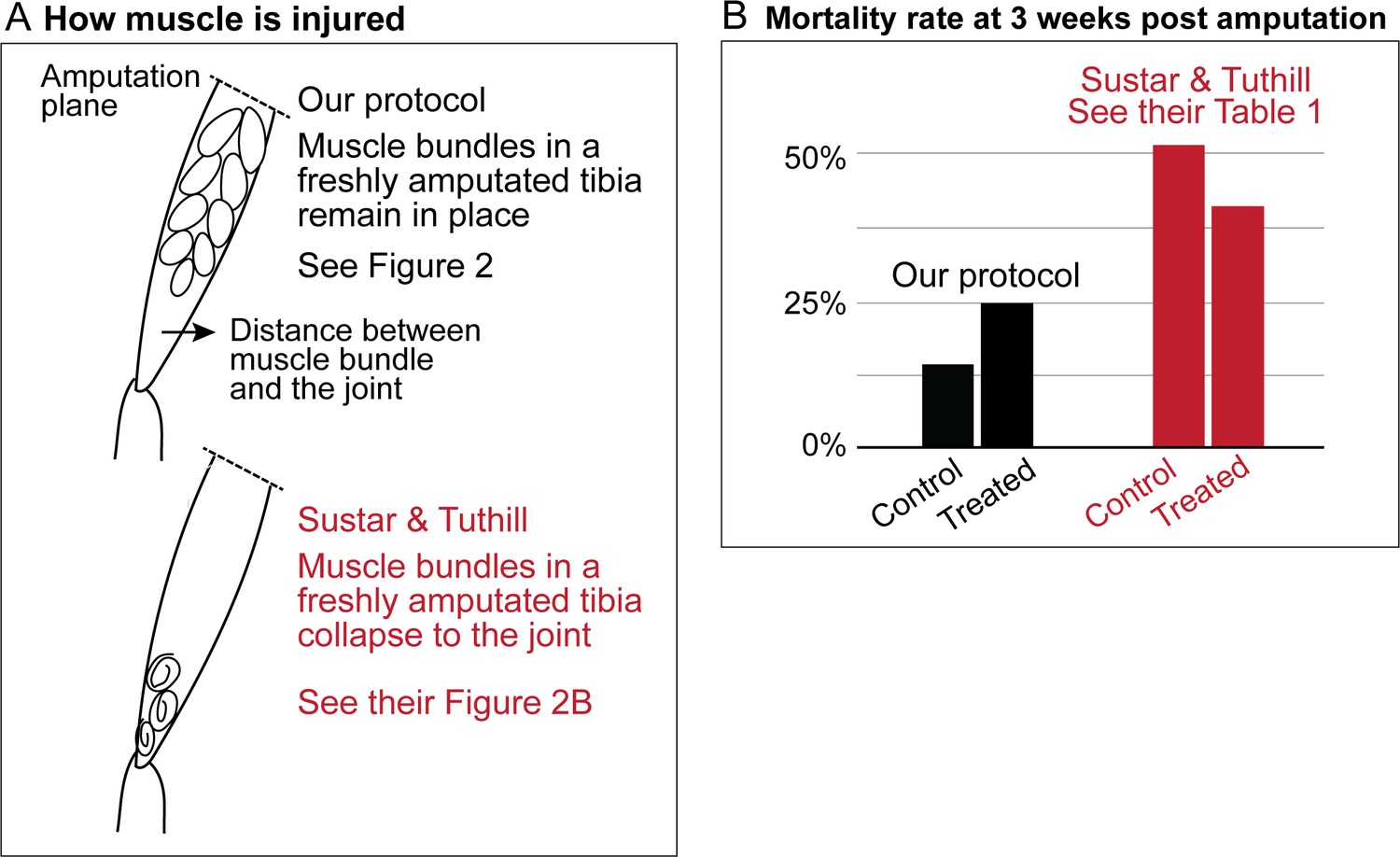
Sustar and Tuthill did not replicate our protocol.
(A) In our amputation method, muscle bundles in the residual tibia remain in place (see Figure 2). By contrast, in the method used by Sustar and Tuthil, muscle bundles collapsed (see their Figure 2B). (B) The lack of stress management in the protocol of Sustar and Tuthill is reflected in the much higher mortality rate in their experiment (as reported in their Table 1).
Figure 1—figure supplement 1
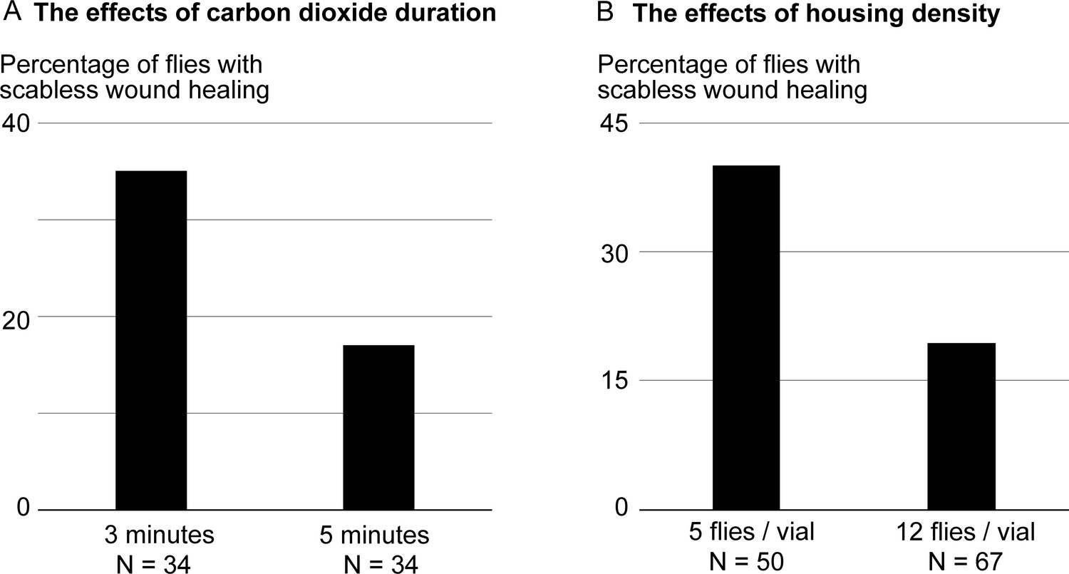
Duration of CO2 exposure and housing density can alter the experimental outcomes.
In these experiments, adult flies were amputated across the tibia, and placed on food supplemented with leucine, glutamine, and insulin. The effects of the treatment were assessed three days after amputation by the absence of scab formation over the amputation site. (A) To perform the amputation, we normally anesthetize the flies using the lowest possible CO2 level, and limit anesthesia duration to one to three minutes. Even at thissub-anesthetic CO2 level, increasing the CO2 exposure time to five minutes is enough to halving the frequency of flies responding to the treatment. (B) After amputation, we normally place up to 6 flies per vial. Increasing the housing density reduces the frequency of flies responding to the treatment.
Figure 2
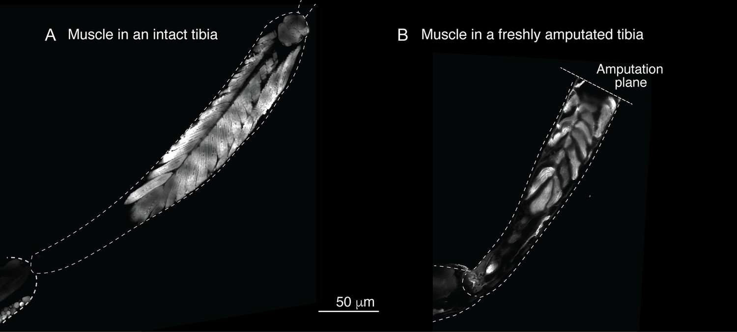
Muscle structure in the limb.
To assess the muscle structure in the limb, we analyzed the Mhc-GFP flies, in which promoter of the muscle-specific myosin heavy chain (Mhc) gene drives GFP expression. In these images, limbs were dissected and imaged using laser-scanning confocal microscopy.
Download links
A two-part list of links to download the article, or parts of the article, in various formats.
Downloads (link to download the article as PDF)
Open citations (links to open the citations from this article in various online reference manager services)
Cite this article (links to download the citations from this article in formats compatible with various reference manager tools)
Response to comment on 'A conserved strategy for inducing appendage regeneration in moon jellyfish, Drosophila, and mice'
eLife 12:e85370.
https://doi.org/10.7554/eLife.85370


