Two RNA-binding proteins mediate the sorting of miR223 from mitochondria into exosomes
- Department of Molecular and Cell Biology, Howard Hughes Medical Institute, University of California, United States
Figures
Figure 1 with 2 supplements
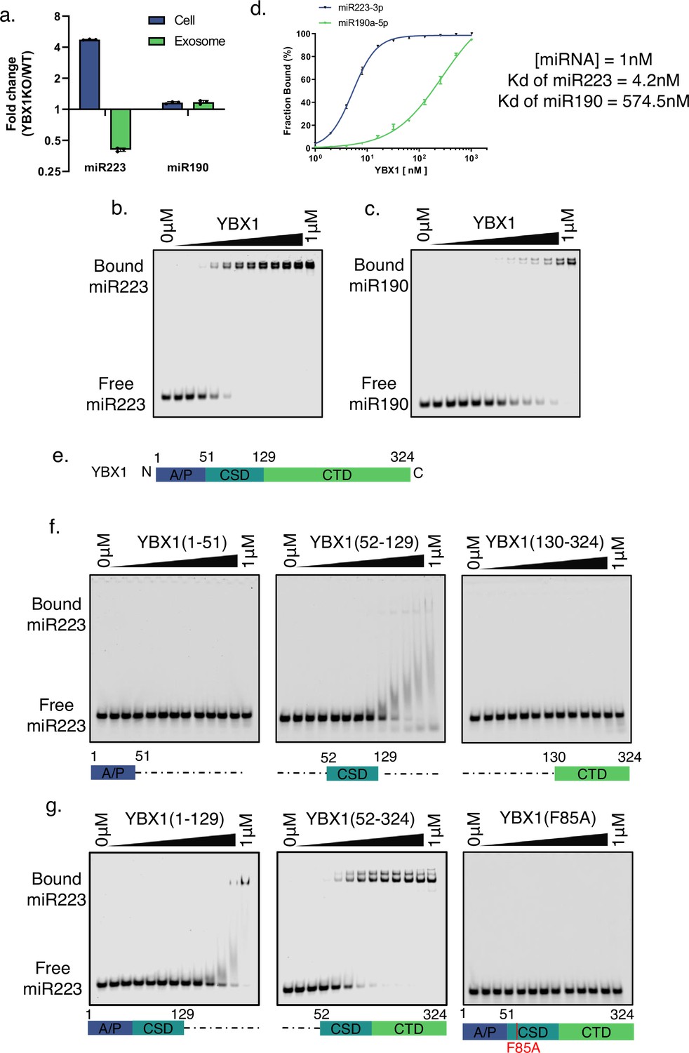
YBX1 directly and specifically binds miR223.
(a) RT-qPCR analysis of fold change of miR-223 and miR-190 in cells and purified exosomes from 293 T WT cells and YBX1 knockout cells. Data are plotted from three independent experiments, each independent experiment with triplicate qPCR reactions; error bars represent standard deviations. (b–c) EMSA assays using 1 nM 5’ fluorescently labeled miR223 or miR190 and purified YBX1. Purified YBX1 was titrated from 500pM to 1 μM. In gel fluorescence was detected. Quantification of (d) shows the calculated Kd. (e) Schematic diagrams of the different domains of YBX1. (f) EMSA assay using 1 nM 5’ fluorescently labeled miR223 and purified YBX1 truncations. [YBX1(1–51) or YBX1(52–129) or YBX1(130–324).] (g) EMSA assay using 1 nM 5’ fluorescently labeled miR223 and purified YBX1 truncations [YBX1(1–129) or YBX1(52–324)] or YBX1(F85A) mutant.
-
Figure 1—source data 1
Uncropped gel images corresponding to Figure 1.
- https://cdn.elifesciences.org/articles/85878/elife-85878-fig1-data1-v2.zip
Figure 1—figure supplement 1

Knockout of YBX1 did not change exosome secretion.
(a) Exosome numbers are measured by Nanosight NS300. Normalized exosome number from 293 T WT and YBX1-KO cells are shown.
Figure 1—figure supplement 2
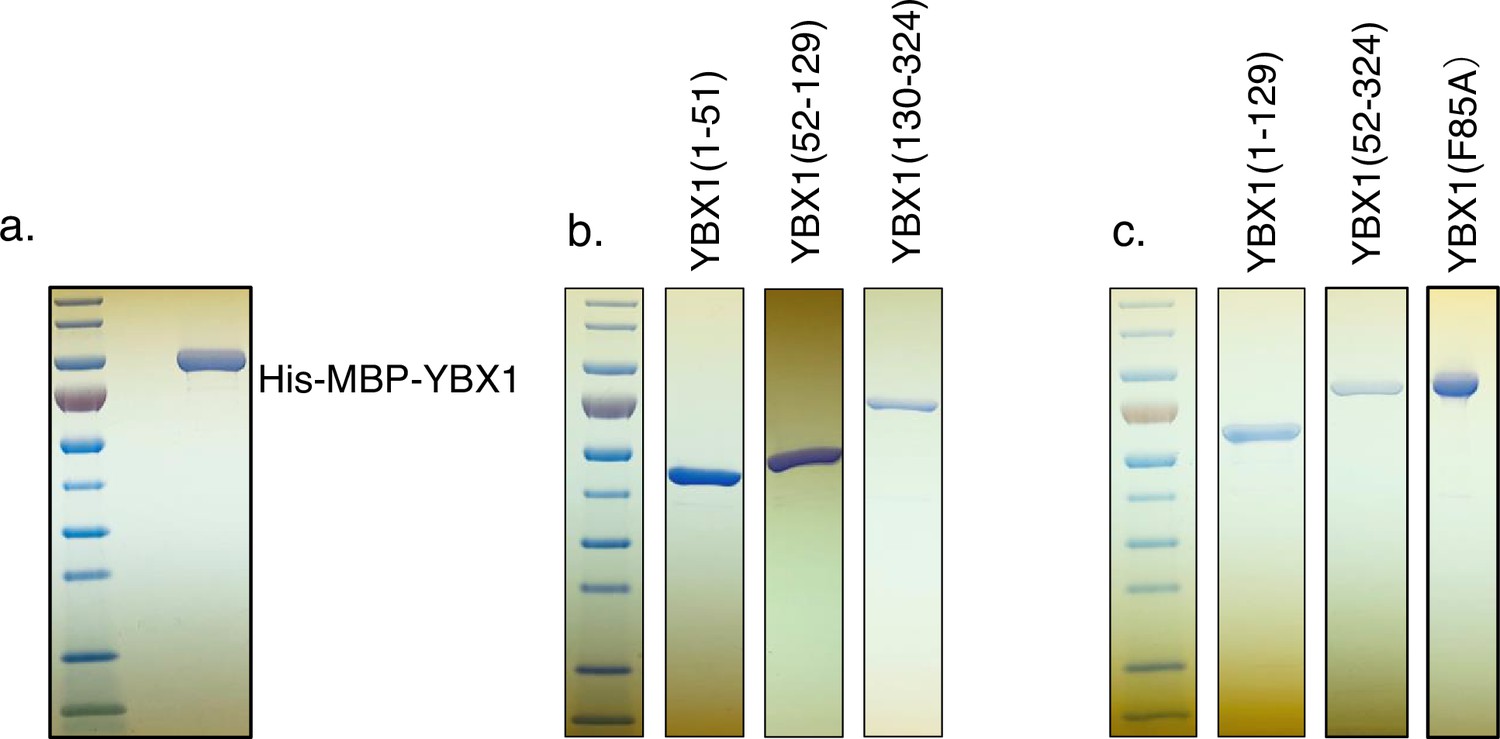
Purified YBX1 full length protein and different truncations and mutation.
(a–c) YBX1 full-length protein or different truncations were overexpressed and purified from insect cells.
-
Figure 1—figure supplement 2—source data 1
Uncropped gel images corresponding to Figure 1—figure supplement 2.
- https://cdn.elifesciences.org/articles/85878/elife-85878-fig1-figsupp2-data1-v2.zip
Figure 2 with 1 supplement

miR223 sequence motif UCAGU binds YBX1.
(a) RNA oligonucleotides corresponding to miR223, miR190 and versions with mutated sorting motif (miR223mut) or mutation to introduce the sorting motif (miR190sort). (b) EMSA assays using 1 nM 5’ fluorescently labeled miR223 WT or miR223mut or miR190 WT or miR190sort and purified YBX1. Purified YBX1 was titrated from 500pM to 1 µM. In gel fluorescence was detected. (c) Binding affinity curves as calculated by EMSA data from (b) (d) Schematic shows exosome purification with buoyant density flotation in a sucrose step gradient from 293T cells overexpressing miR223 WT or mutant or miR190 WT or miR190sort. (e) RT-qPCR analysis of relative abundance of miR223 or miR223mut detected in exosomes compared to cellular level in 293T cells overexpressing miR223 WT or miR223mut. Data are plotted from three independent experiments and error bars represent standard deviations. (f) RT-qPCR analysis of relative abundance of miR190 or miR190sort detected in exosomes compared to cellular level in 293T cells overexpressing miR190 WT or miR190sort. Data are plotted from three independent experiments and error bars represent standard derivations. (g) In vitro packaging assay using 32P 5’end-labeled miR223 and miR223mut. Cell-free packaging of miR223 and miR223mut measured as protected radioactive signal from 32P labeled miR223 and miR223mut. Reactions with or without membrane, cytosol, and 1% Triton X-100, and incubated at 4 or 30 °C are indicated. For the samples containing only cytosol plus membrane at 4 °C, only one-third of the samples were loaded. Each sample was supplemented with 300 mM urea to reduce the background signal. (h) Data quantification showed protected fraction of miR223 and miR223mut as calculated from in vitro packaging data shown in (g).
-
Figure 2—source data 1
Uncropped gel images corresponding to Figure 2.
- https://cdn.elifesciences.org/articles/85878/elife-85878-fig2-data1-v2.zip
Figure 2—figure supplement 1
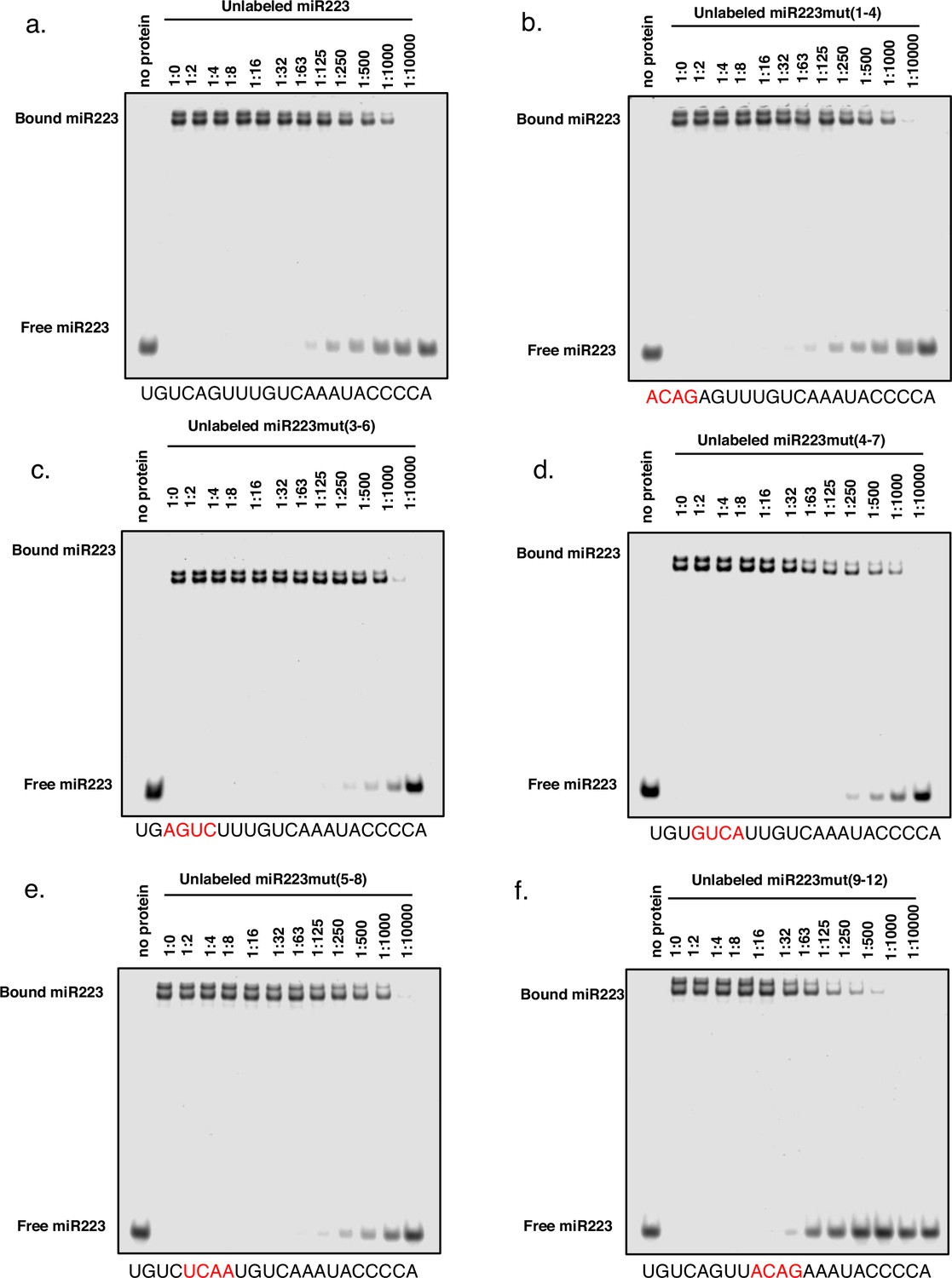
Screening of exosomal sorting motif of miR223.
EMSA competition assay to screen the exosomal sorting motif of miR223. (a–f) EMSA competition assay using 1 nM 5’ fluorescently labeled miR223, 32 nM purified YBX1 protein and unlabeled miR223 or miR223mutants. Unlabeled miR223 or miR223mutants were titrated from 1 nM to 1 μM and are given as the ratio of unlabeled miR223/labeled miR223 (wt and mutant).
-
Figure 2—figure supplement 1—source data 1
Uncropped gel images corresponding to Figure 2—figure supplement 1.
- https://cdn.elifesciences.org/articles/85878/elife-85878-fig2-figsupp1-data1-v2.zip
Figure 3 with 2 supplements
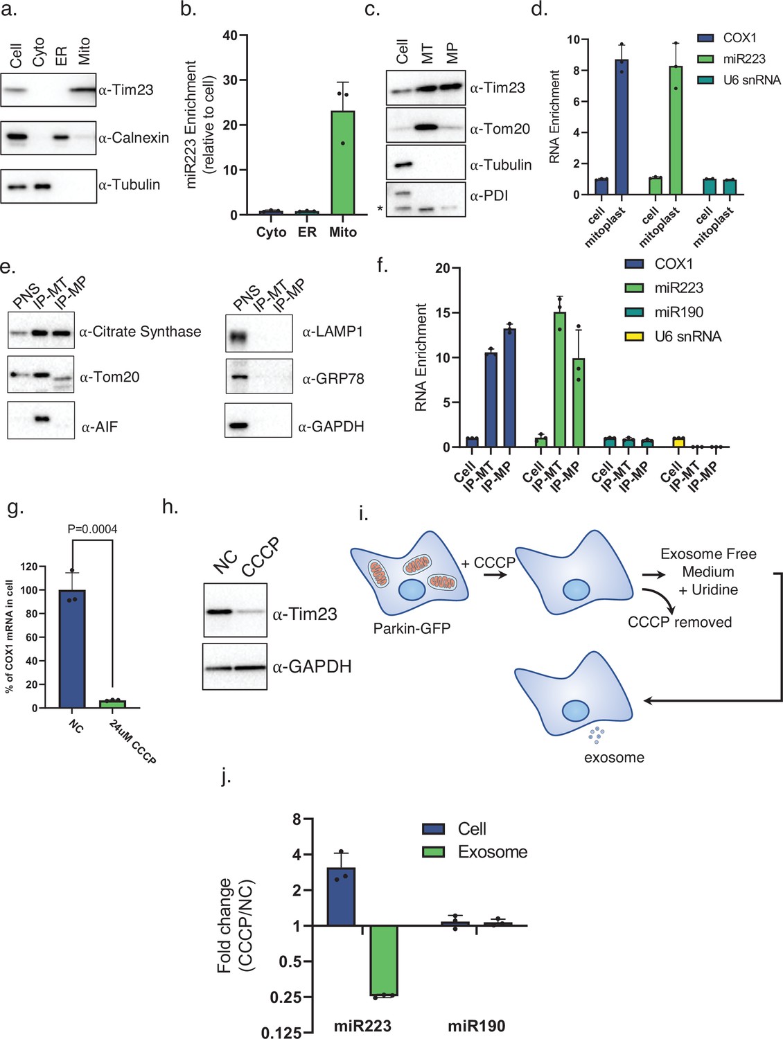
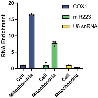
Mitochondria contribute to miR223 enrichment into exosomes.
(a) Immunoblot analysis of protein markers for different subcellular fractions isolated from 293T cells. (b) RT-qPCR analysis of miR223 fold changes of different subcellular fractions isolated from 293T cells relative to cell lysate. (c) Immunoblot analysis of protein markers for mitoplasts purified from 293T cells by Percoll gradient fractionation (MT: mitochondria; MP: mitoplast). (d) RT-analysis of COX1 mRNA, miR223 and U6 snRNA fold changes for mitoplasts purified from 293T cells relative to cell lysate. (e) Immunoblot analysis of protein markers for immunoprecipitated mitochondria and osmotic shock generated mitoplasts. Mitochondria were purified from a 293T 3xHA-EGFP-OMP25 overexpressing cell line using anti-HA magnetic beads. Mitoplasts were purified following mitochondrial immunoprecipitation by osmotic shock, proteinase K and RNase treatment (IP-MT: immunoprecipitated mitochondria; IP-MP: immunoprecipitated mitoplasts). (f) RT-analysis of COX1 mRNA, miR223, miR190 and U6 snRNA fold changes for immunoprecipitaed mitochondria and mitoplasts purified from the 293T 3xHA-EGFP-OMP25 overexpressing cell line. Data are plotted from three independent experiments and error bars represent standard deviations. (g) RT-qPCR analysis of mitochondrial mRNA COX1 in U2OS cells expressing GFP-Parkin treated with or without CCCP. Data are plotted from three independent experiments and error bars represent standard deviations. (h) Immunoblot analysis of mitochondrial marker Tim23 in U2OS cells expressing GFP-Parkin treated with or without CCCP. (i) Schematic of exosome purification from mitochondria depleted GFP-Parkin expressing U2OS cells. (j) RT-qPCR analysis of fold change of miR-223 and miR-190 in cells and purified exosomes from U2OS cells expressing GFP-Parkin which were treated with or without CCCP. Data are plotted from three independent experiments and error bars represent standard deviations.
-
Figure 3—source data 1
Uncropped immunoblot images corresponding to Figure 3.
- https://cdn.elifesciences.org/articles/85878/elife-85878-fig3-data1-v2.zip
Figure 3—figure supplement 1
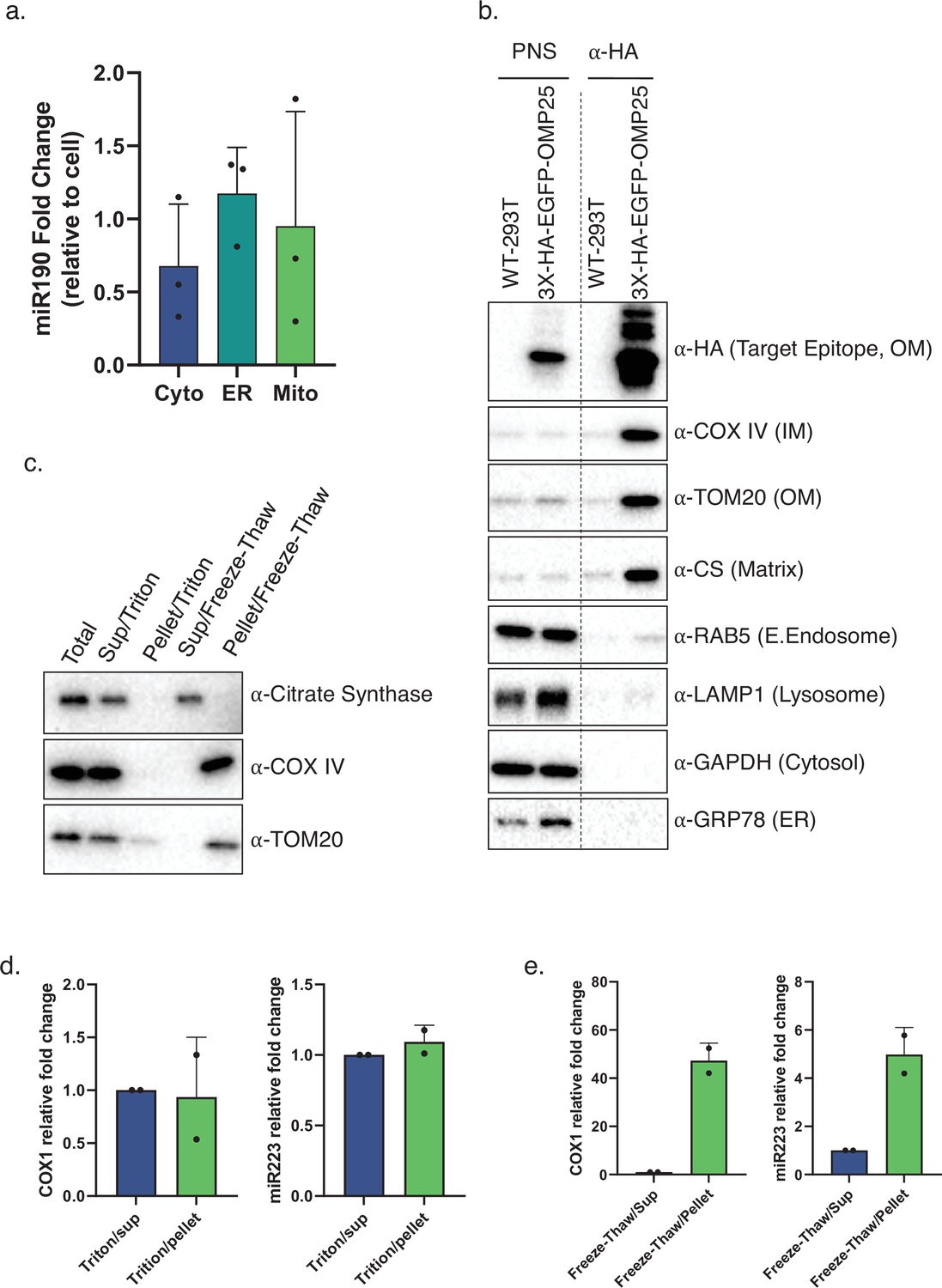
miR223, but not miR190, enriched in mitochondria.
(a) RT-qPCR analysis of miR190 fold changes in subcellular fractions isolated from 293T cells relative to cell lysate. Data are plotted from three independent experiments and error bars represent standard deviations. (b) Mitochondria immunoprecipitation. Mitochondria purified from 293 T WT or 3xHA-EGFP-OMP25 overexpressing cell line using anti-HA magnetic beads. Immunoblot detection of inner membrane marker COX IV, outer membrane marker TOM20 and mitochondrial matrix protein citrate synthase (CS) but not of the early endosome marker RAB5, lysosome marker LAMP1, cytosol marker GAPDH and ER marker GRP78. (c) Purified mitochondria from (b) were treated with 1% Triton X-100 or freeze/thaw using liquid-nitrogen and the supernatant and pellet fractions were collected after centrifugation. Immunoblot analysis of the mitochondrial protein distribution of outer membrane protein TOM20, inner membrane protein COX IV and matrix protein citrate synthase. (d) and (e) RT-qPCR analysis of the distribution of COX1 mRNA and miR223 from (c) Data are plotted from two independent experiments and error bars represent standard deviations.
-
Figure 3—figure supplement 1—source data 1
Uncropped immunoblot images corresponding to Figure 3—figure supplement 1.
- https://cdn.elifesciences.org/articles/85878/elife-85878-fig3-figsupp1-data1-v2.zip
Figure 3—figure supplement 2

Mitochondrial depletion did not change exosome secretion.
(a) CD63-Nluc was stably expressed in the cell lines expressing U2OS-GFP-Parkin. Exosome numbers are measured by luminescence from the conditioned medium of CD63-Nluc cells which were treated with or without CCCP. Normalized exosome production from cells are shown.
Figure 4 with 2 supplements
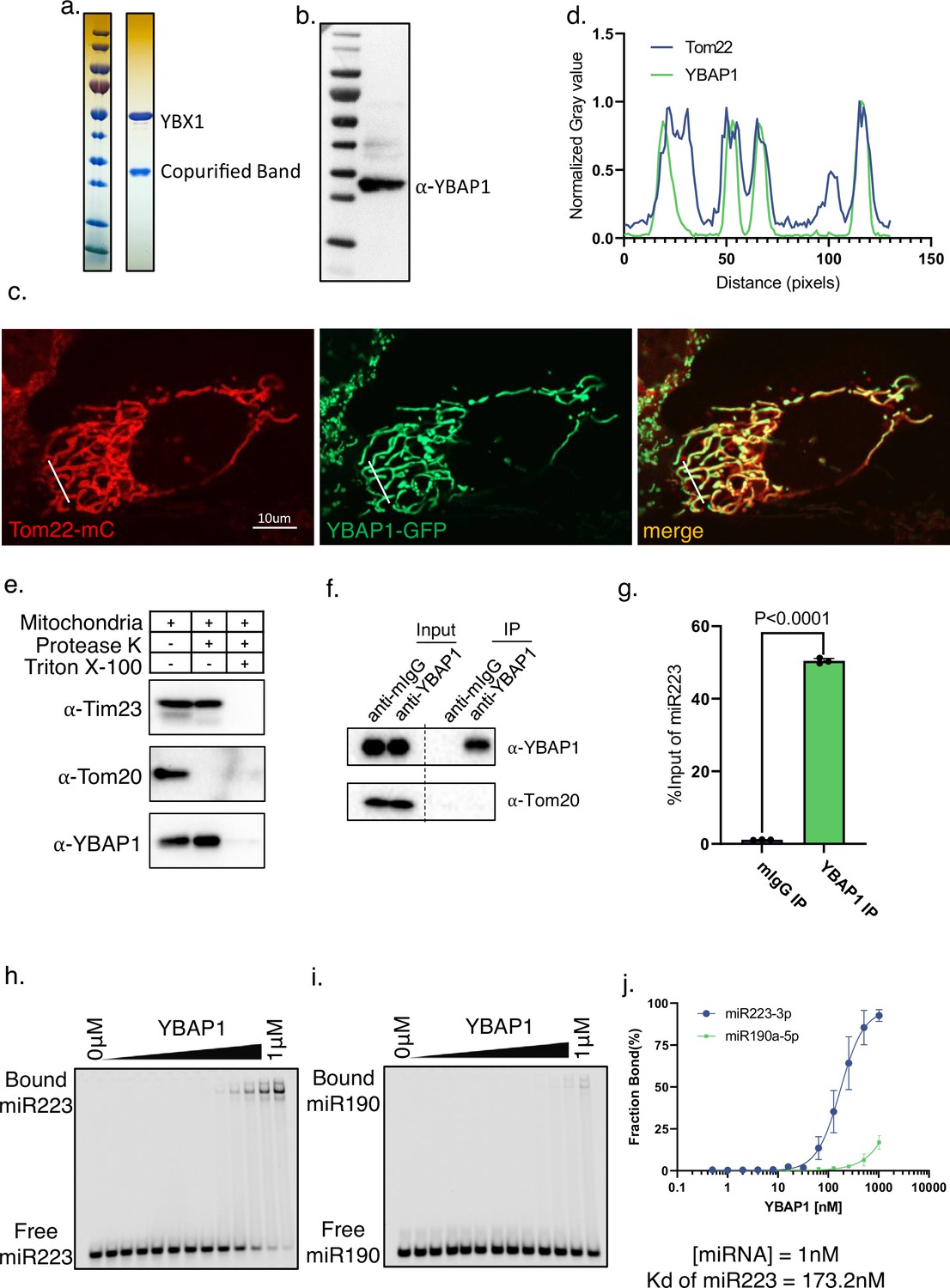
YBAP1 directly and specifically binds miR223.
(a) Strep II-YBX1 was overexpressed in HEK293T cells. Coomassie blue detection of unknown band copurified with YBX1 from 293T cells. (b) Immunoblot identified unknown band was YBAP1. (c) Tom22-mCherry expressing U2OS was transfected with a YBAP1-GFP-expressing plasmid, cultured for 12 hr and observed by confocal microscopy. Scale bar, 10 μm. (d) Quantification of the fluorescence intensity of the different channels indicated by the solid white line of (c). (e) YBAP1 resides in mitochondria. Proteinase K protection assay for YBAP1 using purified mitochondria from 293T cells. Samples were treated with or without proteinase K (10 μg/ml) and or Triton X-100 (0.5%). Immunoblots for Tim23, Tom20, and YBAP1 are shown. (f) Mitochondria were purified for immunoprecipitation with YBAP1 antibody. Immunoblot detection of YBAP1 and Tom20. (g) RT-qPCR analysis of miR223 fold changes of YBAP1 IP samples. Data are plotted from three independent experiments and error bars represent standard deviations. (h–i) EMSA assays using 1 nM 5’ fluorescently labeled miR223 or miR190. Purified YBAP1 was titrated from 500pM to 1 μM. In gel fluorescence was detected. Quantification of (j) shown the calculated Kd.
-
Figure 4—source data 1
Uncropped immunoblot and gel images corresponding to Figure 4.
- https://cdn.elifesciences.org/articles/85878/elife-85878-fig4-data1-v2.zip
Figure 4—figure supplement 1

YBX1 and YBAP1 copurify as a complex from transfected SF9 cells.
(a) YBAP1 purified from insect cells which overexpressed His-MBP-YBAP1. (b) YBX1 and YBAP1 were copurified from insect cells which overexpressed His-MBP-YBX1 with untagged YBAP1.
-
Figure 4—figure supplement 1—source data 1
Uncropped gel images corresponding to Figure 4—figure supplement 1.
- https://cdn.elifesciences.org/articles/85878/elife-85878-fig4-figsupp1-data1-v2.zip
Figure 4—figure supplement 2

YBAP1 does not share the same miR223-binding motif as YBX1.
(a) EMSA assays using 1 nM 5’ fluorescently labeled miR223 WT or miR223mut or miR190 WT or miR190sort and purified YBAP1. Purified YBAP1 was titrated from 500pM to 1 µM. In gel fluorescence was detected. (b) Binding affinity curves as calculated by EMSA data from (a).
-
Figure 4—figure supplement 2—source data 1
Uncropped gel images corresponding to Figure 4—figure supplement 2.
- https://cdn.elifesciences.org/articles/85878/elife-85878-fig4-figsupp2-data1-v2.zip
Figure 5 with 1 supplement
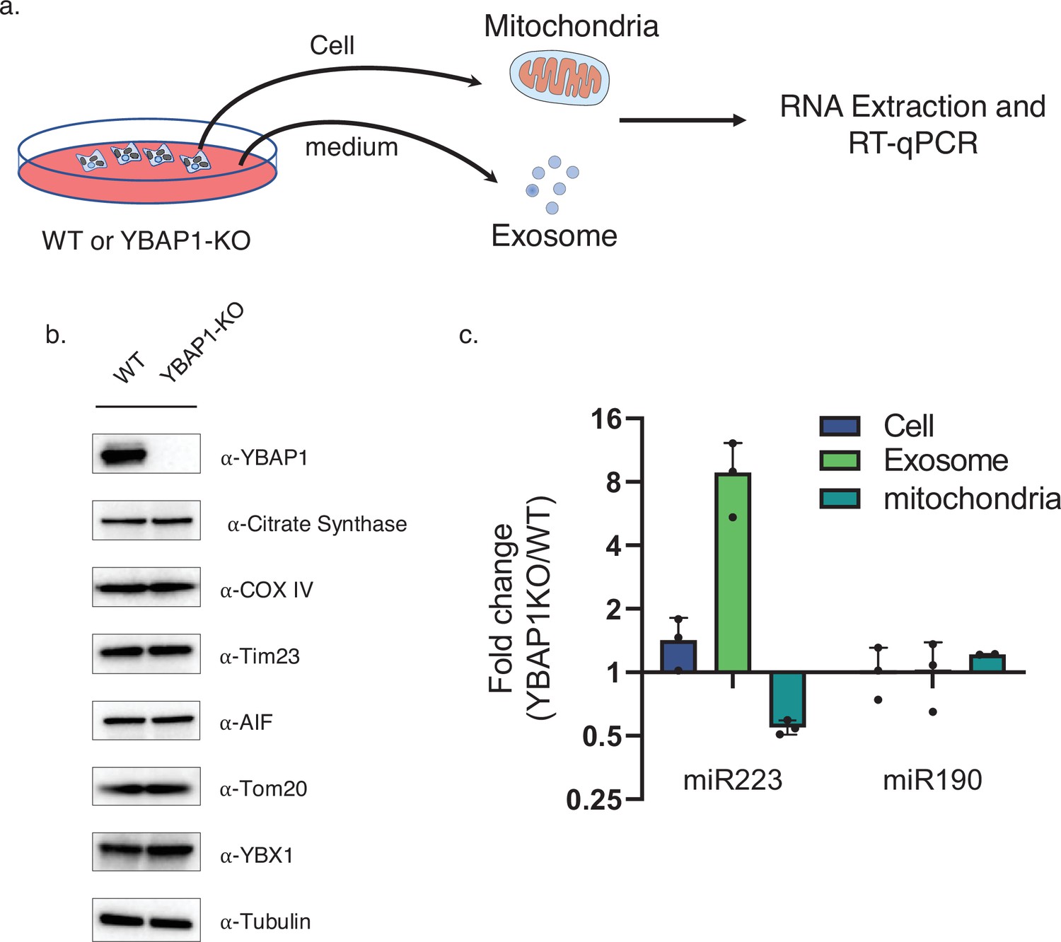
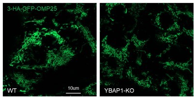
YBAP1 sequesters miR223 which is released and secreted in YBAP1 KO cells.
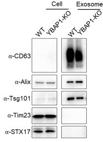
(a) Schematic shows exosome and mitochondria purification from 293 T WT cells and YBAP1 knock out cells for RT-qPCR analysis. (b) Analysis of 293 T WT and CRISPR/Cas9 genome-edited cells by immunoblot for YBAP1, YBX1 and mitochondrial markers (c) RT-qPCR analysis of miR223 enrichment in mitochondria purified from 293 T WT cells and YBAP1 KO cells relative to cell lysate. Data are plotted from three independent experiments and error bars represent standard deviations. (d) RT-qPCR analysis of miR223 and miR190 fold change in cells, purified mitochondria and exosomes from 293 T WT cells and YBAP1 KO cells. Data are plotted from three independent experiments and error bars represent standard deviations.
-
Figure 5—source data 1
Uncropped immunoblot images corresponding to Figure 5.
- https://cdn.elifesciences.org/articles/85878/elife-85878-fig5-data1-v2.zip
Figure 5—figure supplement 1

Knockout of YBAP1 did not change exosome secretion.
(a) CD63-Nluc was stably expressed in the 293 T WT and YBAP1-KO cell lines. Exosome production was measured by luminescence of CD63-Nluc in the conditioned medium of cells which were treated with or without CCCP. Normalized exosome production from 293 T WT and YBAP1-KO cells are shown.
Figure 6
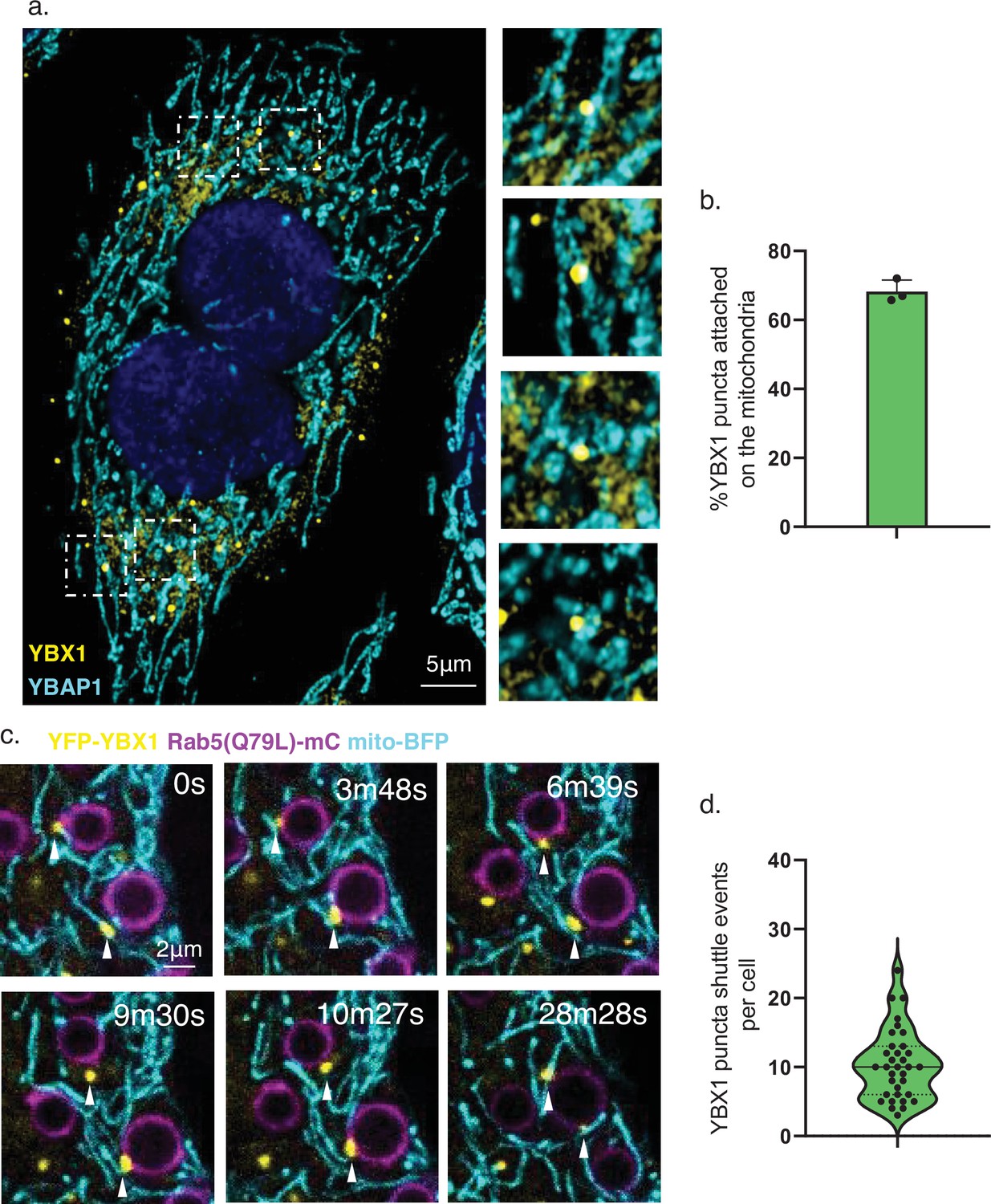
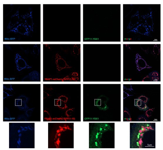
YBX1 puncta relocalize from mitochondria to endosomes.
(a) YBX1 puncta on the mitochondria. U2OS cells were stained with anti-YBX1 and anti-YBAP1 antibodies and observed by confocal microscopy. The right panel shows enlarged regions of interest from the left panel. Scale bar, 5 μm. (b) The statistics are of the percentage of YBX1 puncta detected in proximity to mitochondria. N=30 cells. (c) YBX1 puncta relocalize from mitochondria to endosomes. U2OS cells overexpressed YFP-YBX1, Rab5(Q79L)-mCherry and mito-BFP. Time-lapse images were acquired with a Zeiss LSM900 confocal microscope. Scale bar, 2 μm. (d) The statistics are of YBX1 puncta shuttle events per cell. The data was represented as violin plots. N=34 cells.
Figure 7
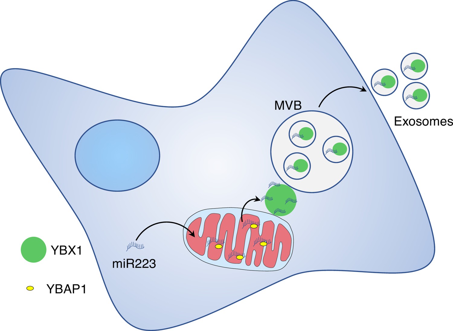
Diagram representing a model of miR223 sorting from mitochondria into exosomes.
Stages in the transfer of miR223 from mitochondria. Cytosolic miR223 is enriched in mitochondria where it may be sequestered by a weak interaction with YBAP1. Cytoplasmic YBX1 interacts more tightly with miR223 which may drive the removal of miR223 from mitochondria. YBX1 in RNA granules may accumulate miR223 removed from mitochondria. YBX1 puncta may give rise to small particles carrying miR223 for uptake into endosomes and secretion in exosomes.
Author response image 1

Author response image 2
Author response image 3
Author response image 4
Author response image 5
Tables
Key resources table
| Reagent type (species) or resource | Designation | Source or reference | Identifiers | Additional information |
|---|---|---|---|---|
| Cell line (Spodoptera frugiperda) | Sf9 | Other | Cell culture facility at UC Berkeley | |
| Cell line (Homo sapiens) | HEK 293T cells | Other | Cell culture facility at UC Berkeley | |
| Cell line (Homo sapiens) | HEK 293T-YBX1 KO cells | Other | Obtained by CRISPR-Cas9 in Schekman Lab | |
| Cell line (Homo sapiens) | HEK 293T-YBAP1 KO | This study | Obtained by CRISPR-Cas9 in Schekman Lab | |
| Cell line (Homo sapiens) | HEK 293T-3xHA-EGFP-OMP25 | This study | Obtained by overexpression of pLJM1-3XHA-EGFP-OMP25 in Schekman lab | |
| Cell line (Homo sapiens) | U-2OS cells | Other | Cell culture facility at UC Berkeley | |
| Cell line (Homo sapiens) | U-2OS Parkin-GFP cells | This study | Obtained by overexpression of Parkin-GFP in Schekman lab | |
| Recombinant DNA reagent | pFastBac His6 MBP N10 TEV LIC cloning vector (4 C) | Addgene | RRID: Addgene_30116 | N/A |
| Recombinant DNA reagent | Tom22-mCherry (plasmid) | This study | Gift of Dr Li Yu lab | |
| Recombinant DNA reagent | His-MBP-YBX1 (plasmid) | This study | To express YBX1 in insect cells. Plasmid maintained in Schekman lab | |
| Recombinant DNA reagent | His-MBP-YBX1(1–51) (plasmid) | This study | To express YBX1(1–51) in insect cells. Plasmid maintained in Schekman lab | |
| Recombinant DNA reagent | His-MBP-YBX1(52–129) (plasmid) | This study | To express YBX1(52–129) in insect cells. Plasmid maintained in Schekman lab | |
| Recombinant DNA reagent | His-MBP-YBX1(130–324) (plasmid) | This study | To express YBX1(1–51) in insect cells. Plasmid maintained in Schekman lab | |
| Recombinant DNA reagent | His-MBP-YBX1(1–129) (plasmid) | This study | To express YBX1(1–129) in insect cells. Plasmid maintained in Schekman lab | |
| Recombinant DNA reagent | His-MBP-YBX1(F85A) (plasmid) | This study | To express YBX1(F85A) in insect cells. Plasmid maintained in Schekman lab | |
| Recombinant DNA reagent | His-MBP-YBAP1 (plasmid) | This study | To express YBAP1 in insect cells. Plasmid maintained in Schekman lab | |
| Recombinant DNA reagent | Mito-BFP | This study | Gift of Dr. Samantha Lewis lab | |
| Recombinant DNA reagent | mCherry-Rab5(Q79L) (plasmid) | Addgene | RRID: Addgene_35138 | |
| Recombinant DNA reagent | pLJM1-3XHA-EGFP-OMP25 | This study | To express 3xHA-EGFP-OMP25 in HEK293T cells. Plasmid maintained in Schekman lab | |
| Antibody | Anti-YBX1 (Rabbit polyclonal) | Abcam | RRID: AB_1950384 | WB 1:1000 |
| Antibody | Anti-YBAP1 (Mouse monoclonal) | Santa Cruz | RRID: AB_10611471 | WB 1:1000 |
| Antibody | Anti-YBAP1(Rabbit polyclonal) | Thermo Fisher Scientific | RRID: AB_2638956 | WB 1:1000 |
| Antibody | Anti-Tim23 (Mouse monoclonal) | BD Biosciences | RRID: AB_398754 | WB 1:1000 |
| Antibody | Anti-Tom20 (Mouse monoclonal) | Abcam | RRID: AB_945896 | WB 1:1000 |
| Antibody | Anti-Calnexin (Rabbit polyclonal) | Abcam | RRID: AB_2069006 | WB 1:2000 |
| Antibody | Anti-HA (Rabbit monoclonal) | Cell Signaling | RRID: AB_1549585 | WB 1:1000 |
| Antibody | Anti-COX IV (Rabbit Monoclonal) | Cell signaling | RRID: AB_2085424 | WB 1:1000 |
| Antibody | Anti-Citrate Synthase (Rabbit monoclonal) | Cell signaling | RRID: AB_2665545 | WB 1:1000 |
| Antibody | Anti-Rab5 (Rabbit monoclonal) | Cell signaling | RRID: AB_2300649 | WB 1:1000 |
| Antibody | Anti-LAMP1 (Rabbit monoclonal) | Cell signaling | RRID: AB_2687579 | WB 1:1000 |
| Antibody | Anti-GRP78 (Rabbit polyclonal) | Abcam | RRID: AB_2119834 | WB 1:3000 |
| Antibody | Anti-GADPH (Rabbit monoclonal) | Cell signaling | RRID: AB_561053 | WB 1:5000 |
| Antibody | Anti-alpha Tubulin (Mouse monoclonal) | Abcam | RRID: AB_2241126 | WB 1:5000 |
| Antibody | Anti-beta Actin (Mouse monoclonal) | Abcam | RRID: AB_449644 | WB 1:5000 |
| Software, algorithm | Image Studio Lite | LICOR | https://www.licor.com/bio/image-studio-lite/ | |
| Software, algorithm | FIJI | NIH | RRID: SCR_002285 | https://fiji.sc/ |
| Software, algorithm | Prism 9 | GraphPad | RRID: SCR_002798 | https://www.graphpad.com |
Additional files
Download links
A two-part list of links to download the article, or parts of the article, in various formats.
Downloads (link to download the article as PDF)
Open citations (links to open the citations from this article in various online reference manager services)
Cite this article (links to download the citations from this article in formats compatible with various reference manager tools)
Two RNA-binding proteins mediate the sorting of miR223 from mitochondria into exosomes
eLife 12:e85878.
https://doi.org/10.7554/eLife.85878


