What happens to the inhibitory control functions of the right inferior frontal cortex when this area is dominant for language?
- Neuropsychology and Functional Neuroimaging; Jaume I University, Spain
Figures
Figure 1
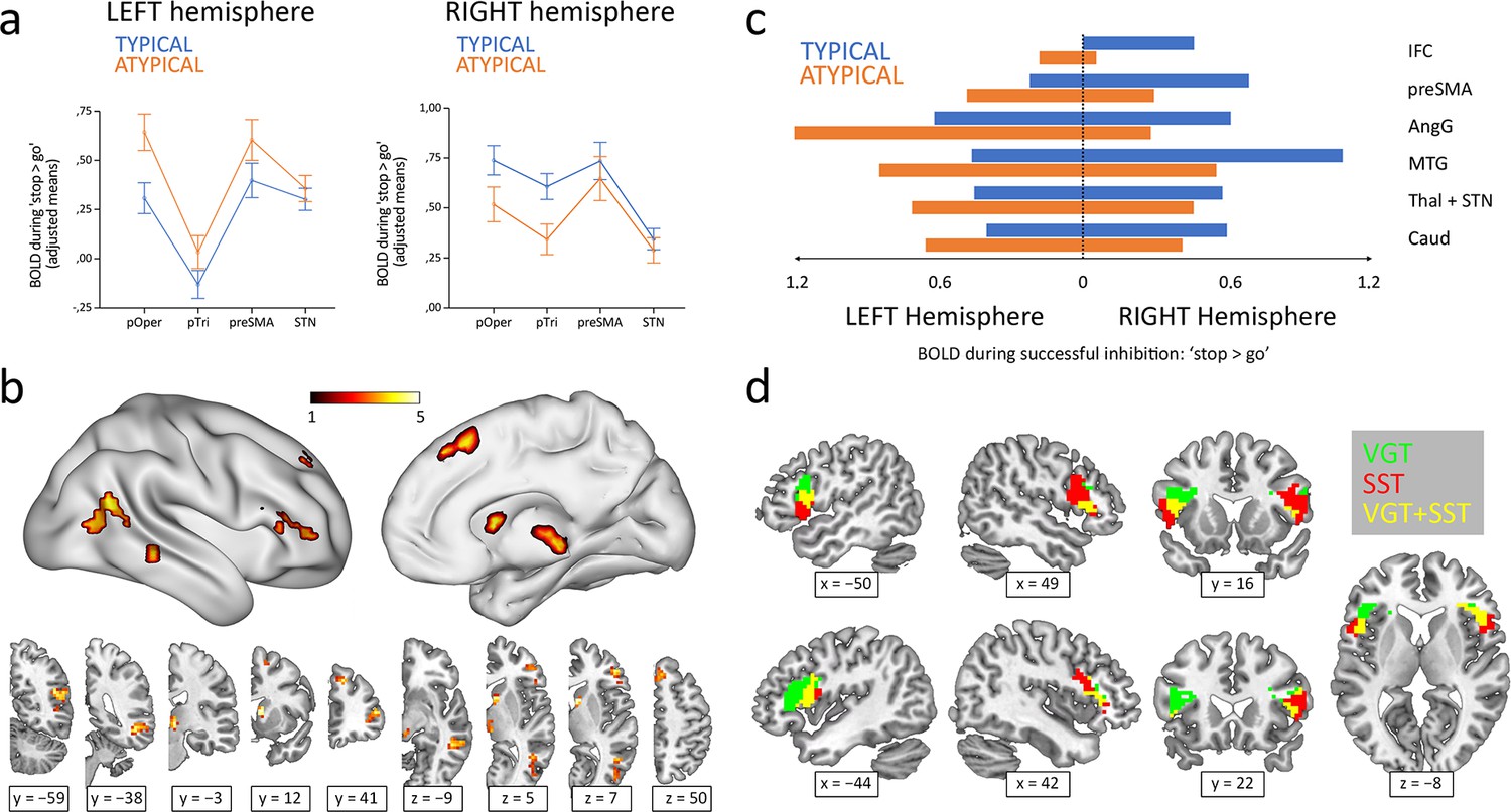
Hemispheric lateralization of inhibitory control according to language lateralization.
(a) ROI analysis of the main components of the inhibitory control network. Graphs depict adjusted mean BOLD signal during successful inhibitions on the stop-signal task (‘stop >go’ contrast) for both hemispheres and both groups (n=50 typical and 36 atypical). All four structures showed significant Hemisphere × Group interactions in a repeated-measures MANOVA (p<0.05). Error bars represent one standard error. pOper = pars opercularis, pTri = pars triangularis, preSMA = presupplementary motor area, and STN = subthalamic nucleus. (b) Voxel-wise whole-brain analysis of functional asymmetry. Significance maps (voxel-wise p<0.001; FWE cluster-corrected at p<0.05; color bar represents t value) are displayed in three-dimensional reconstructions plus coronal and transversal slices using MNI space. (c) Mean BOLD values of the significant regions found in the voxel-wise whole-brain analysis. Graphic depicts BOLD values for every region, hemisphere, and group. IFC = inferior frontal cortex, preSMA = presupplementary motor area, AngG = angular gyrus, MTG = middle temporal gyrus, Thal = thalamus, STN = subthalamic nucleus, Caud = caudate. (d) Functional overlap between language production and inhibitory control in the IFC of ambilateral participants. Overlapping maps for inhibition (voxel-wise p<0.001; FWE cluster-corrected at p<0.05) and language (voxel-wise p<0.05; uncorrected) are displayed in coronal, sagittal and transversal slices using MNI space. VGT = verb generation task, SST = stop-signal task.
Figure 2
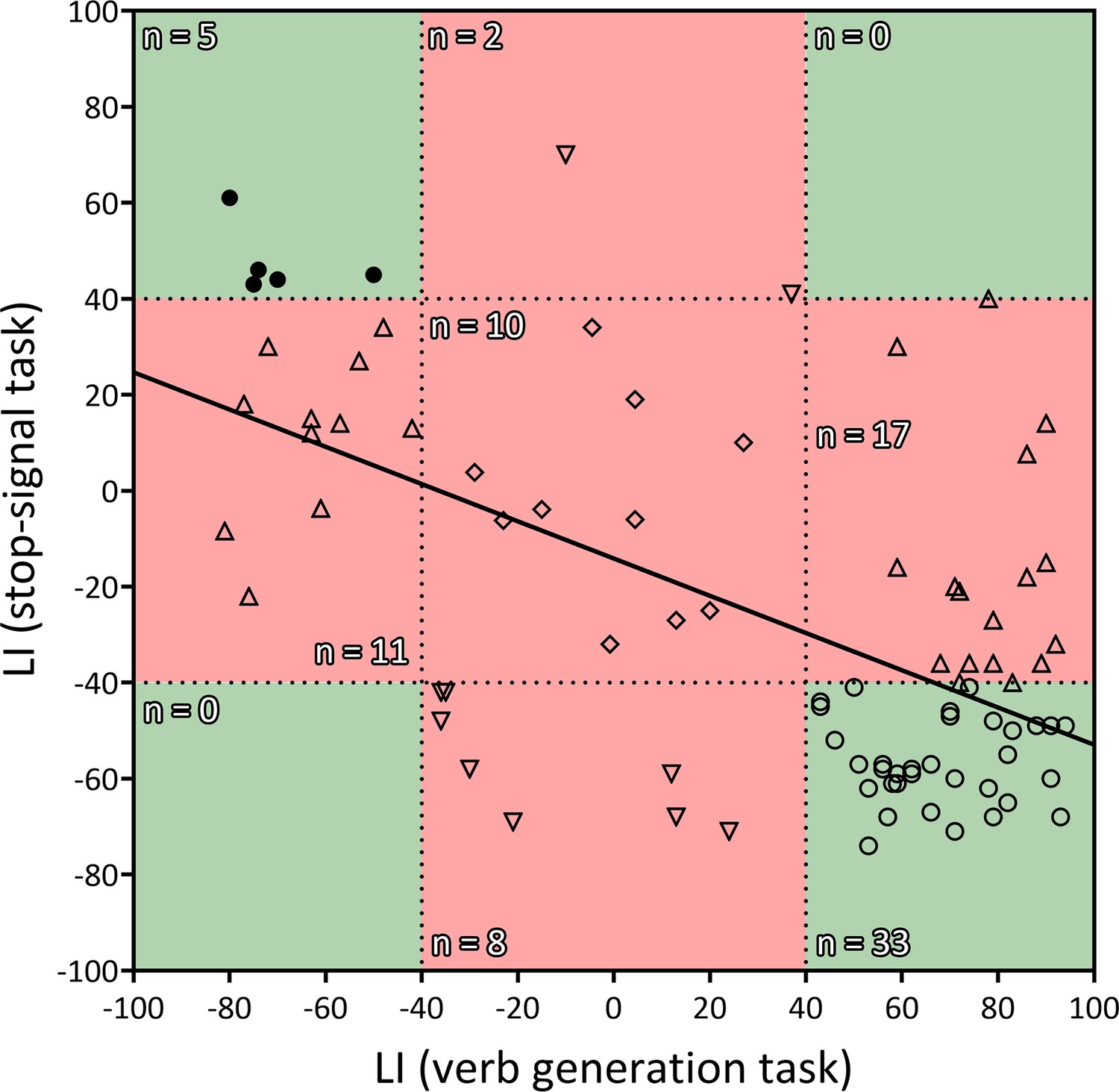
Correlation between the LIs of the verb generation and stop-signal tasks.
r = −0.583, two-tailed p<0.001, R2=0.339. Negative values indicate rightward lateralization, whereas positive values indicate leftward lateralization. Segregated and ambilateral phenotypes are also depicted according to the background color. The green area corresponds to segregated individuals (both functions strongly lateralized), and the red area corresponds to integrated individuals (at least one function ambilaterally controlled). Numbers inside each quadrant denote the number of individuals (n) contained in it. Each individual datapoint is symbolized according to its functional organization: 〇=typical segregation; ⬤=reversed segregation; △=ambilateral inhibition; ▽=ambilateral language; ◇=ambilateral language and inhibition.
Tables
Table 1
Spearman’s partial correlations between task LIs and neuroanatomical plus behavioral variables.
| Verb generation task LI | Stop-signal task LI | |||
|---|---|---|---|---|
| Spearman’s ρ | p | Spearman’s ρ | p | |
| Callosal genu volume | −0.249 | 0.021* | 0.251 | 0.02* |
| Callosal body volume | −0.258 | 0.017* | 0.132 | 0.224 |
| Callosal splenium volume | −0.249 | 0.021* | 0.118 | 0.279 |
| Pars Opercularis VMHC | −0.062 | 0.584 | 0.148 | 0.192 |
| Pars Triangularis VMHC | −0.119 | 0.295 | 0.258 | 0.022* |
| preSMA VMHC | 0.052 | 0.647 | 0.037 | 0.748 |
| STN VMHC | 0.033 | 0.772 | 0.217 | 0.054 |
| ‘Go’ reaction time | 0.125 | 0.251 | −0.041 | 0.71 |
| ‘Go’ accuracy | −0.076 | 0.489 | 0.039 | 0.724 |
| SSRT | 0.173 | 0.111 | −0.038 | 0.726 |
| Reading length accuracy | −0.237 | 0.028* | 0.214 | 0.047* |
| Reading familiarity accuracy | −0.232 | 0.032* | 0.301 | 0.005** |
| Reading length time | 0.054 | 0.622 | −0.184 | 0.068 |
| Reading familiarity time | 0.091 | 0.403 | −0.198 | 0.091 |
| SPQ | −0.194 | 0.078 | 0.247 | 0.023* |
| AQ | −0.247 | 0.023* | 0.075 | 0.498 |
-
General intelligence and age were included as covariates of no interest. Callosal volume correlations were additionally corrected for total intracranial volume.
Additional files
Download links
A two-part list of links to download the article, or parts of the article, in various formats.
Downloads (link to download the article as PDF)
Open citations (links to open the citations from this article in various online reference manager services)
Cite this article (links to download the citations from this article in formats compatible with various reference manager tools)
What happens to the inhibitory control functions of the right inferior frontal cortex when this area is dominant for language?
eLife 12:RP86797.
https://doi.org/10.7554/eLife.86797.3


