Pulsed ultrasound promotes secretion of anti-inflammatory extracellular vesicles from skeletal myotubes via elevation of intracellular calcium level
- Department of Rehabilitation Science, Kobe University Graduate School of Health Sciences, Japan
- School of Life Sciences and Technology, ShanghaiTech University, China
- Department of Pathology, Nanjing Medical University, China
- Department of Health and Nutrition , Shubun University, Japan
- John B. Little Center for Radiation Sciences, Harvard University T.H. Chan School of Public Health, United States
Figures
Figure 1
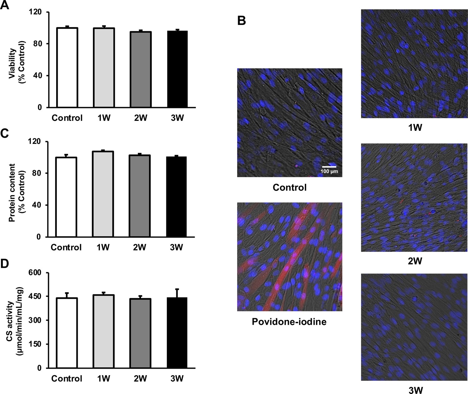
The cytotoxicity of ultrasound (US) irradiation on myotubes was investigated.
Viability of myotubes was assessed by (A) MTT assay and (B) Zombie Red staining at 24 hr after US irradiation. (red: Zombie Red, blue: DAPI). (C) Total protein content was measured by the Bradford method at 24 hr after US irradiation. (D) Energy metabolism in C2C12 myotubes was measured by citrate synthase assay at 24 hr after US irradiation. The US intensities of 1.0 W/cm2, 2.0 W/cm2, and 3.0 W/cm2 were tested. Data are expressed as mean ± SEM. n=4.
Figure 2
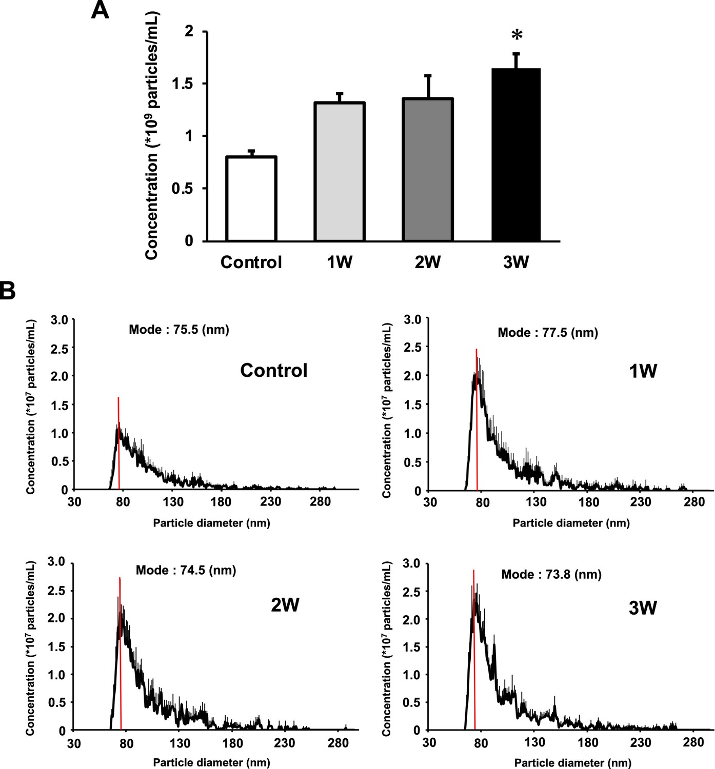
Characterization of extracellular vesicles (EVs) from ultrasound (US)-treated/untreated myotubes.
EVs were isolated using ExoQuick reagent 12 hr after US irradiation. (A) EV concentration in each group was quantified by a qNano system. (B) Size distribution of EVs in each group was investigated by a qNano system. The mode value was indicated as a red line. The US intensities of 1.0 W/cm2, 2.0 W/cm2, and 3.0 W/cm2 were tested. Data are expressed as mean ± SEM. *p<0.01, vs. control. n=6.
Figure 3
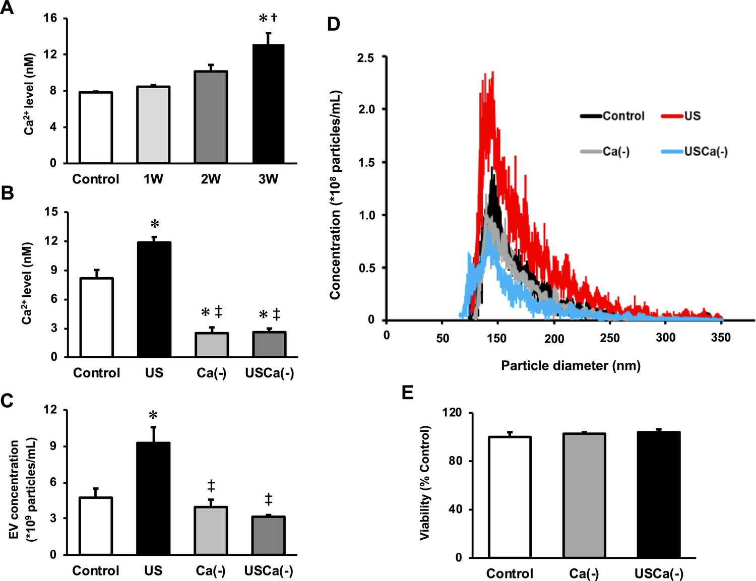
Ca2+ mediates the promotive effect of ultrasound (US) on extracellular vesicle (EV) release from myotubes.
(A) Intracellular Ca2+ levels were measured after US irradiation. The US intensities of 1.0 W/cm2, 2.0 W/cm2, and 3.0 W/cm2 were tested. (B) Cell culture with Ca2+-free medium decreased the intracellular Ca2+ level and canceled the facilitating effect of US on Ca2+ uptake by myotubes. (C) Cell culture with Ca2+-free medium inhibited the facilitating effect of US on EV release from myotubes. (D) Size distribution of EVs in each group. EV concentration and size distribution were quantified by a qNano system. (E) Cytotoxicity of cell culture with Ca2+-free medium was investigated by MTT assay. Control: untreated; US: 3.0 W/cm2 US treatment; Ca(-): Ca2+-free culture; USCa(-): 3.0 W/cm2 US treatment and Ca2+-free culture. Data are expressed as mean ± SEM. *p<0.01 vs. control, †p<0.05 vs 1W, ‡p<0.01 vs. US. n=4.
Figure 4
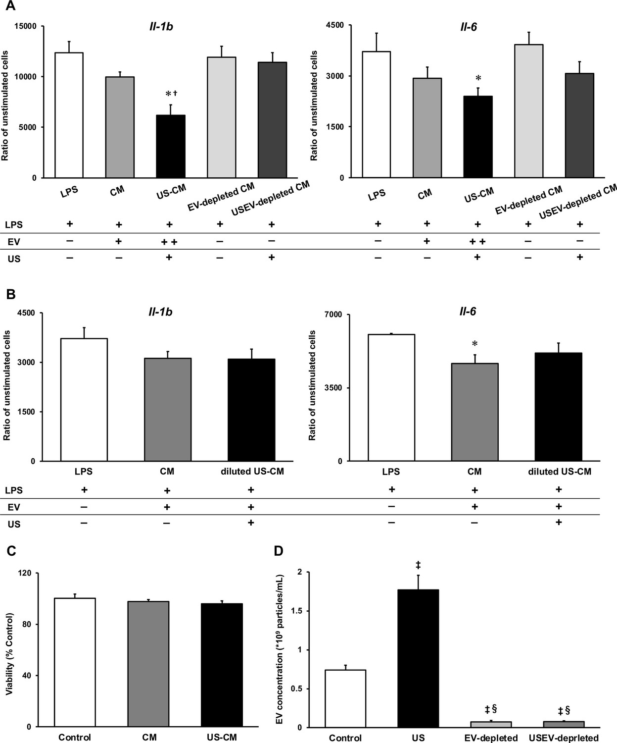
Anti-inflammatory effect of extracellular vesicles (EVs) from ultrasound (US)-treated myotubes on bone marrow-derived macrophages (BMDMs).
(A) The mRNA expression levels of Il-1b and Il-6 were measured by qPCR. Lipopolysaccharide (LPS): LPS-treated BMDMs; CM: BMDMs treated with C2C12 conditioned medium and LPS; US-CM: BMDMs treated with US-irradiated C2C12 conditioned medium and LPS; EV-depleted CM: BMDMs treated with EV-depleted C2C12 conditioned medium and LPS; USEV-depleted: BMDMs treated with EV-depleted C2C12 (US-irradiated) conditioned medium and LPS. (B) When the concentration of EVs are equated, the enhancement of anti-inflammatory effect of EVs by US was not observed. LPS: LPS-treated BMDMs; CM: BMDMs treated with myotube EVs and LPS; diluted US-CM: BMDMs treated with US-EVs at the same concentration as the EV group and LPS. (C) Cytotoxicity of C2C12 conditioned medium on BMDMs was investigated by MTT assay. (D) EV concentration in each condition was measured by a qNano system. Data are expressed as mean ± SEM. *p<0.05 vs. LPS, †p<0.01 vs. CM, ‡p<0.01 vs. control, §p<0.01 vs. US. n=4.
Figure 5
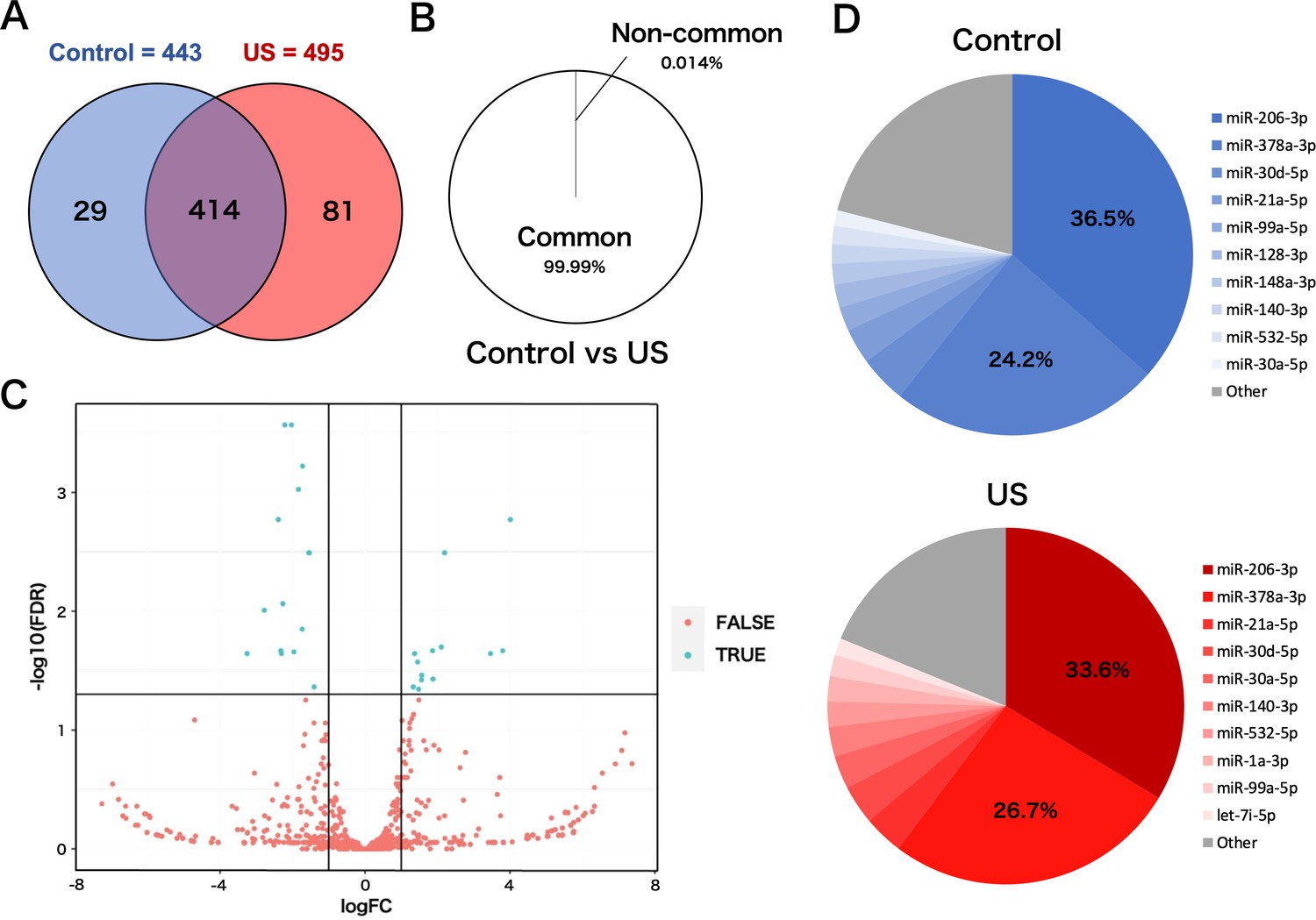
miRNA-sequencing analysis in extracellular vesicles (EVs) from ultrasound (US)-treated/untreated C2C12 myotubes.
(A) miRNA characterization in EVs from US-treated/untreated myotubes. (B) Percentage of miRNAs that were common to the control and US groups and those that were not. (C) Volcano plot of differentially expressed RNAs in the control group vs. US group. Blue dots represent miRNAs with statistically significant difference and red dots show miRNAs with no statistically significant difference between the control group vs. US group. (D) Top 10 abundant miRNAs and their proportion to total miRNA content in each group. n=3.
Figure 6
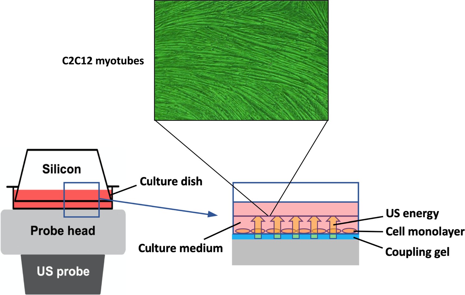
In vitro ultrasound (US) irradiation system.
The culture dish was placed on the probe of a US transducer (SONICTIZER SZ-100, Minato Medical Science Co., Ltd., Japan). US waves are sent out from the probe placed under the culture dish, and surplus energy is absorbed by the silicon.
Tables
Table 1
Upregulated miRNAs in myotube-derived extracellular vesicles by ultrasound irradiation.
| miRNA | logFC | FDR |
|---|---|---|
| miR-193a-3p | 4.012954262 | 0.000742983 |
| miR-138-5p | 3.800680285 | 0.00141537 |
| miR-223-3p | 3.462174268 | 0.008790418 |
| miR-362-3p | 2.195592968 | 0.009437513 |
| miR-34a-5p | 2.105459261 | 0.009437513 |
| miR-675-3p | 1.876128033 | 0.009966738 |
| miR-106b-5p | 1.862814288 | 0.009966738 |
| miR-30b-5p | 1.566332803 | 0.011740976 |
| miR-188-5p | 1.559409694 | 0.015175451 |
| miR-133b-3p | 1.48161012 | 0.016332293 |
| miR-1a-3p | 1.450599776 | 0.016622751 |
| miR-30a-5p | 1.367918225 | 0.019014654 |
| miR-27b-3p | 1.330089099 | 0.01987041 |
Table 2
Downregulated miRNAs in myotube-derived extracellular vesicles by ultrasound irradiation.
| miRNA | logFC | FDR |
|---|---|---|
| miR-128-2-5p | –3.254069169 | 0.000118723 |
| miR-184-3p | –2.779741068 | 0.000118723 |
| miR-615-3p | –2.393817511 | 0.000263853 |
| miR-344d-3p | –2.324403579 | 0.00041415 |
| miR-344d-4p | –2.308193263 | 0.000742983 |
| miR-1964-3p | –2.267802775 | 0.00141537 |
| miR-320-3p | –2.212812155 | 0.00141537 |
| miR-128-3p | –2.031468208 | 0.003795561 |
| miR-351-3p | –1.963143876 | 0.004305251 |
| miR-1198-5p | –1.733517014 | 0.009437513 |
| miR-501-3p | –1.72302403 | 0.009643704 |
| miR-222-3p | –1.547695372 | 0.009966738 |
| let-7a-5p | –1.538094973 | 0.009966738 |
| miR-423-5p | –1.404879603 | 0.019014654 |
Additional files
-
Supplementary file 1
Lists of specific miRNAs in ultrasound (US)-treated/untreated groups.
miRNA-sequencing analysis in extracellular vesicles from US-treated/untreated C2C12 myotubes was conducted.
- https://cdn.elifesciences.org/articles/89512/elife-89512-supp1-v1.docx
-
Supplementary file 2
Myotube contraction by electrical stimulation.
Myotubes were electrically stimulated (30 mA at 1 Hz for 15 ms at 985 ms intervals) with an electrical pulse generator (ITO Co., Ltd, Saitama, Japan) to confirm differentiation. Separately plated cells were used to confirm contraction and the cells used for the experiments were not electrically stimulated.
- https://cdn.elifesciences.org/articles/89512/elife-89512-supp2-v1.pptx
-
Supplementary file 3
Sequences for qPCR primers used in this study.
- https://cdn.elifesciences.org/articles/89512/elife-89512-supp3-v1.docx
-
MDAR checklist
- https://cdn.elifesciences.org/articles/89512/elife-89512-mdarchecklist1-v1.docx
Download links
A two-part list of links to download the article, or parts of the article, in various formats.
Downloads (link to download the article as PDF)
Open citations (links to open the citations from this article in various online reference manager services)
Cite this article (links to download the citations from this article in formats compatible with various reference manager tools)
Pulsed ultrasound promotes secretion of anti-inflammatory extracellular vesicles from skeletal myotubes via elevation of intracellular calcium level
eLife 12:RP89512.
https://doi.org/10.7554/eLife.89512.3


