Figures
Figure 1 with 1 supplement
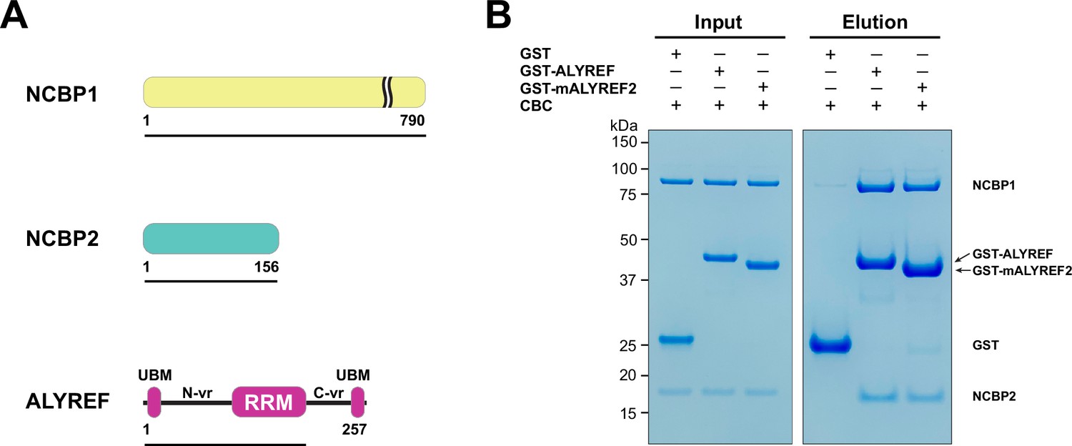
ALYREF directly binds to CBC.
(A) Schematics of the NCBP1 and NCBP2 subunits of the human CBC and ALYREF. ALYREF contains a central RRM domain. The RRM domain is connected to two conserved motifs (UBMs) at both termini through variable regions (N-vr and C-vr). Protein constructs used in GST pull-down assays in panel (B) (NCBP1, residues 1–790; NCBP2, residues 1–156; ALYREF, residues 1–183) are indicated by black lines under the respective proteins. (B) ALYREF directly interacts with the CBC. In vitro GST pull-down assays were performed with purified recombinant human CBC and GST-tagged ALYREF or the corresponding construct of mouse ALYREF2 (mALYREF2, residues 1–155). Results are representative of three technical repeats.
-
Figure 1—source data 1
Original file for the gels in Figure 1B.
- https://cdn.elifesciences.org/articles/91432/elife-91432-fig1-data1-v1.zip
Figure 1—figure supplement 1

Human ALYREF and mouse ALYREF2 are conserved.
Sequence alignment of ALYREF and mALYREF2 (Robert and Gouet, 2014). The DDX39B binding motifs (N-UBM and C-UBM), the WxHD motif, and the RRM domain are indicated.
Figure 2 with 5 supplements
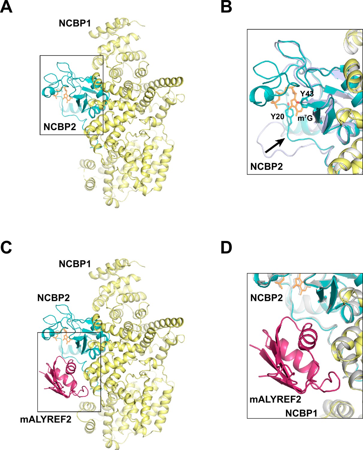
Cryo-EM structures of CBC and CBC-mALYREF2.
(A) Overall architecture of the CBC complex. NCBP1 and NCBP2 are colored in yellow and teal, respectively. The cap analog is shown as orange sticks. (B) Comparison of the CBC cryo-EM structure to the unliganded CBC crystal structure (PDB ID 1N54). The cryo-EM structure is colored as in (A). The unliganded CBC crystal structure is colored in light blue. Arrow indicates the conformational change in the N-terminal extension of NCBP2 upon cap analog binding. (C) Overall architecture of the CBC-mALYREF2 complex. NCBP1, NCBP2, and mALYREF2 are colored in yellow, teal, and red, respectively. The RRM domain of mALYREF2 binds to the CBC. (D) Comparison of the CBC-mALYREF2 and the CBC cryo-EM structures. CBC structure is colored in gray.
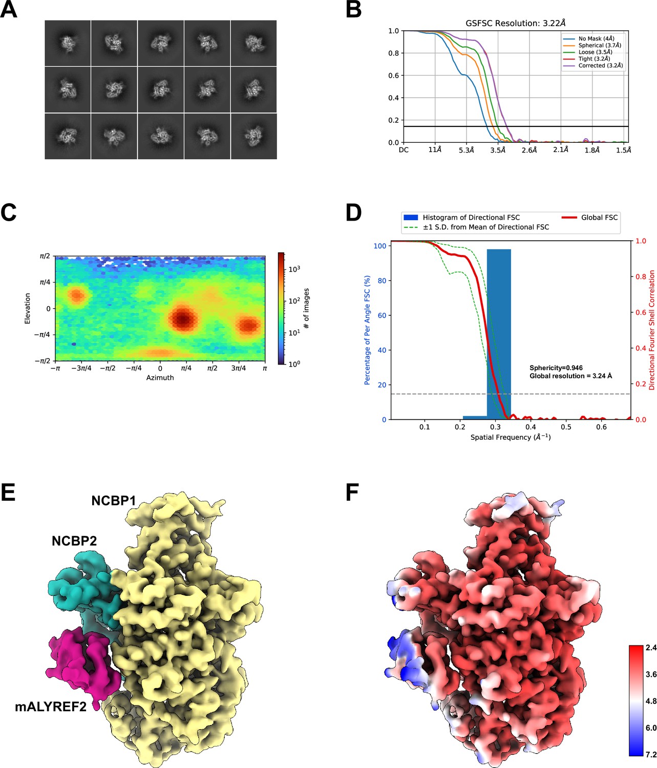
Figure 2—figure supplement 1
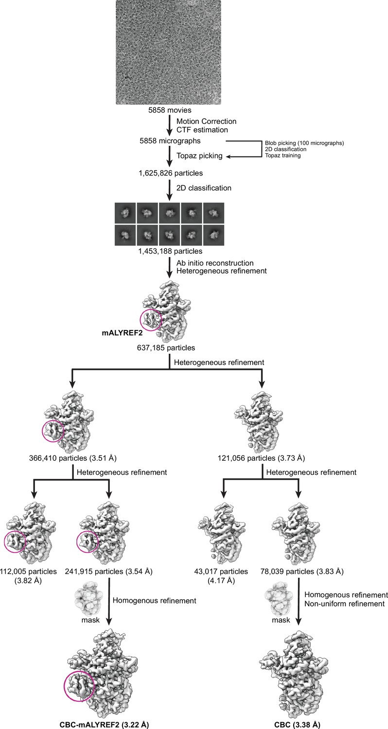
Workflow for cryo-EM data processing.
Data were processed in CryoSPARC. A set of 241,915 particles yielded a reconstruction of a CBC-mALYREF2 map at 3.22 Å resolution. A set of 78,039 particles yielded a reconstruction of a CBC map at 3.38 Å resolution.
Figure 2—figure supplement 2
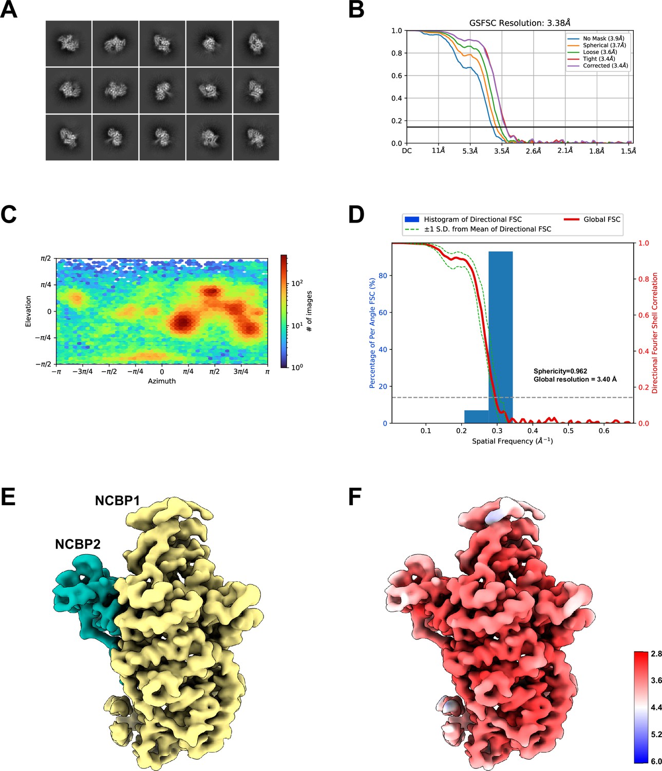
Cryo-EM reconstruction of the CBC.
(A) Selective 2D class averages from the final particle set of the CBC. (B) The gold-standard FSC curves of the final reconstruction of the CBC. (C) Angular distribution of the final reconstruction of the CBC computed in CryoSPARC. (D) 3DFSC analysis of the CBC reconstruction. The sphericity and global resolution values are noted within the graph window. (E) Cryo-EM map of the CBC complex. NCBP1 and NCBP2 are colored in yellow and teal, respectively. (F) Local resolution of the CBC cryo-EM map.
Figure 2—figure supplement 3
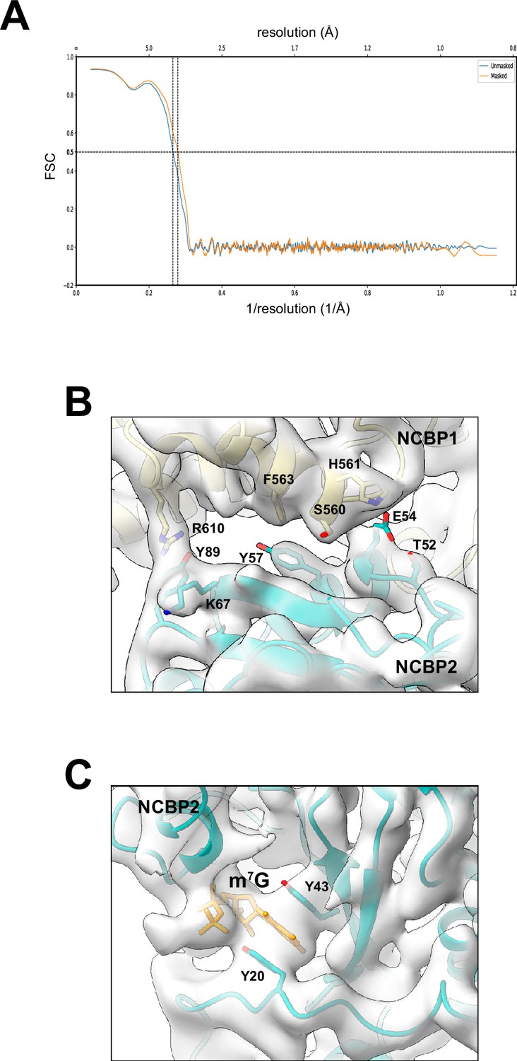
Structural model of the CBC.
(A) Map versus model FSC curves with or without mask calculated using Phenix. A resolution of 3.6 Å is estimated at FSC = 0.5. (B, C) Electron density map and structural model of CBC at the interface between NCBP1 and NCBP2 (B) and near m7G (C).
Figure 2—figure supplement 4
Cryo-EM reconstruction of the CBC-mALYREF2 complex.
(A) Selective 2D class averages from the final particle set of CBC-mALYREF2. (B) The gold-standard Fourier shell correlation (FSC) curves of the final reconstruction of CBC-mALYREF2. (C) Angular distribution of the final reconstruction of CBC-mALYREF2 computed in CryoSPARC. (D) 3DFSC analysis of the CBC-mALYREF2 reconstruction. The sphericity and global resolution values are noted within the graph window. (E) Cryo-EM map of the CBC-mALYREF2 complex. NCBP1, NCBP2, and mALYREF2 are colored in yellow, teal, and red, respectively. (F) Local resolution of the CBC-mALYREF2 cryo-EM map.
Figure 2—figure supplement 5
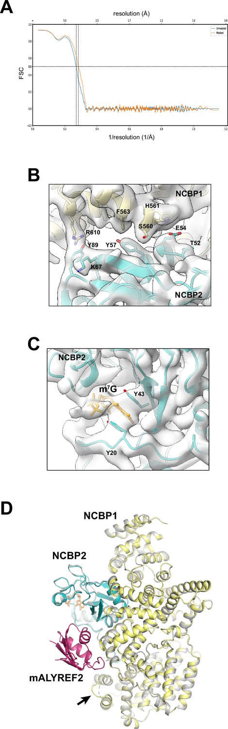
Structural model of the CBC-mALYREF2 complex.
(A) Map versus model Fourier shell correlation (FSC) curves with or without mask calculated using Phenix. A resolution of 3.5 Å is estimated at FSC = 0.5. (B, C) Electron density map and structural model of CBC-mALYREF2 at the interface between NCBP1 and NCBP2 (B) and near m7G (C). (D) Comparison of the CBC-mALYREF2 (colored as in Figure 2C) and the CBC (gray) cryo-EM structures. A loop (residues 38–45) of NCBP1, indicated by the arrow, becomes ordered upon mALYREF2 binding.
Figure 3 with 1 supplement
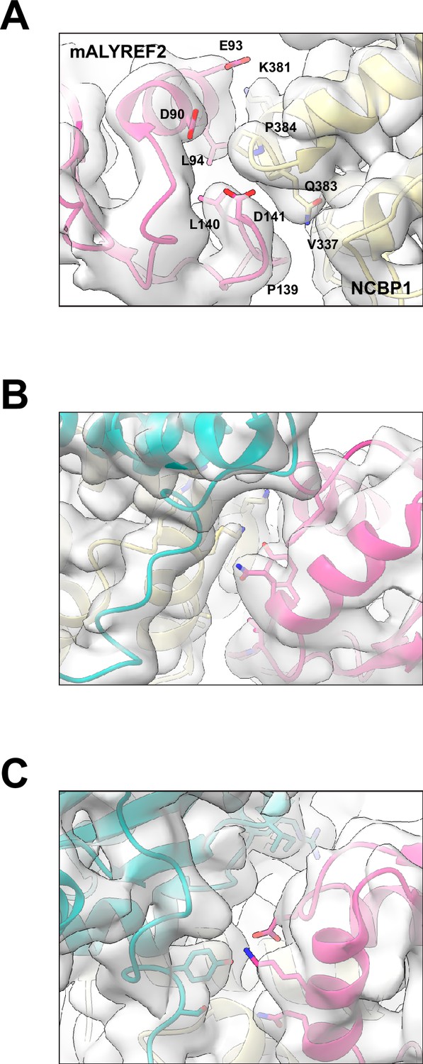
ALYREF binds to both NCBP1 and NCBP2.
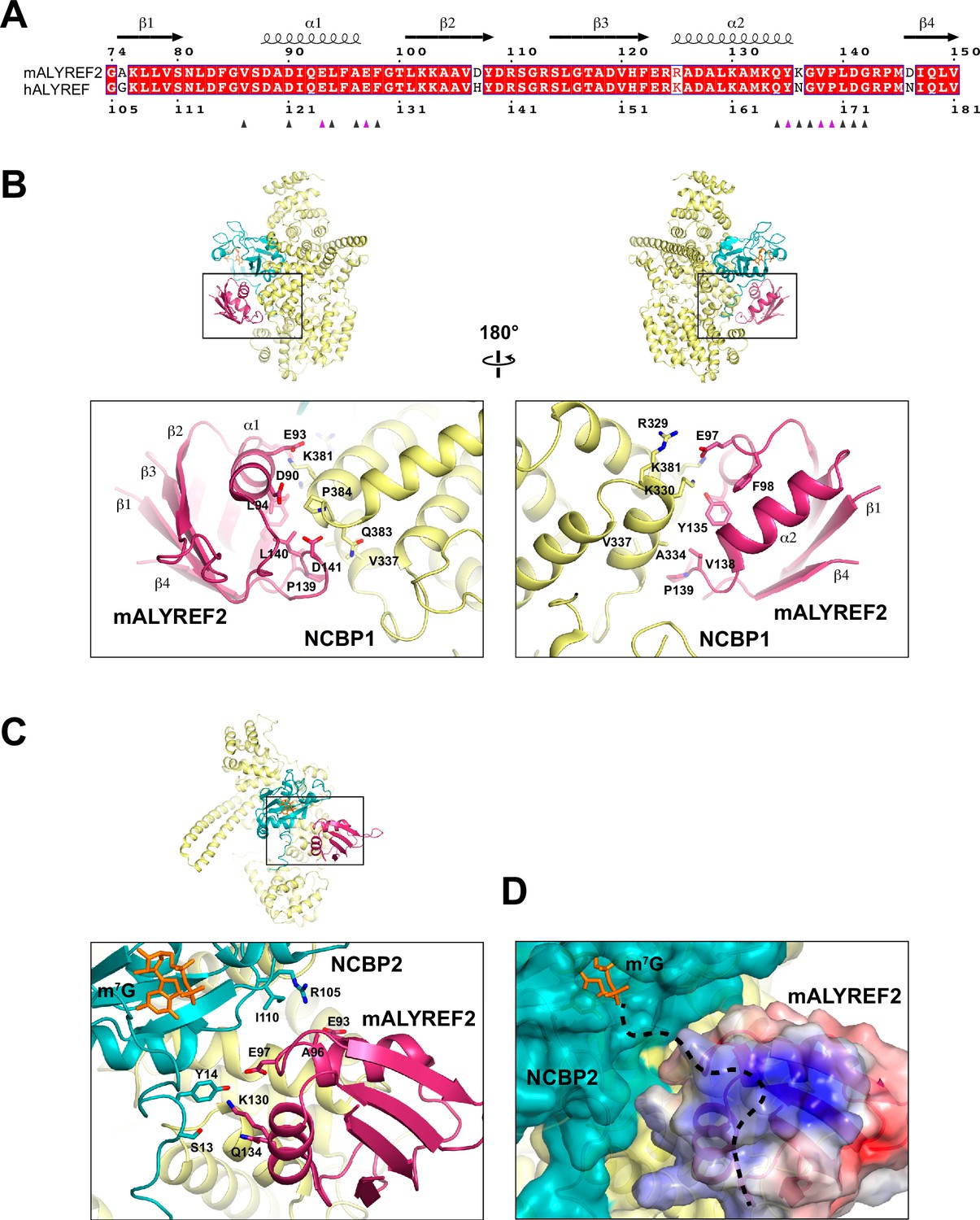
(A) Sequence alignment of the RRM domain of mALYREF2 and ALYREF (Robert and Gouet, 2014). The residues that interface with the CBC are indicated by triangles below the sequence. The triangles colored in purple correspond to residues subjected to mutagenesis in Figure 4. (B) Details of the interaction between NCBP1 and mALYREF2. As a reference, an overall model is shown on the top to indicate the zoomed-in area. (C) Details of the interaction between NCBP2 and mALYREF2. (D) mALYREF2 features a positively charged surface near the cap analog bound to NCBP2. The surface of mALYREF2 is colored according to the electrostatic potential, ranging from red (–5 kBT/e) to blue (+5 kBT/e). Dotted line indicates a putative RNA binding path.
Figure 3—figure supplement 1
Electron density maps at the CBC-mALYREF2 interfaces.
Figure 4
Dissection of the ALYREF and CBC interfaces.
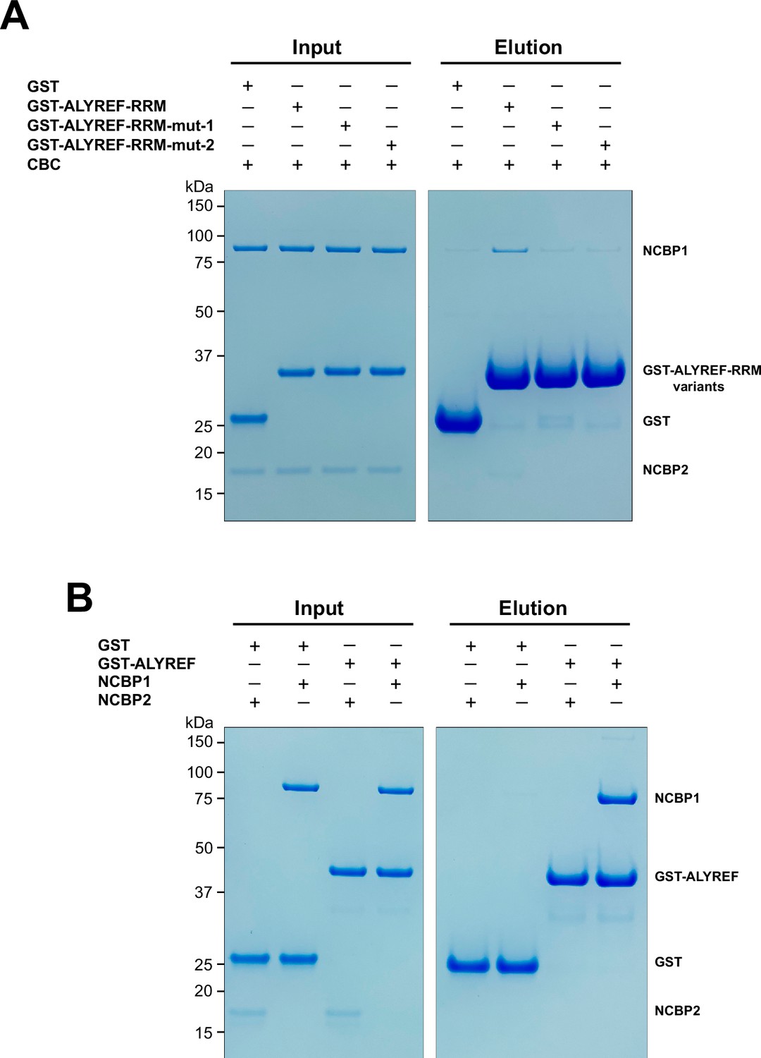
(A) Mutations of key interface residues on ALYREF reduced CBC binding. In vitro GST pull-down assays were performed with purified recombinant human CBC and GST-tagged ALYREF-RRM wild type or mutants (mut-1, Y166R/V169R/P170R; mut-2, E124R/E128R). (B) NCBP1 is sufficient to interact with ALYREF. In vitro GST pull-down assays were performed with purified recombinant GST-tagged ALYREF and individual CBC subunits. Results are representative of three technical repeats.
-
Figure 4—source data 1
Original file for the gels in Figure 4A.
- https://cdn.elifesciences.org/articles/91432/elife-91432-fig4-data1-v1.zip
-
Figure 4—source data 2
Original file for the gels in Figure 4B.
- https://cdn.elifesciences.org/articles/91432/elife-91432-fig4-data2-v1.zip
Figure 5 with 1 supplement
Functional implications for ALYREF and CBC in 5′ cap-dependent mRNP export.
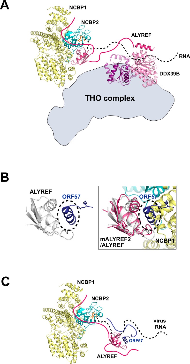
(A) ALYREF links the TREX complex to the CBC. The RRM domain of ALYREF recognizes the CBC at the 5′ end of mRNA. The UBM of ALYREF binds to the DDX39B subunit of TREX; their complex is represented using their yeast orthologs Yra1 and Sub2, respectively (PDB ID 5SUP). DDX39B in turn associates with the THO subcomplex of TREX. (B) HVS ORF57 binds to the RRM domain of ALYREF and interferes with the CBC-ALYREF interaction. (Left) NMR structure of the ALYREF-ORF57 complex (PDB ID 2YKA). (Right) The CBC-ALYREF structure is overlayed with the ALYREF-ORF57 structure. (C) Proposed model of viral mRNA export mediated by herpes viral ORF57 homologs. On viral transcripts, ALYREF associates with the CBC and ORF57 via the WxHD motif and the RRM domain, respectively. Both ALYREF and ORF57 feature RNA binding regions to form contacts with the RNA. ALYREF recruits the other TREX complex components to facilitate the nuclear export of viral mRNAs.
Figure 5—figure supplement 1

HSV-1 ICP27 binds to the RRM domain of ALYREF and interferes with the CBC-ALYREF interaction.
(A) NMR structure of the ALYREF-ICP27 complex (PDB ID 2KT5). (B) The CBC-ALYREF structure is overlayed with the ALYREF-ICP27 structure.
Figure 6
ALYREF and CBC in splicing and export.
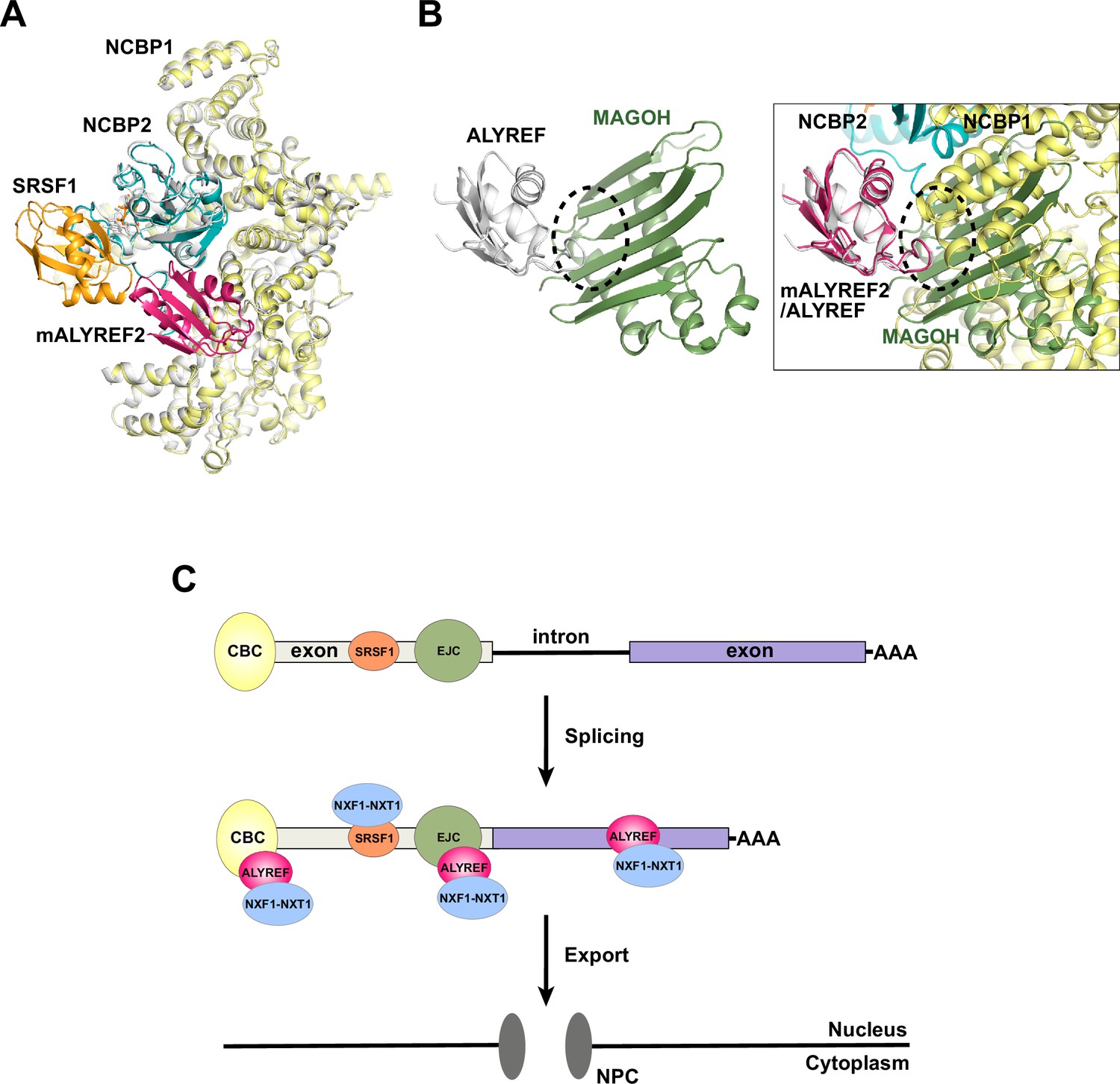
(A) Overlay of the CBC-ALYREF structure with the CBC-SRSF1 structure (PDB ID 7ABG). CBC-ALYREF is colored as in Figure 2B. CBC-SRSF1 is colored with the CBC in white and SRSF1 in orange. (B) ALYREF interaction with the CBC and the EJC is mutually exclusive. (Left) ALYREF binds to the MAGOH subunit of the EJC (PDB ID 7ZNJ). (Right) The CBC-ALYREF structure is overlayed with the ALYREF-MAGOH structure. (C) Proposed model of the mRNP export receptor NXF1-NXT1 recruitment by CBC-ALYREF and other factors during mRNA maturation.
Tables
Table 1
Cryo-EM data collection, refinement, and validation statistics.
| CBC-mALYREF2(EMDB EMD-40739)(PDB 8SRR) | CBC(EMDB EMD-40780)(PDB 8SUY) | |
|---|---|---|
| Data collection and processing | ||
| Microscope/camera | Glacios/Falcon 4i | |
| Voltage (kV) | 200 | |
| Electron exposure (e–/Å2) | 52 | |
| Defocus range (μm) | –1.0 to –2.0 | |
| Pixel size (Å) | 0.732 | |
| Box size (pixels) | 288 | |
| Initial particle images (no.) | 1,625,826 | |
| Final particle images (no.) | 241,915 | 78,039 |
| Map resolution (masked, Å) | 3.22 | 3.38 |
| Fourier shell correlation (FSC) threshold | 0.143 | 0.143 |
| Refinement | ||
| Model resolution (masked, Å) | 3.5 | 3.6 |
| FSC threshold | 0.5 | 0.5 |
| Model composition | ||
| Protein residues | 975 | 895 |
| Ligands | 1 | 1 |
| B factors (Å2) | ||
| Protein | 162.9 | 180.1 |
| Ligand | 157.5 | 163.2 |
| r.m.s. deviations | ||
| Bond lengths (Å) | 0.003 | 0.003 |
| Bond angles (°) | 0.417 | 0.413 |
| Validation | ||
| MolProbity score | 1.34 | 1.48 |
| Clashscore | 6.24 | 7.37 |
| Poor rotamers (%) | 0.7 | 0.9 |
| Ramachandran plot | ||
| Favored (%) | 98.0 | 97.6 |
| Allowed (%) | 2.0 | 2.4 |
| Disallowed (%) | 0.0 | 0.0 |
Additional files
Download links
A two-part list of links to download the article, or parts of the article, in various formats.
Downloads (link to download the article as PDF)
Open citations (links to open the citations from this article in various online reference manager services)
Cite this article (links to download the citations from this article in formats compatible with various reference manager tools)
Cryo-EM structure of the CBC-ALYREF complex
eLife 12:RP91432.
https://doi.org/10.7554/eLife.91432.3


