Multiple origins of dorsal ecdysial sutures in trilobites and their relatives
- Research Center of Paleobiology, Yuxi Normal University, China
- Key Laboratory for Palaeobiology and MEC International Joint Laboratory for Palaeoenvironment, Institute of Palaeontology, Yunnan University, China
- Management Committee of the Chengjiang Fossil Site World Heritage, China
- Museum of Comparative Zoology and Department of Organismic and Evolutionary Biology, Harvard University, United States
- Centre for Ecology and Conservation, University of Exeter, United Kingdom
- Department of Zoology, University of Cambridge, Downing Street, United Kingdom
- Institute of Geology, Chinese Academy of Geological Sciences, China
- State Key Laboratory of Palaeobiology and Stratigraphy, Nanjing Institute of Geology and Paleontology, China
Figures
Figure 1
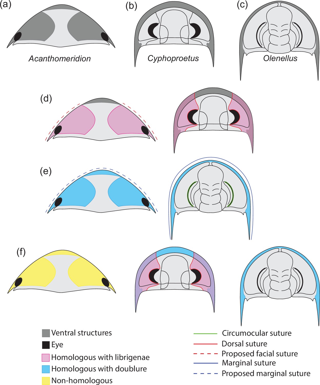
Hypotheses of homology in sutures between Acanthomeridion and trilobites.
(a) Cephalic morphology of Acanthomeridion serratum. (b) Cephalic morphology of a proetid trilobite (modified from Daley and Drage, 2016). (c) Cephalic morphology of a redlichid trilobite (modified from Whittington, 1989). (d) Ventral plates and librigenae as homologous with dorsal suture. (e) Ventral plates and doublure as homologous with marginal suture. (f) Non-homology between ventral plates of Acanthomeridion, librigenae, and doublure.
Figure 2
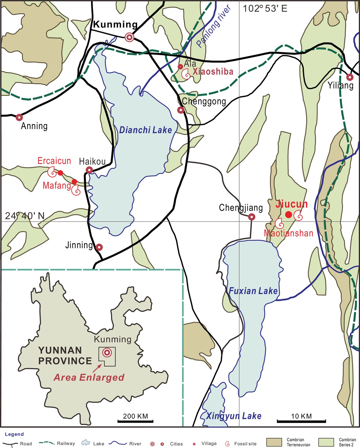
Sites yielding Acanthomeridion from the Cambrian Stage 3 Chengjiang Biota (red circles).
All the specimens of A. serratum used here are collected from Jiucun town, Chengjiang County.
Figure 3 with 6 supplements
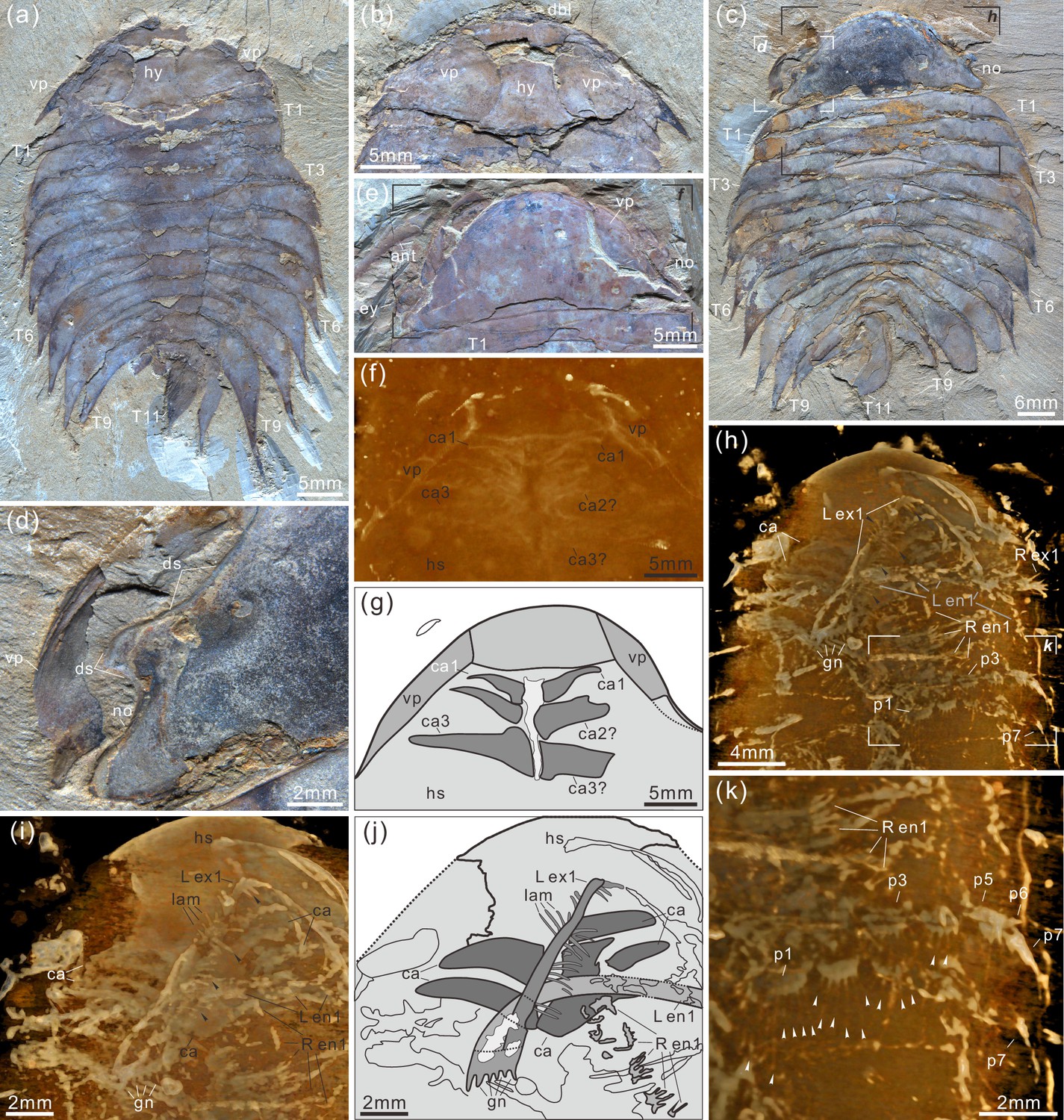
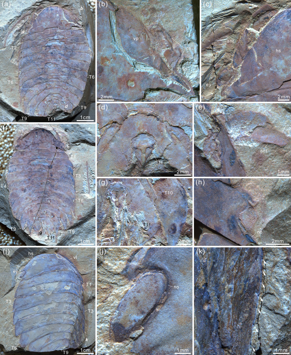
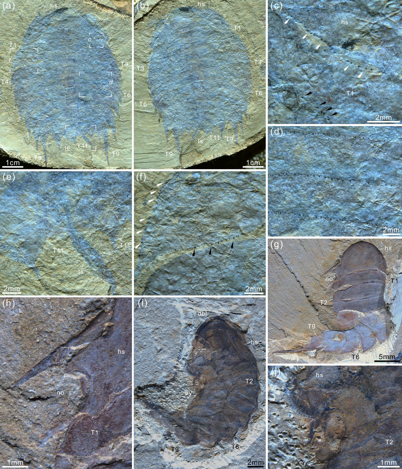
Acanthomeridion serratum from the Cambrian Stage 3 Chengjiang Biota.
(a, b) CJHMD 00,052 a/b, respectively, an individual with hypostome, ventral plates, and 11 thoracic tergites. (c, d) CJHMD 00,053 a, showing ventral plate, dorsal sutures, and 11 thoracic tergites. (e-g) YRCP 0016 a, showing the ventral plates, and three post-antennal limbs. (h-k) Showing the post-antennal appendages under head, gnathobases of trunk limbs, stick-like exopodites with bristles (black arrows), and endopodites with long spines (white arrows). (f) Micro-CT image of YRCP 0016 a; (h, i, k) Micro-CT images of CJHMD 00,053 a. Abbreviations: ant, antenna; can, post-antennal appendage n beneath head; dbl, doublure; ds, dorsal suture; en, endopodites; ex, exopodites; ey, eye; hs, head shield; hy, hypostome; L, left; lam, lamellae; no, notch; pn, podomere n; R, right; Tn, tergite n; ts, terminal spine; vp, ventral plate.
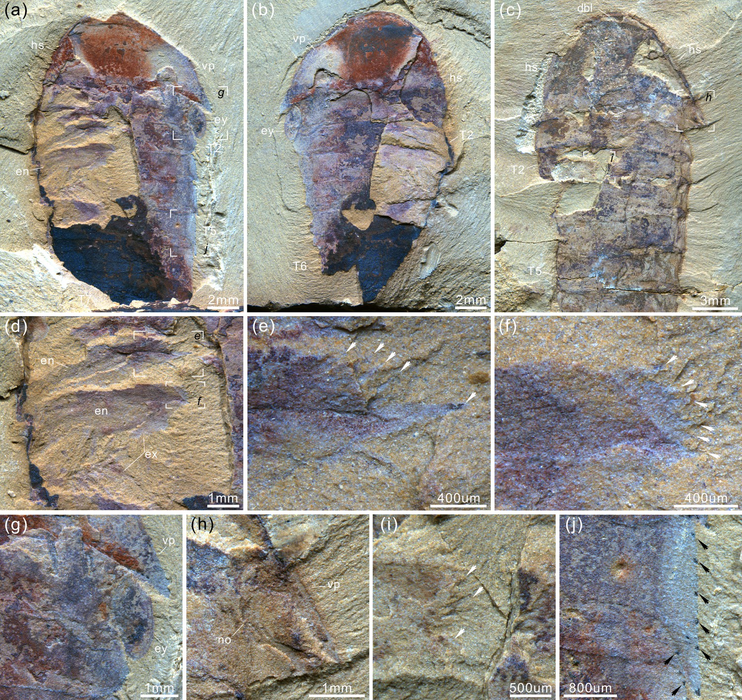
Figure 3—figure supplement 1
Acanthomeridion serratum from the Cambrian Stage 3 Chengjiang Biota.
(a) CJHMD 00052b articulated specimen preserved hypostome, librigena-like ventral plates, and 11 tergites with pleural spines. (b) CJHMD 00053b complete specimen with head, ventral plate, and 11 tergites. (c, d) Marginal spines (arrows) on the tergites. Abbreviations: ant, antenna; ca, post-antennal appendage beneath head; dbl, doublure; ds, dorsal suture; en, endopod; ex, exopod; ey, eye; es, eyestalk; gut, digestive tract; hs, head shield; hy, hypostome; lam, lamellae; no, notch; pn, podomere n; R, right; Tn, tergite n; ts, terminal spine; vp, ventral plate.
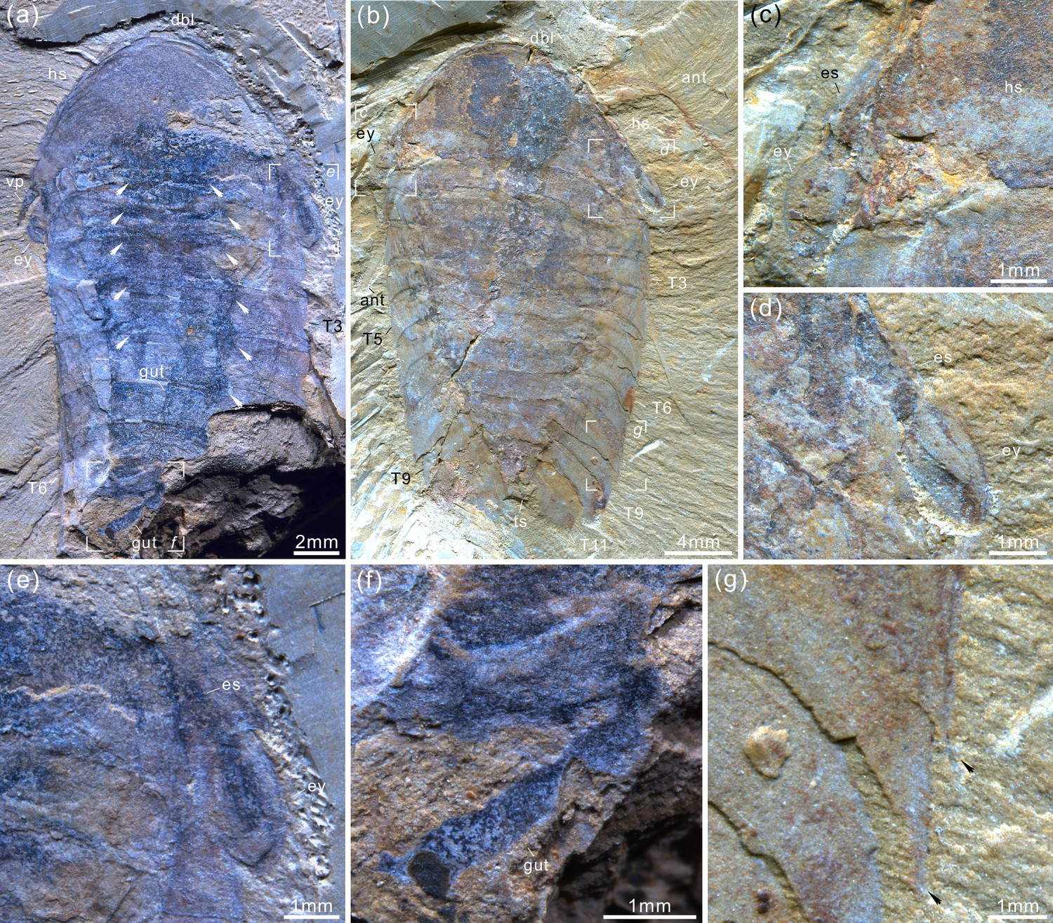
Figure 3—figure supplement 2
Acanthomeridion serratum from the Cambrian Stage 3 Chengjiang Biota.
(a) YRCP 0016 a, articulated individual with antenna, ventral plates, and 11 tergites. (b) Details of ventral plate and notch. (c) Close-up of antenna and stalk eye. (d, g) Terminal spine and its joint. (e) attachment of antenna. (f) Counterpart of label (a), YRCP 0016b. (h) Detail of notch. (i-k) YRCP 0017, preserved head, compound eye, nine tergites, and marginal spines on tergite. For abbreviations, see Figure 3—figure supplement 1.
Figure 3—figure supplement 3
Acanthomeridion serratum from the Cambrian Stage 3 Chengjiang Biota.
(a) CJHMD 00,056 a, articulated specimen preserved ventral plates, gnathobases of limbs, endopods, and exopod. (b) CJHMD 00056b. (c) CJHMD 00057, showing the anterior sclerite, head, gnathobases, and five tergites. (d) details of gnathobases, endopods, and exopod. (e, f) Close-up of gnathobases. (g, h) Details of elliptical eye and ventral plate. (i) Close-up of gnathobases. (j) Details of marginal spines on tergite. For abbreviations, see Figure 3—figure supplement 1.
Figure 3—figure supplement 4
Acanthomeridion serratum from the Cambrian Stage 3 Chengjiang Biota.
(a) CJHMD 00058, articulated specimen with head, spine of ventral plate, stalked eyes, gut, midgut diverticulae (white arrows), and six tergites. (b) YRCP 0018, complete individual with long antennae, compound eyes, and 11 tergites. (c–e) Details of stalked eyes and the eyestalks. (f) Close-up of the gut. (g) Pleural spines of T7 and T8. For abbreviations, see Figure 3—figure supplement 1.
Figure 3—figure supplement 5
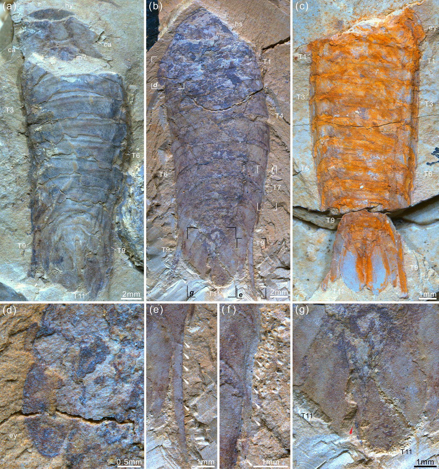
Acanthomeridion serratum from the Cambrian Stage 3 Chengjiang Biota.
(a) CJHMD 00059, articulated individual with hypostome, cephalic appendages, possible mouth, and 11 tergites. (b) YRCP 0019, complete individual with stalked eyes, long spines on T9, and 11 tergites. (c) YRCP 0020, articulated specimen with compound eyes and 11 tergites. (d) Elliptical eye. (e) Marginal spines (white arrows) on the long pleural spine of T9. (f) Marginal spines (white arrows) on the pleural spine of T7. (g) Paddle-like structure (red arrow). For abbreviations, see Figure 3—figure supplement 1.
Figure 3—figure supplement 6
Acanthomeridion serratum from the Cambrian Stage 3 Chengjiang Biota.
(a) CJHMD 00,060 a, complete individual with head, 11 tergites, well-developed pleural spines, and terminal spine. (b) Counterpart of CJHMD 00060b. (c) Marginal spines on the posterior margin of head (white arrows) and T1 (black arrows). (d) Marginal spines on the posterior margin of tergites. (e) Pleural spines and marginal spines of T11, terminal spine. (f) Marginal spines on the posterior and lateral margins of tergite. (g, h) CJHMD 00061, articulated juvenile individual with head, notch, and nine tergites. (i, j) CJHMD 00062, juvenile individual with stalked eyes. For abbreviations, see Figure 3—figure supplement 1.
Figure 4

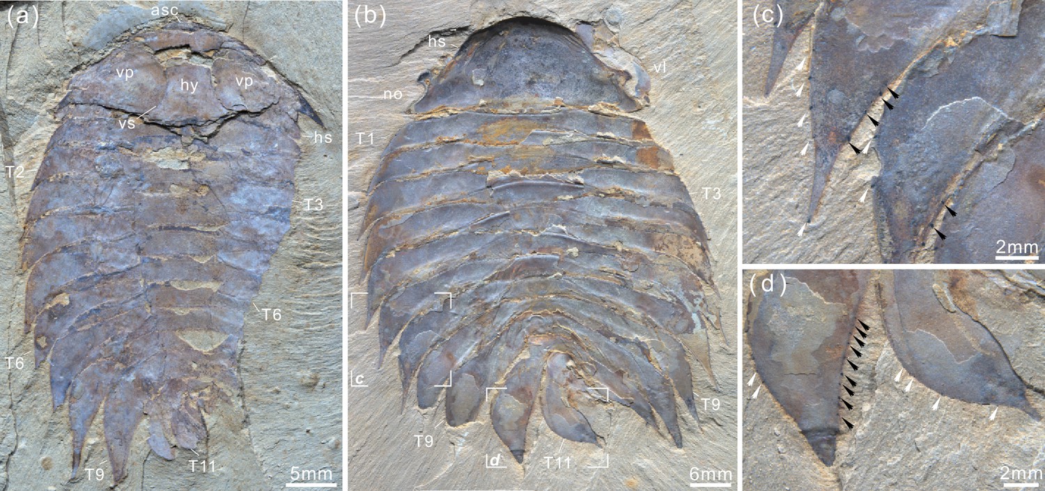
Acanthomeridion serratum from the Cambrian Stage 3 Chengjiang Biota.
(a) CJHMD 00054, complete individual with head, eye, and 11 tergites. (b) Micro-CT image of panel (a) showing the stick-like exopods. Abbreviations: dbl, doublure; ex, exopod; ey, eye; es, eyestalk; hs, head shield; Tn, tergite n.
Figure 5
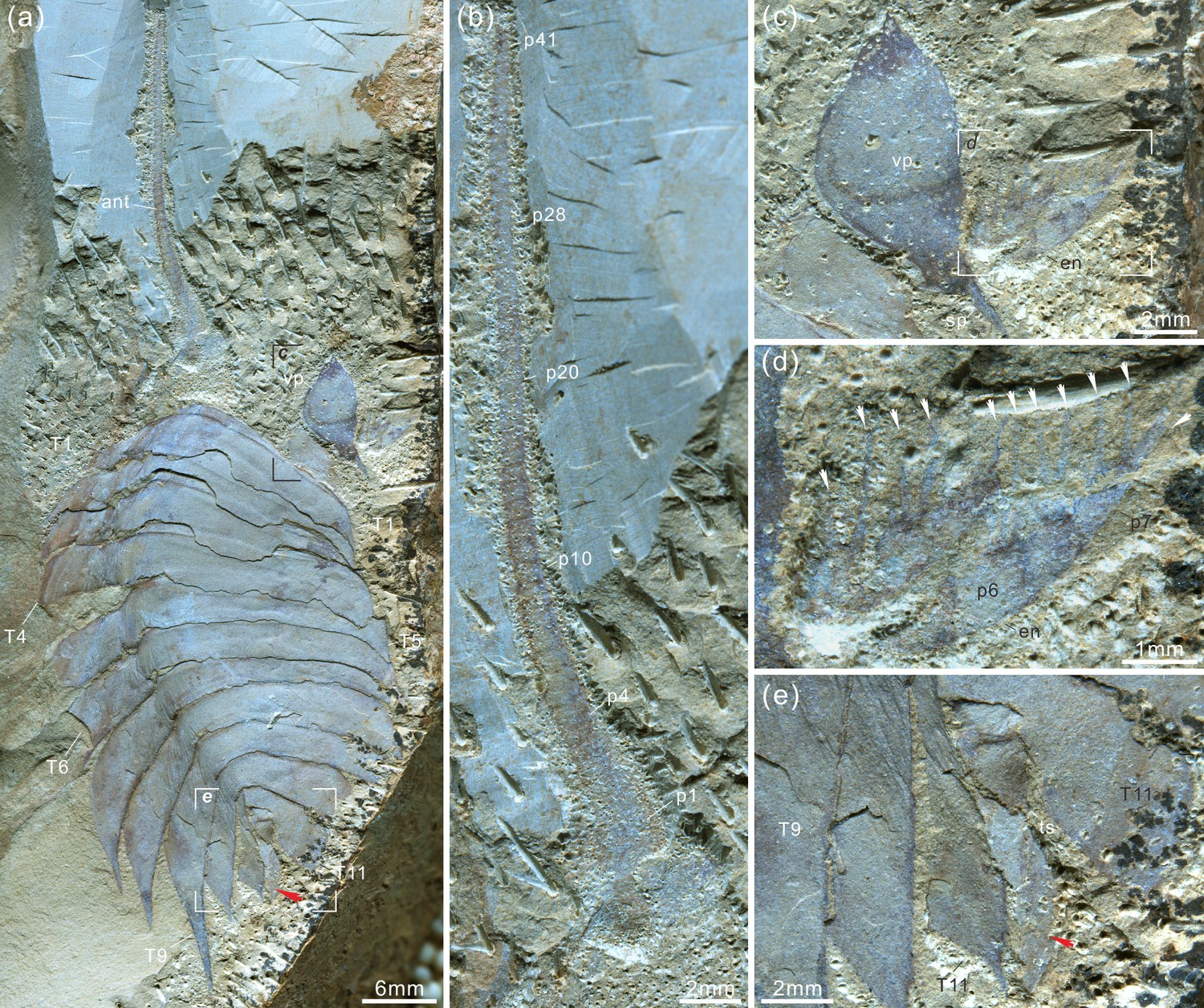
Acanthomeridion serratum from the Cambrian Stage 3 Chengjiang Biota.
(a–e) CJHMD 00055, showing the antenna, ventral plate, endopodites with long spines (arrows in d), 11 tergites, and paddle-like structure (red arrow). (a) Overview of whole specimen. (b) Detail of long left antenna. (c) Close-up of ventral plate. (d) Details of long spines (arrows) of right endopodites. (e) Close-up of paddle-like structure (red arrow). Abbreviations same as Figures 3 and 4.
Figure 6

Ontogenetic series (a–r) of Acanthomeridion and their ventrally curling pleurae (s, t).
(a–r) Showing the individuals from smallest to largest with same scale bar. (s) Lateral view of (o), note the right curling pleurae and left flat pleurae. (t) Lateral view of (f), showing the left curling pleurae.
Figure 7
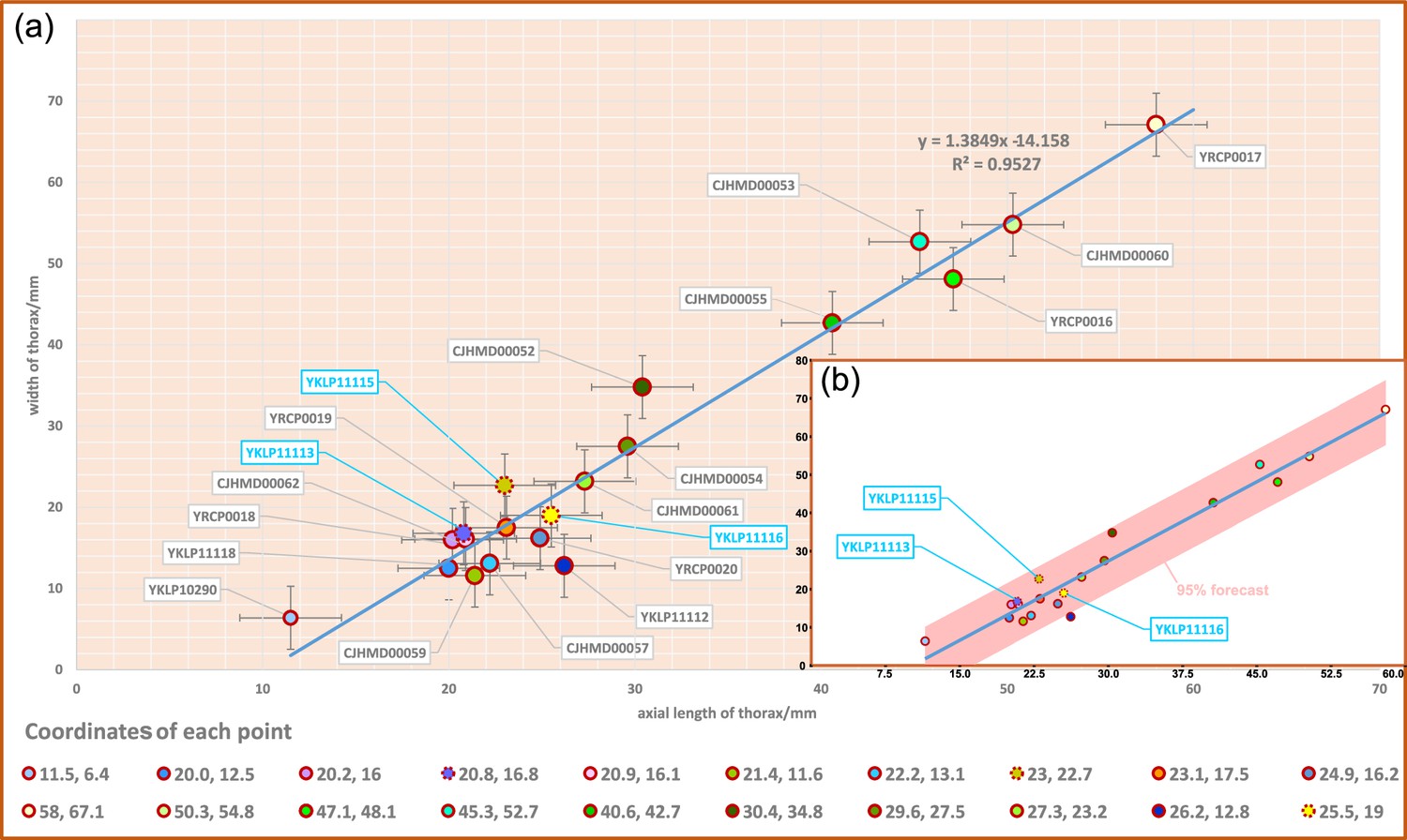
Scatterplot of axial length and width of thorax of Acanthomeridion serratum.
(a) Scatterplot of raw data. (b) Data with a 95% forecast shown in pink. Specimens previously assigned to A. anacanthus indicated by blue lettering.
Figure 8
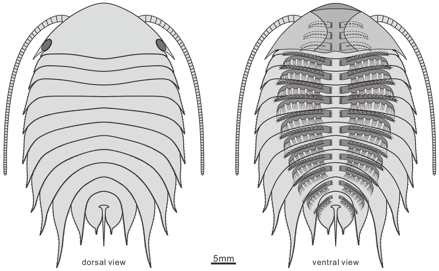
Reconstruction of Acanthomeridion serratum in dorsal and ventral view.
Only the protopodite of the head appendages has been observed. There appear to be three post-antennal limbs with the fourth pair below the cephalic-thoracic boundary.
Figure 9
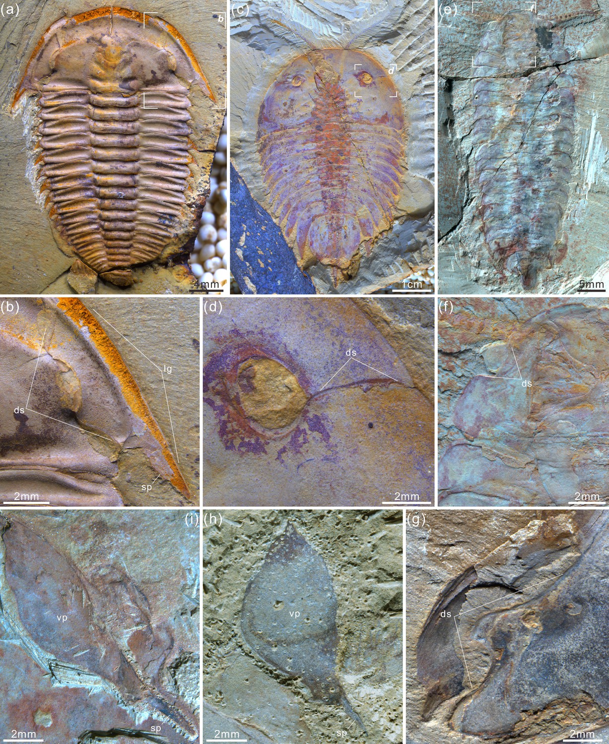
Four representative artiopodans from early Cambrian with their dorsal sutures, free cheeks, and ventral plates.
(a, b) Trilobite Wutingaspis tingi from Chengjiang Biota, note its free cheeks and dorsal or facial sutures. (c, d) Xandarellid artiopodan Xandarella spectaculum from Chengjiang Biota bearing the distinctive dorsal sutures. (e, f) Holotype of the protosuturan artiopodan Zhiwenia coronata from Xiaoshiba Biota developing dorsal sutures. (g) The left dorsal suture of Acanthomeridion serratum from Chengjiang Biota, showing the morphological and positional similarities to that of W. tingi (a, b), X. spectaculum (c, d), and Z. coronata (e, f). (h, i) Right ventral plates of A. serratum from Chengjiang Biota bearing a terminal spine, which is similar to free cheek of trilobite like W. tingi (a, b).
Figure 10 with 3 supplements
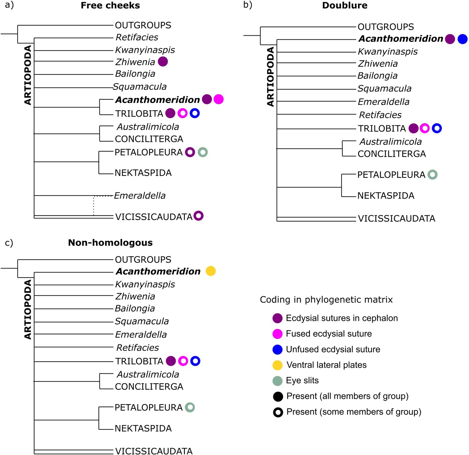
Simplified results of phylogenetic analyses.
Comparison of results from matrices with different coding strategies, with coding of key characters for terminals within the analysis indicated by colored circles. (a) Ventral plates are considered homologous to free cheeks. (b) Ventral plates are considered homologous to trilobite doublure. (c) Ventral plates are not considered homologous to any artiopodan character.
Figure 10—figure supplement 1
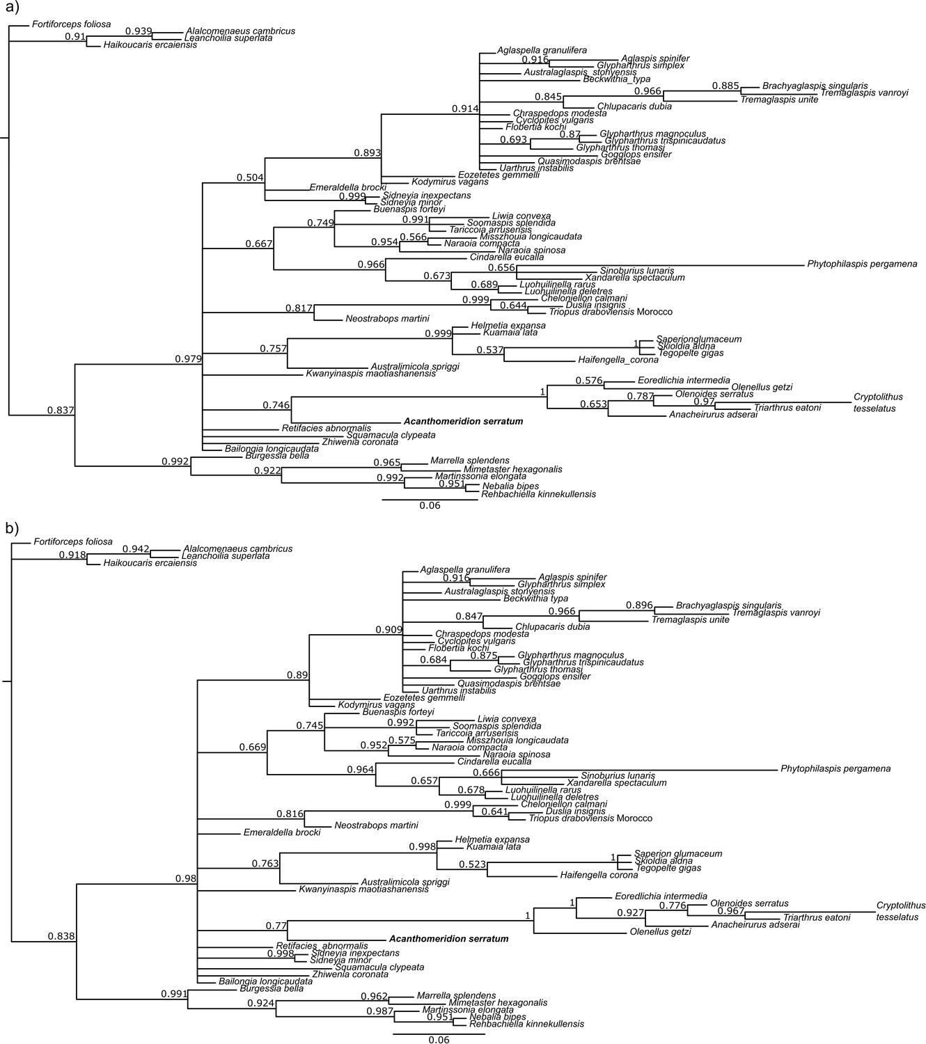
Results of phylogenetic analyses using Bayesian inference, with the ventral plates of Acanthomeridion treated as homologous to the librigenae of trilobites.
(a) Unconstrained analysis. (b) Clade comprising Anacheirurus adserai, Cryptolithus tesselatus, Eoredlichia intermedia, Olenoids serratus, Triarthrus eatoni constrained. Values at nodes are posterior probabilities.
Figure 10—figure supplement 2
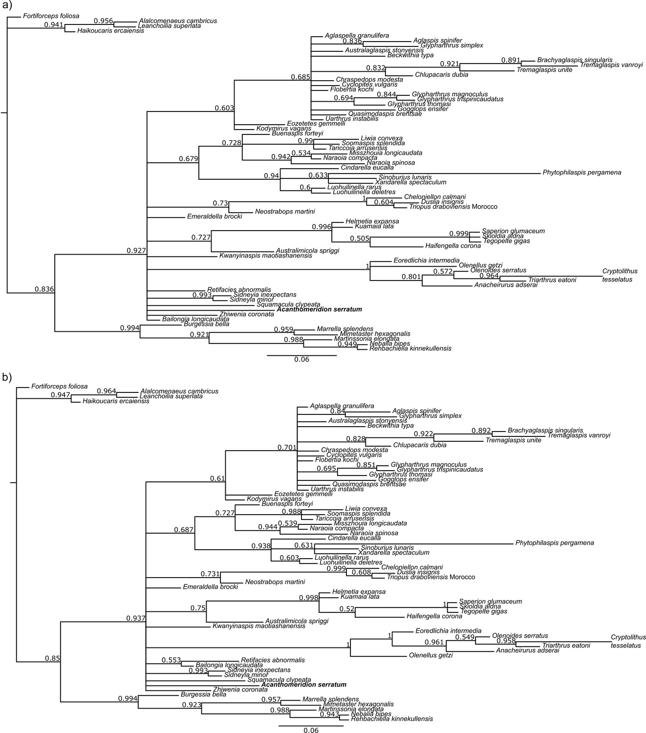
Results of phylogenetic analyses using Bayesian inference, with the ventral plates of Acanthomeridion treated as homologous to cephalic doublure of trilobites.
(a) Unconstrained analysis. (b) Clade comprising Anacheirurus adserai, Cryptolithus tesselatus, Eoredlichia intermedia, Olenoids serratus, Triarthrus eatoni constrained. Values at nodes are posterior probabilities.
Figure 10—figure supplement 3
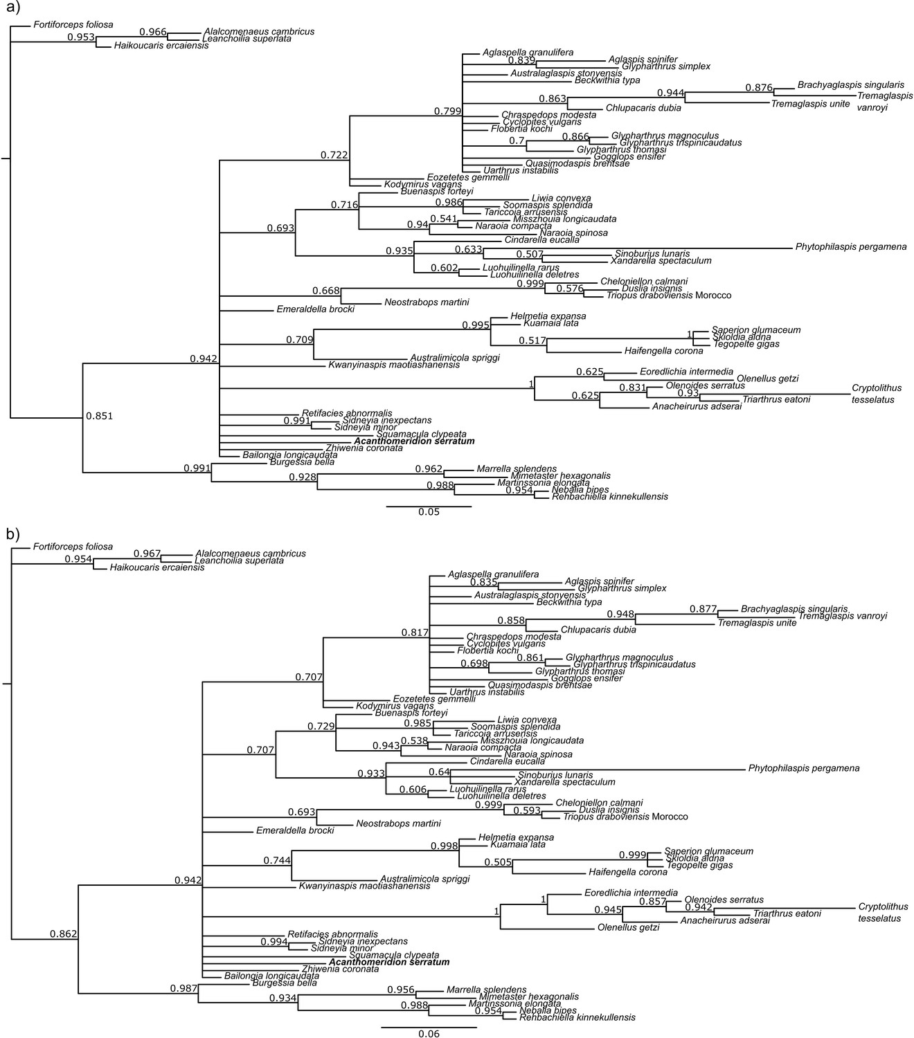
Results of phylogenetic analyses using Bayesian inference, with the ventral plates of Acanthomeridion treated as non homologous to any cephalic feature of trilobites.
(a) Unconstrained analysis. (b) Clade comprising Anacheirurus adserai, Cryptolithus tesselatus, Eoredlichia intermedia, Olenoids serratus, Triarthrus eatoni constrained. Values at nodes are posterior probabilities.
Figure 11
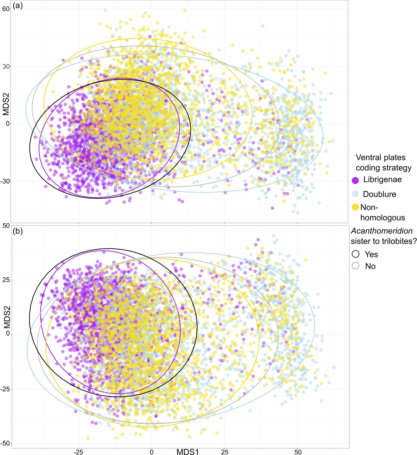
Treespace visualization of Bayesian Inference phylogenetic analyses.
Multidimensional treespace plotted by bipartition relating to coding strategies (color of point). (a) Analyses were unconstrained. (b) Trilobites were constrained so that Olenellus getzi was resolved as the earliest diverging member of the group. Area included within a 95% confidence interval (CI) indicated by points inside the line of different colors. Area where Acanthomeridion is recovered sister to trilobites (95% CI) also shown by area within black line. Where Acanthomeridion is not recovered sister to trilobites (95% CI), shown by area within gray line. Note the overlap in the areas of the black and purple lines.
Tables
Table 1
Number of trees in posterior sample supporting Acanthomeridion as sister to trilobites / number of trees in the posterior sample total.
Results tabulated for each coding strategy, with unconstrained and constrained analyses (constrained analyses where a monophyletic group of all trilobites except for Olenellus getzi was forced, in order to recover trilobite relationships compatible with Paterson et al., 2019).
| Unconstrained | Constrained | |
|---|---|---|
| Homologous to free cheeks | 7832/10504 (75%) | 8083/10504 (77%) |
| Homologous to doublure | 1496/12004 (12%) | 1836/10504 (17%) |
| Not homologous | 761/12004 (6%) | 926/10504 (9%) |
Additional files
Download links
A two-part list of links to download the article, or parts of the article, in various formats.
Downloads (link to download the article as PDF)
Open citations (links to open the citations from this article in various online reference manager services)
Cite this article (links to download the citations from this article in formats compatible with various reference manager tools)
Multiple origins of dorsal ecdysial sutures in trilobites and their relatives
eLife 12:RP93113.
https://doi.org/10.7554/eLife.93113.4


