An atlas of brain-bone sympathetic neural circuits in mice
- Center for Translational Medicine and Pharmacology, Icahn School of Medicine at Mount Sinai, United States
- Department of Medicine and of Pharmacological Sciences, Icahn School of Medicine at Mount Sinai, United States
- Department of Psychiatry, Icahn School of Medicine at Mount Sinai, United States
Figures
Figure 1
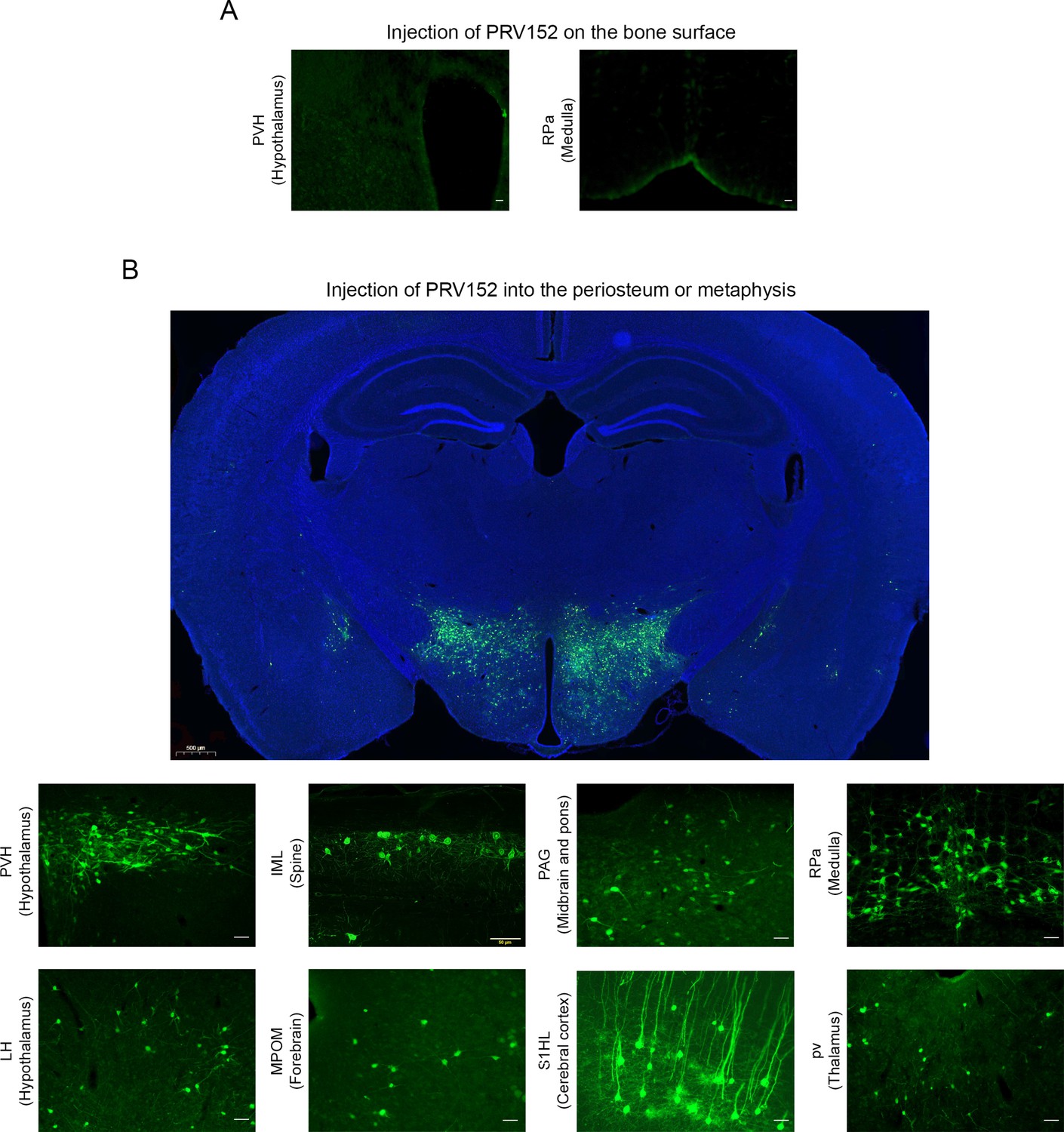
PRV152 transneuronal viral tract tracing.
(A) As a control for viral injection, no expresses enhanced green fluorescent protein (EGFP) signal was detected in the paraventricular nucleus (PVH), known to possess main sympathetic pre-autonomic neurons, and the RPa, when PRV152 was placed on the bone surface. (B) By contrast, PRV152 injections into the periosteum or metaphyseal bone resulted in positive EGFP immunoreactivity in the PVH. In addition, we found PRV152-infected neurons in the intermediolateral cell column (IML) of the spinal cord at T13-L2 levels, suggesting specific bone-sympathetic nervous system (SNS) ganglia-IML-brain route of infection which are in concordance with our previous findings where PRV152 individually infected the classic SNS spinal cord neurons. Also shown are representative microphotographs illustrating PRV152 immunolabeling in the PAG (midbrain and pons), RPa (medulla), LH (hypothalamus), MPOM (forebrain), S1HL (cerebral cortex), and pv (thalamus). PVH, paraventricular hypothalamic nucleus; PAG, periaqueductal gray; RPa, raphe pallidus; LH, lateral hypothalamus; MPOM, medial preoptic nucleus, medial part; S1HL, primary somatosensory cortex, hindlimb region; pv, periventricular fiber system. Scale bar = 50 µm. Also shown is a representative low-magnification image at the hypothalamus neuroanatomical level (scale bar = 500 µm).
Figure 2
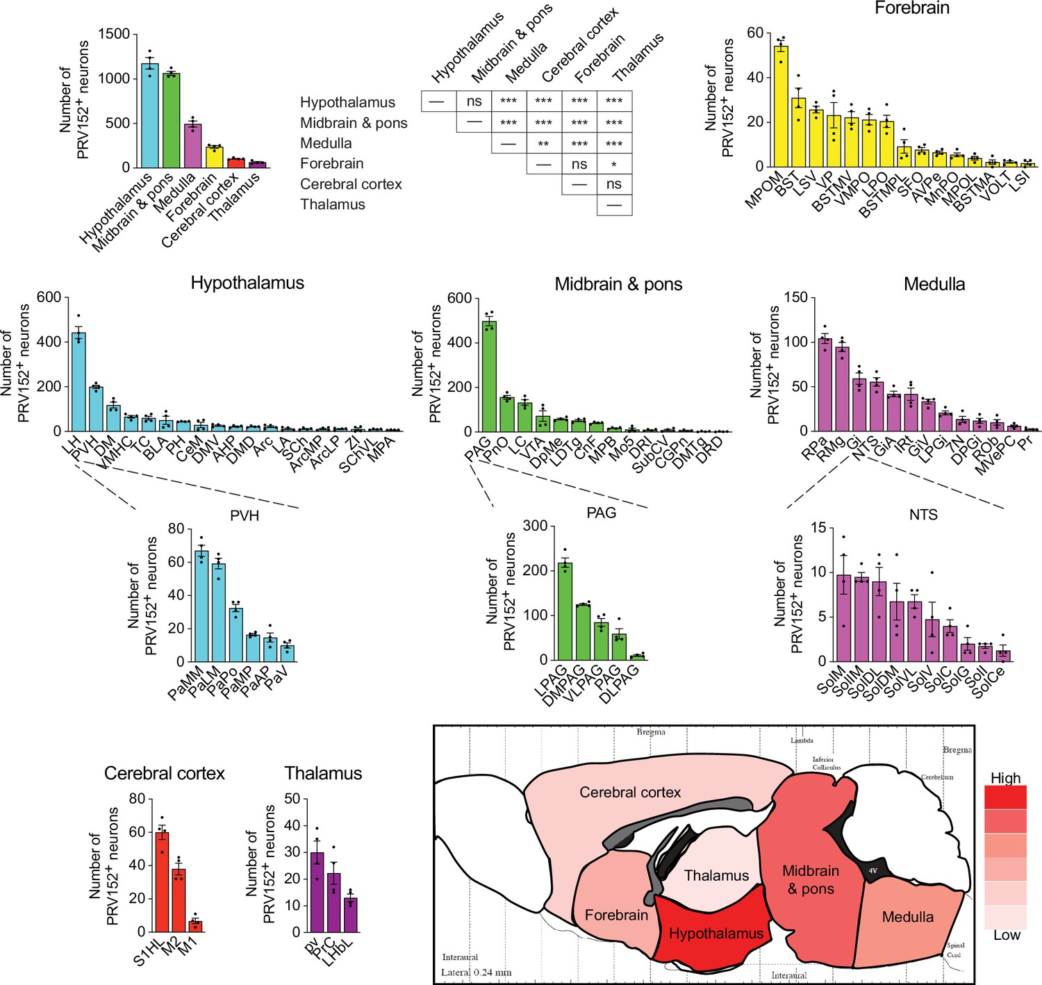
PRV152 immunolabeling in brain regions, sub-regions, and nuclei.
Numbers, and heat map representation of PRV152-labeled neurons in brain regions, namely, hypothalamus, midbrain and pons, medulla, forebrain, cerebral cortex, and thalamus, as well as their sub-regions and nuclei, following viral injections into bone. n=4. Statistics: Mean ± SEM, two-tailed Student’s t-test, *p<0.05, **p<0.01, ***p<0.001, ns (no significance).
-
Figure 2—source data 1
Contains the numerical data used to generate the figures.
- https://cdn.elifesciences.org/articles/95727/elife-95727-fig2-data1-v2.xlsx
Figure 3
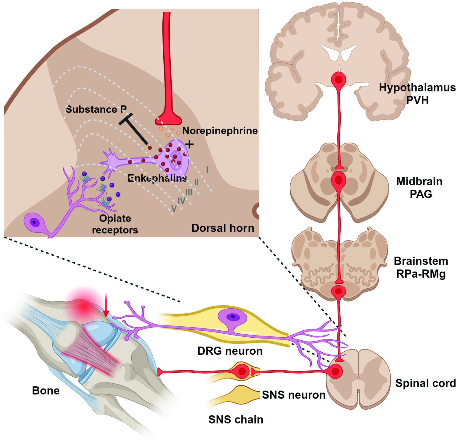
Diagrammatic outline of the sympathetic nervous system (SNS) brain–bone neuroaxis relevant to pain.
The central SNS brain-bone circuit starts in the hypothalamic paraventricular nucleus (PVH) known to home SNS pre-autonomic neurons projecting to the SNS neurons of the periaqueductal gray (PAG) in the midbrain. From the PAG the SNS outflow is further relayed to the raphe pallidus-raphe magnus (RPa-RMg) neurons that are terminated in the dorsal horn of spinal gray matter, where they regulate the release of enkephalins that inhibit pain sensation by attenuating substance P (SP) release. In turn, opiates produce antinociception via the µ-opiate receptors, in part, through modulation of responses to SP. Neurons in the RMg are involved in the central modulation of noxious stimuli, therefore, the RMg-PAG could be the part of the ascending hierarchical circuit relating to the perception of bone pain.
Additional files
Download links
A two-part list of links to download the article, or parts of the article, in various formats.
Downloads (link to download the article as PDF)
Open citations (links to open the citations from this article in various online reference manager services)
Cite this article (links to download the citations from this article in formats compatible with various reference manager tools)
An atlas of brain-bone sympathetic neural circuits in mice
eLife 13:e95727.
https://doi.org/10.7554/eLife.95727


