PPI-hotspotID for detecting protein–protein interaction hot spots from the free protein structure
- Institute of Biomedical Sciences, Academia Sinica, Taiwan
Figures
Figure 1
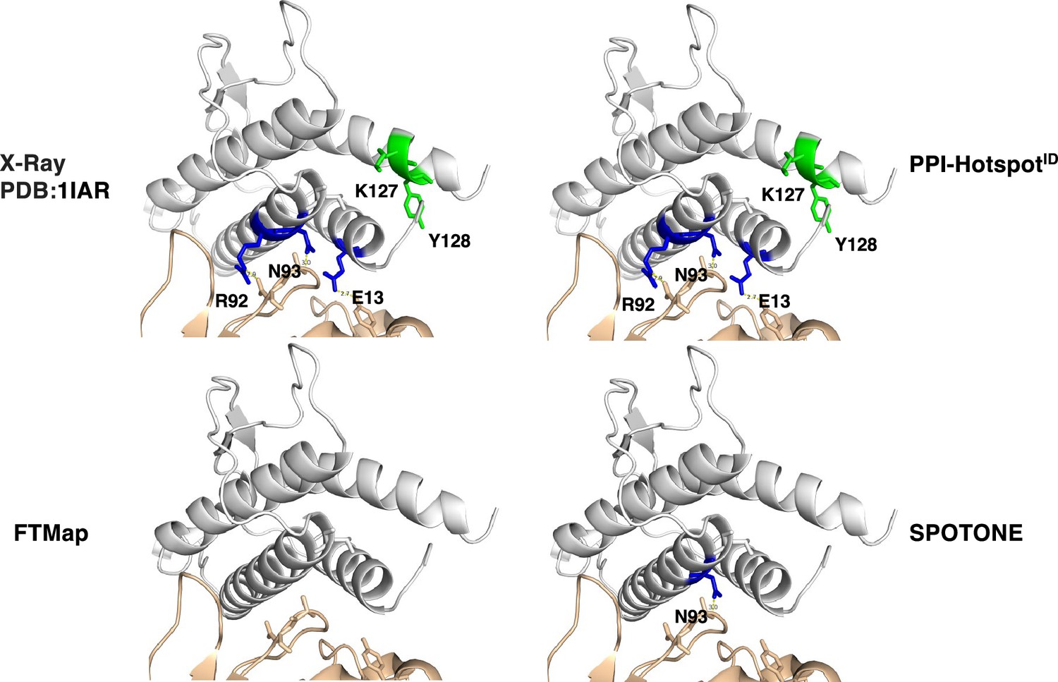
Interface vs. noninterface PPI-hot spots.
(Top left) The X-ray structure (PDB: 1IAR) of interleukin-4 (gray) in complex with interleukin-4 receptor subunit alpha (wheat) with five PPI-hot spots; interface PPI-hot spots (E13, R92, and N93) are in blue and the noninterface ones (K127 and Y128) are in green. The PPI-hot spot numberings are based on the interleukin-4 free structure (PDB: 1BBN). The correct predictions of PPI-hotspotID (top right), FTMap (bottom left), and SPOTONE (bottom right) are mapped to the corresponding residues of the complex structure.
Figure 2 with 1 supplement
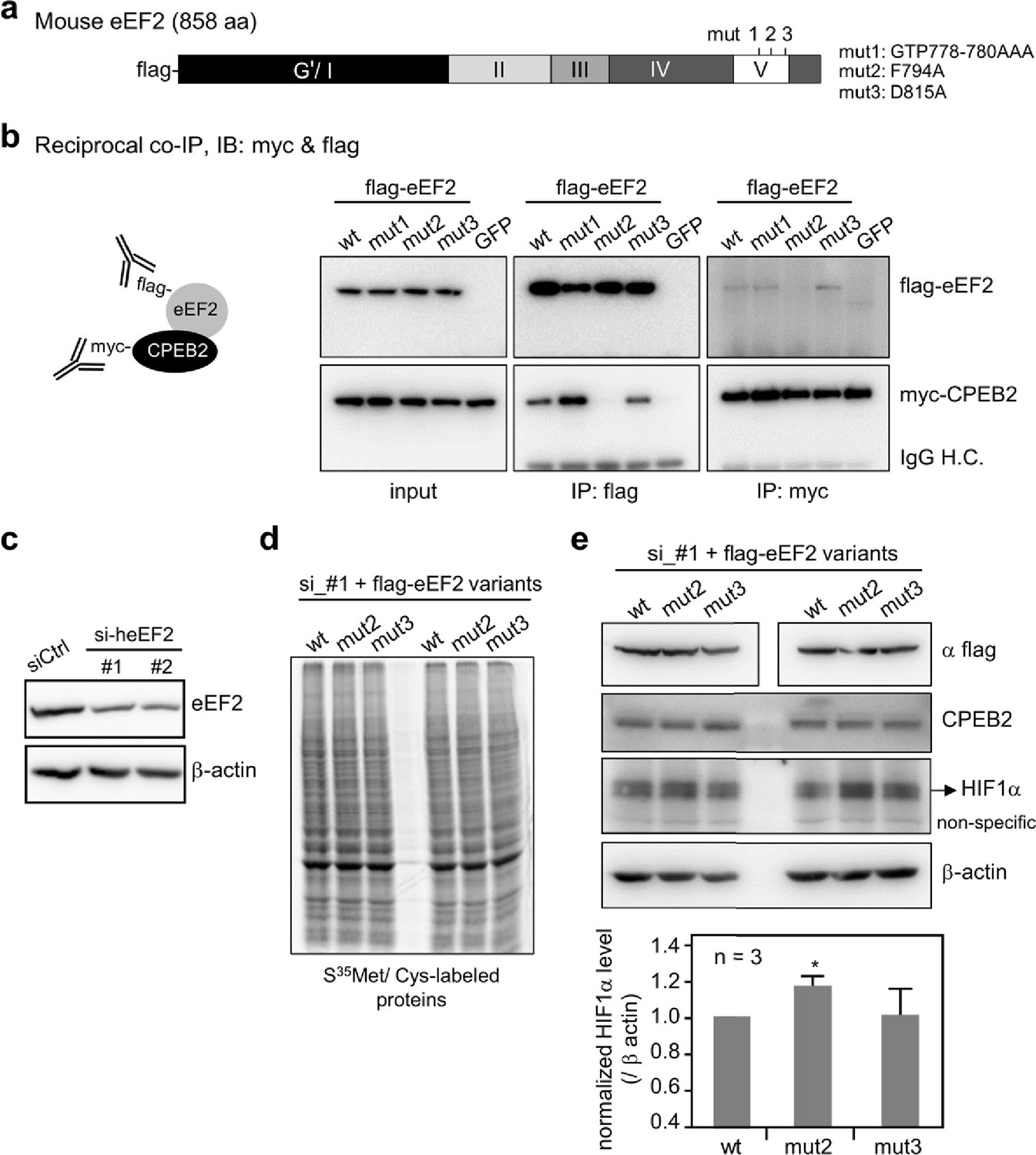
Evaluation of the predicted CPEB2-interacting amino acid residues in eEF2.
(a) Salient features of mouse eEF2, showing the various domains and the mutated amino acids in domain V. mut 1, G778A, T779A, and P780A; mut 2, F794A; mut 3, D815A. (b) Reciprocal co-immunoprecipitation (co-IP). The 293T cells expressing myc-CPEB2 along with wt or mutant flag-eEF2 or control GFP were harvested and then precipitated with flag or myc IgG. The precipitated substances were used for western blotting with myc and flag antibodies. IP, immunoprecipitation; IB, immunoblotting; IgG H.C., IgG heavy chain. (c) HeLa cells transfected with the plasmid expressing shRNA against human eEF2 (siheEF2) were harvested after 4-day puromycin selection for western blotting. HeLa cells transfected with the eEF2 knockdown plasmid along with flag-tagged wt or mutant mouse eEF2 after 4-day selection with puromycin and G418 were used for (d) S35 -met/cys-labeling of synthesized proteins or (e) western blotting with the denoted antibodies. The normalized HIF-1α protein level (HIF-1α/β-actin signal) was calculated and expressed as mean ± SEM from three independent experiments. Two-tailed Student’s t-test, *<0.05.
-
Figure 2—source data 1
Containing uncropped images of the membranes for Figure 2b, c and e and phosphoimager file for Figure 2d.
- https://cdn.elifesciences.org/articles/96643/elife-96643-fig2-data1-v1.docx
-
Figure 2—source data 2
Containing the original files of the full raw unedited blots.
- https://cdn.elifesciences.org/articles/96643/elife-96643-fig2-data2-v1.zip
-
Figure 2—source data 3
Listing primer sequences.
- https://cdn.elifesciences.org/articles/96643/elife-96643-fig2-data3-v1.docx
Figure 2—figure supplement 1
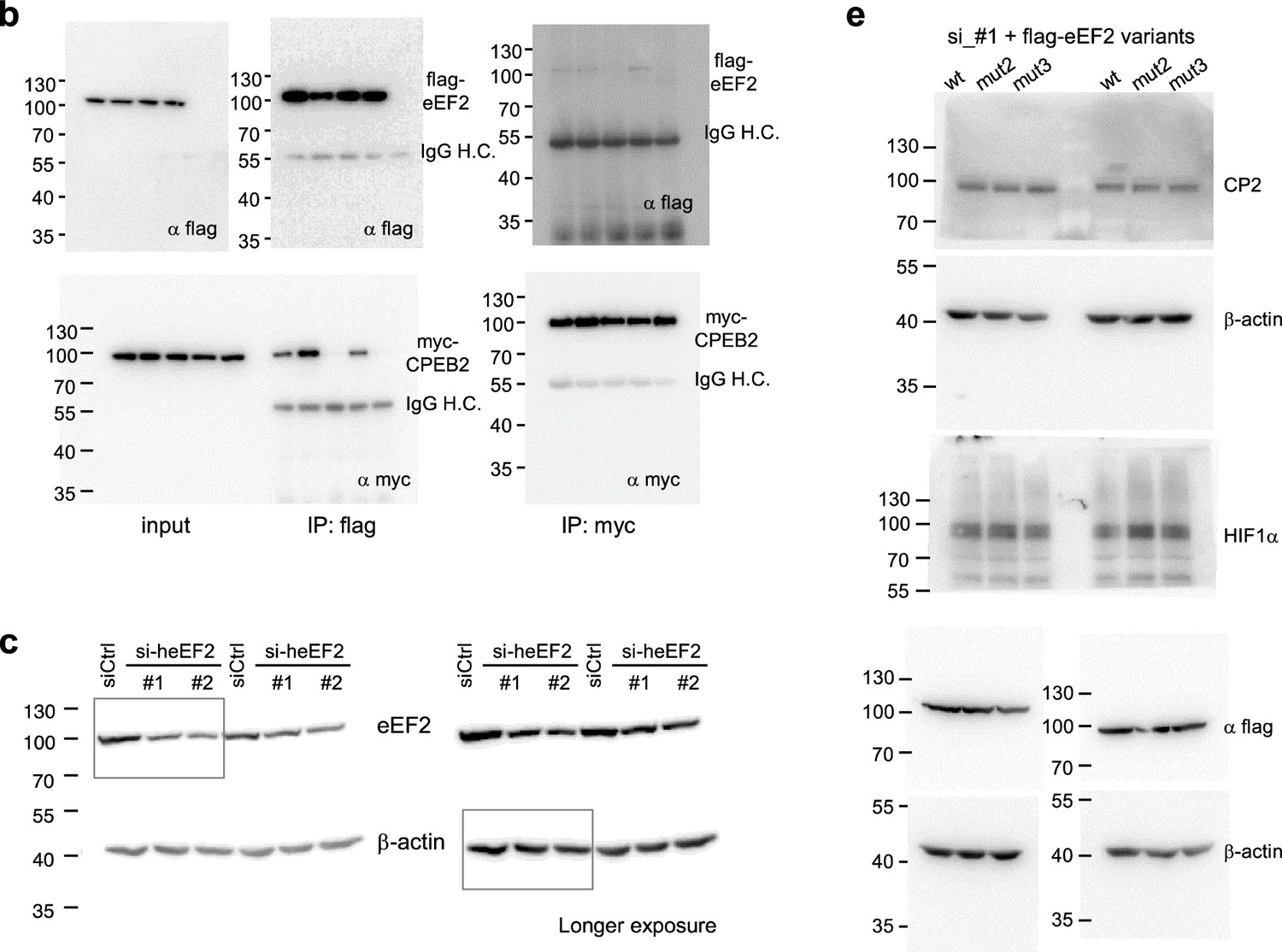
Uncropped immunoblot images.
The uncropped images for Figure 2b, c, and e are shown with indicated molecular weight marker.
Figure 3
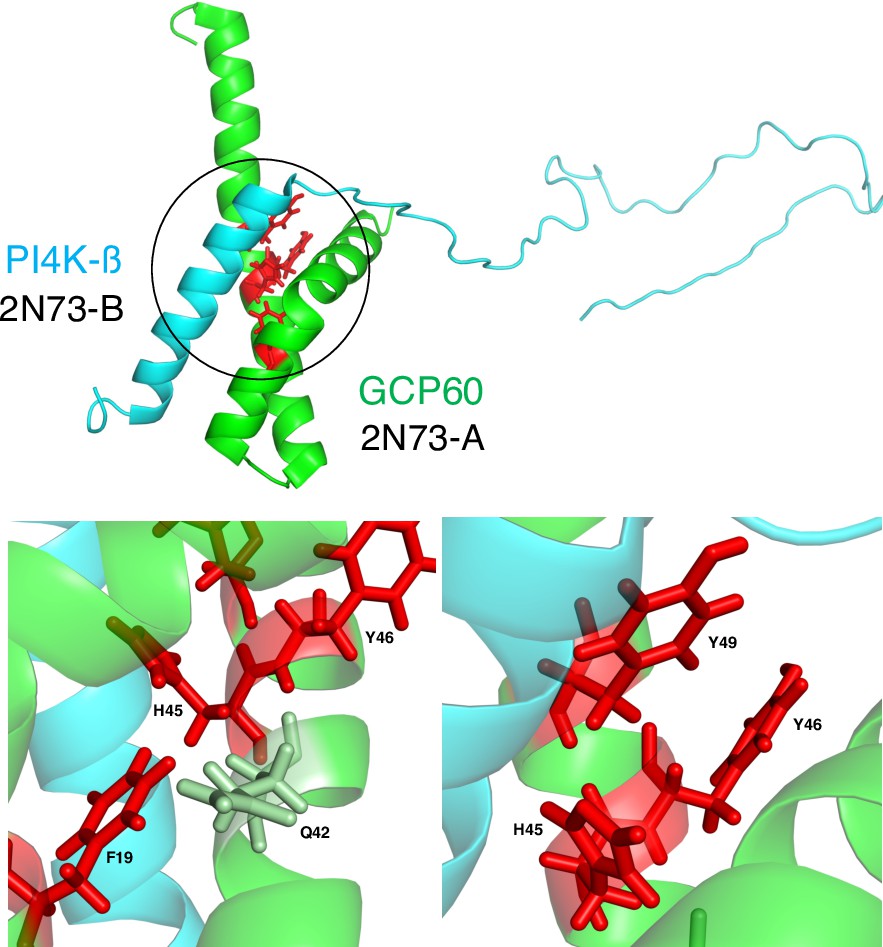
Interface and noninterface PPI-hot spots of Golgi resident protein (GCP60).
(Top) The structure (PDB 2N73) (Wright et al., 2024) of GCP60 (green) in complex with PI4K-β (cyan) with the GCP60–PI4K-β interface encircled. (Bottom) The four experimentally known PPI-hot spots of GCP60 are shown in red. H45 and Y49 form hydrogen bonds across the interface with PI4K-β. Although F19 and Y46 do not directly contact PI4K-β, F19 is in van der Waals (vdW) contact with Q42, which in turn forms vdW contacts with H45, whereas Y46 is in vdW contact with both H45 and Y49.
Figure 4
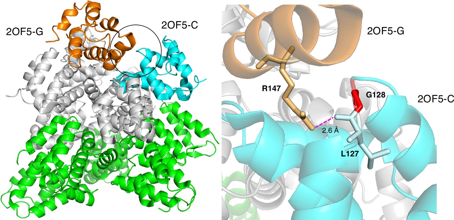
Based on the free CRADD X-ray structure (PDB 2O71-A), PPI-hotspotID predicted G128 as a PPI-hot spot for CRADD–CRADD interactions.
(Left) The structure (PDB 2OF5) (Park et al., 2007) of seven CRADD proteins in complex with fuve PIDD proteins. The circle shows the CRADD–CRADD interface between chains C (cyan) and G (orange), whereas the other five CRADD chains are in gray, and the five PIDD proteins are in green. (Right) G128 (red) in CRADD (chain C) participates indirectly in CRADD–CRADD interactions via a backbone – side chain hydrogen bond between its neighbor, L127, and R147 in another CRADD (chain G).
Tables
Table 1
Performance of the PPI-hotspotID vs. FTMap and SPOTONE.
| Method | PPI-hotspotID | FTMap | SPOTONE | |
|---|---|---|---|---|
| TP | 278 | 30 | 40 |  |
| FN | 136 | 384 | 374 | |
| TN | 417 | 487 | 481 | |
| FP | 87 | 17 | 23 | |
| Sensitivity | 0.67 | 0.07 | 0.10 | |
| Precision | 0.76 | 0.64 | 0.64 | |
| F1 | 0.71 (0.66)* | 0.13 | 0.17 | |
| Specificity | 0.83 | 0.97 | 0.95 |
-
Each method was tested using the same dataset comprising 414 experimentally known PPI-hot spots (TP + FN) and 504 PPI-nonhot spots (TN + FP).
-
TP = true positive; FP = false positive; TN = true negative; FN = false negative.
-
*
The F1 score in parentheses corresponds to the validation F1 score.
Table 2
Performance of AlphaFold-Multimer, PPI-hotspotID, and their combination for 48 ‘unsolved’ complex structures.
| Method | AlphaFold2-Multimer | PPI-hotspotID | AlphaFold2-Multimer+PPI-hotspotID |
|---|---|---|---|
| TP | 37 | 52 | 63 |
| FN | 53 | 38 | 27 |
| TN | 35 | 29 | 24 |
| FP | 10 | 16 | 21 |
| Sensitivity | 0.41 | 0.58 | 0.70 |
| Precision | 0.79 | 0.77 | 0.75 |
| F1 | 0.54 | 0.66* | 0.72 |
| Specificity | 0.78 | 0.64 | 0.53 |
-
Each method was tested using the same dataset comprising 90 experimentally known PPI-hot spots (TP+FN) and 45 PPI-nonhot spots (TN+FP) in 48 protein complexes with no known structures.
-
TP = true positive; FP = false positive; TN = true negative; FN = false negative.
-
*
No validation F1 score is provided since AutoGluon was used to train an ensemble of machine-learning models on a dataset that excludes the 48 ‘unsolved’ complex structures (see text).
Additional files
-
MDAR checklist
- https://cdn.elifesciences.org/articles/96643/elife-96643-mdarchecklist1-v1.pdf
-
Source data 1
Dataset of experimentally confirmed PPI-hot spots and PPI-nonhot spots with free protein structures.
- https://cdn.elifesciences.org/articles/96643/elife-96643-data1-v1.xlsx
-
Source data 2
Dataset of experimentally confirmed PPI-hot spots with both free and bound protein structures.
- https://cdn.elifesciences.org/articles/96643/elife-96643-data2-v1.xlsx
Download links
A two-part list of links to download the article, or parts of the article, in various formats.
Downloads (link to download the article as PDF)
Open citations (links to open the citations from this article in various online reference manager services)
Cite this article (links to download the citations from this article in formats compatible with various reference manager tools)
PPI-hotspotID for detecting protein–protein interaction hot spots from the free protein structure
eLife 13:RP96643.
https://doi.org/10.7554/eLife.96643.3


