Cyclin-dependent kinase 5 (Cdk5) activity is modulated by light and gates rapid phase shifts of the circadian clock
- Department of Biology, University of Fribourg, Switzerland
- Department of Endocrinology, Metabolism, and Cardiovascular System, Section of Medicine, University of Fribourg, Switzerland
- Zentrum für Experimentelle Neurologie, Department of Neurology, Inselspital, Bern University Hospital, University of Bern, Switzerland
- Department of Biomedical Research, University of Bern, Switzerland
Figures
Figure 1 with 1 supplement

Knock-down of Cdk5 in the suprachiasmatic nucleus (SCN) shortens period and reduces phase delays but not phase advances.
(a) Examples of double-plotted wheel-running actograms of control (scr) and Cdk5 knock-down (shCdk5) male mice. Animals were kept under a 12 hr light/12 hr dark cycle (white and gray areas, respectively) (LD). After 8–10 days they received a 15 min light pulse at the indicated zeitgeber times (ZT) (yellow stars). After the light pulse animals were released into constant darkness (DD). This light pulse assessment is termed Aschoff type II. (b) Circadian period (τ) of shCdk5 mice (red) is significantly shorter compared to scr controls (blue). τ scr = 23.21 ± 0.08 hr, τ shCdk5=22.47 ± 0.09 hr. All values are mean ± SEM, unpaired t-test with Welch’s correction, n=6, ***p<0.001. (c) Quantification of phase shifts (φ) after a 15 min light pulse at ZT10, ZT14, and ZT22. The phase shift at ZT14 is strongly reduced in shCdk5 animals (red) compared to scr controls (blue). scr: φ ZT10: –1.93±1.43 min, φ ZT14: –105.24±1.54 min, φ ZT22: 34.30±2.97 min, shCdk5: φ ZT10: –2.60±1.72 min, φ ZT14: –11.80±2.81 min, φ ZT22: 35.88±5.68 min. All values are mean ± SEM, unpaired t-test with Welch’s correction, n=5–6, ****p<0.0001. (d) Examples of double-plotted wheel-running actograms of control (scr) and Cdk5 knock-down (shCdk5) mice. Animals were kept under DD. After 8–10 days they received a 15 min light pulse at the indicated circadian times (CT) (orange stars). This light pulse assessment is termed Aschoff type I. (e) Circadian period of shCdk5 mice (red) is significantly shorter compared to scr controls (blue). τ scr = 23.20±0.05 hr, τ shCdk5=22.48±0.09 hr. All values are mean ± SEM, unpaired t-test with Welch’s correction, n=5, ***p<0.001. (f) Quantification of phase shifts after a 15 min light pulse at CT10, CT14, and CT22. The phase shift at CT14 is strongly reduced in shCdk5 animals (red) compared to scr controls (blue). scr: φ ZT10: 0.12±3.31 min, φ ZT14: –121.52±8.18 min, φ ZT22: 48.40±3.43 min, shCdk5: φ ZT10: –1.68±2.78 min, φ ZT14: –46.60±5.84 min, φ ZT22: 54.16±3.19 min. All values are mean ± SEM, unpaired t-test with Welch’s correction, n=5, ***p<0.001.
Figure 1—figure supplement 1
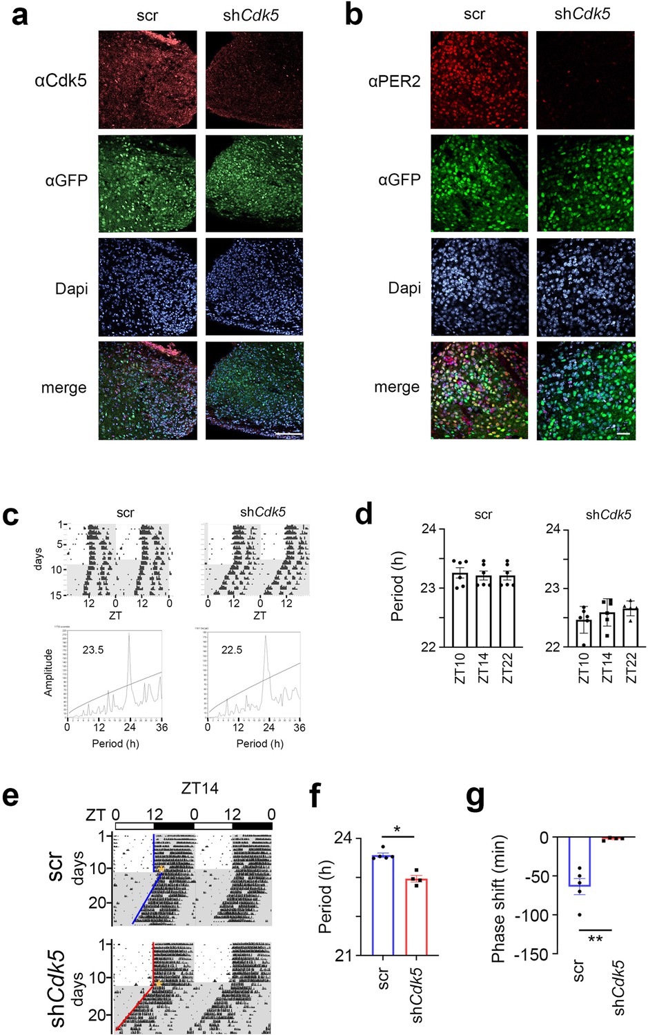
Validation of knock-down of Cdk5 expression.
(a) Representative coronal sections (40 µm) of the suprachiasmatic nucleus (SCN) region after injection of adeno-associated viruses (AAVs) carrying either scrambled (scr) or shCdk5 shRNA. Slices were stained with DAPI to stain nuclei (blue), and anti-GFP (green) and anti-Cdk5 (red) antibodies. GFP shows those cells infected by the virus. Cdk5 was efficiently down-regulated in the SCN by shCdk5 but not by scr shRNA (red), which was as efficiently delivered as shCdk5 (green). Scale bar: 100 µm. (b) Same as (a) but stained with anti-PER2 (red). PER2 was efficiently down-regulated in the SCN by shCdk5 but not by scr shRNA, which was as efficiently delivered as shCdk5 (green). Scale bar: 30 µm. (c) Examples of double-plotted actograms of control (scr) and Cdk5 knock-down (shCdk5) animals (top panels). Mice were kept under a 12 hr light/12 hr dark cycle for 8–10 days before they were released into constant darkness (DD). In DD period was determined using χ2-periodogram analysis (bottom panels). (d) Period was determined after a light pulse at zeitgeber time (ZT) 10, ZT14, or ZT22. τ control ZT10: 23.26±0.09 hr, ZT14: 23.21±0.08 hr, ZT22: 23.21±0.08 hr; τ shCdk5 ZT10: 22.47±0.09 hr, ZT14: 22.59±0.1 hr, ZT22: 22.66±0.06 hr. Values are the mean ± SEM (n=6). Period was not significantly altered by the light pulse itself neither in control (scr) not in shCdk5 animals. Brown-Forsythe ANOVA test, p>0.05, n=6. (e) Examples of double-plotted wheel-running actograms of control (scr) and Cdk5 knock-down (shCdk5) female mice. Animals were kept under a 12 hr light/12 hr dark cycle (white and gray areas, respectively) (LD). After 10–12 days they received a 15 min light pulse at ZT14 (yellow stars). After the light pulse animals were released into constant darkness (DD). (f) Circadian period (τ) of female shCdk5 mice (red) is significantly shorter compared to female scr controls (blue). τ scr = 23.53±0.05 hr, τ shCdk5=22.95±0.08 hr. All values are mean ± SEM, Mann-Whitney test, n=5 scr, n=4–5, *p<0.02. (g) Quantification of phase shifts (φ) after a 15 min light pulse at ZT14. The phase shift at ZT14 is strongly reduced in shCdk5 animals (red) compared to scr controls (blue). scr: φ ZT14: –63.8±11.2 min, shCdk5: φ ZT14: –2.5±0.13 min. All values are mean ± SEM, unpaired t-test with Welch’s correction, n=4–5, **p<0.01.
Figure 2 with 1 supplement
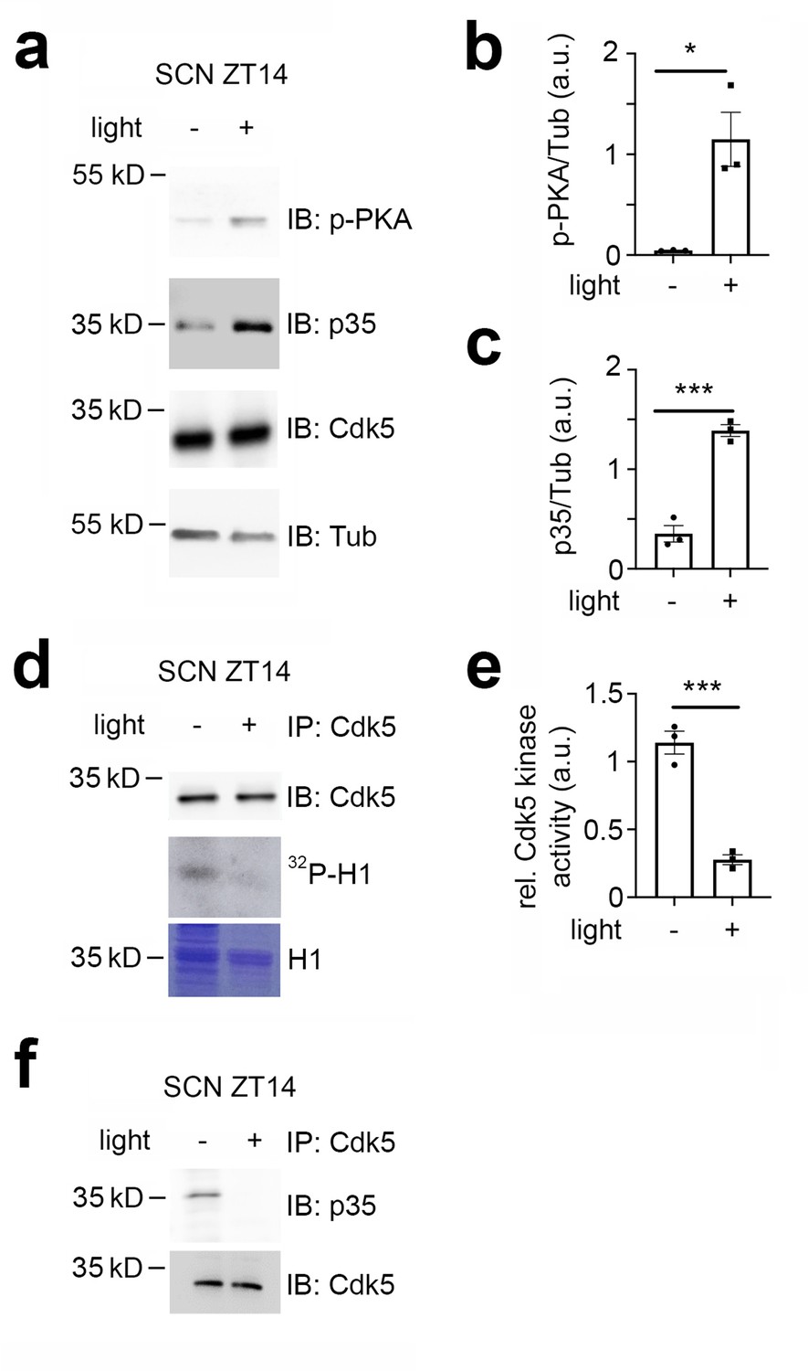
Cdk5 activity is modulated by light in the early night.
Immuno-western blotting (IB), immunoprecipitation (IP), and Cdk5 kinase activity assays from suprachiasmatic nucleus (SCN) tissue extracts harvested 30 min after light (+) and no light (-) given at zeitgeber time (ZT) 14. (a) Western blot depicting the amounts of phospho PKA (p-PKA), p35 co-activator, Cdk5, and tubulin (Tub, control) before and after light pulse at ZT14. (b) Quantification of p-PKA relative to tubulin. Values are the mean ± SEM. Unpaired t-test, n=3, *p<0.05. (c) Quantification of p35 co-activator of Cdk5. Values are the mean ± SEM. Unpaired t-test, n=3, ***p<0.001. (d) Cdk5 kinase activity assay. IP of SCN extracts with antibodies against Cdk5 showing the presence of Cdk5 (upper panel) and total protein with Coomassie blue staining (lower panel) as a control for the presence of H1. The middle panel depicts histone H1 phosphorylated by Cdk5, visualized as 32P-histone H1 (32P-H1). (e) Quantification of Cdk5 kinase activity relative to H1 levels. Values are the mean ± SEM. Unpaired t-test, n=3, ***p<0.001. (f) Co-immunoprecipitation of p35 with Cdk5 before and after a light pulse.
Figure 2—figure supplement 1
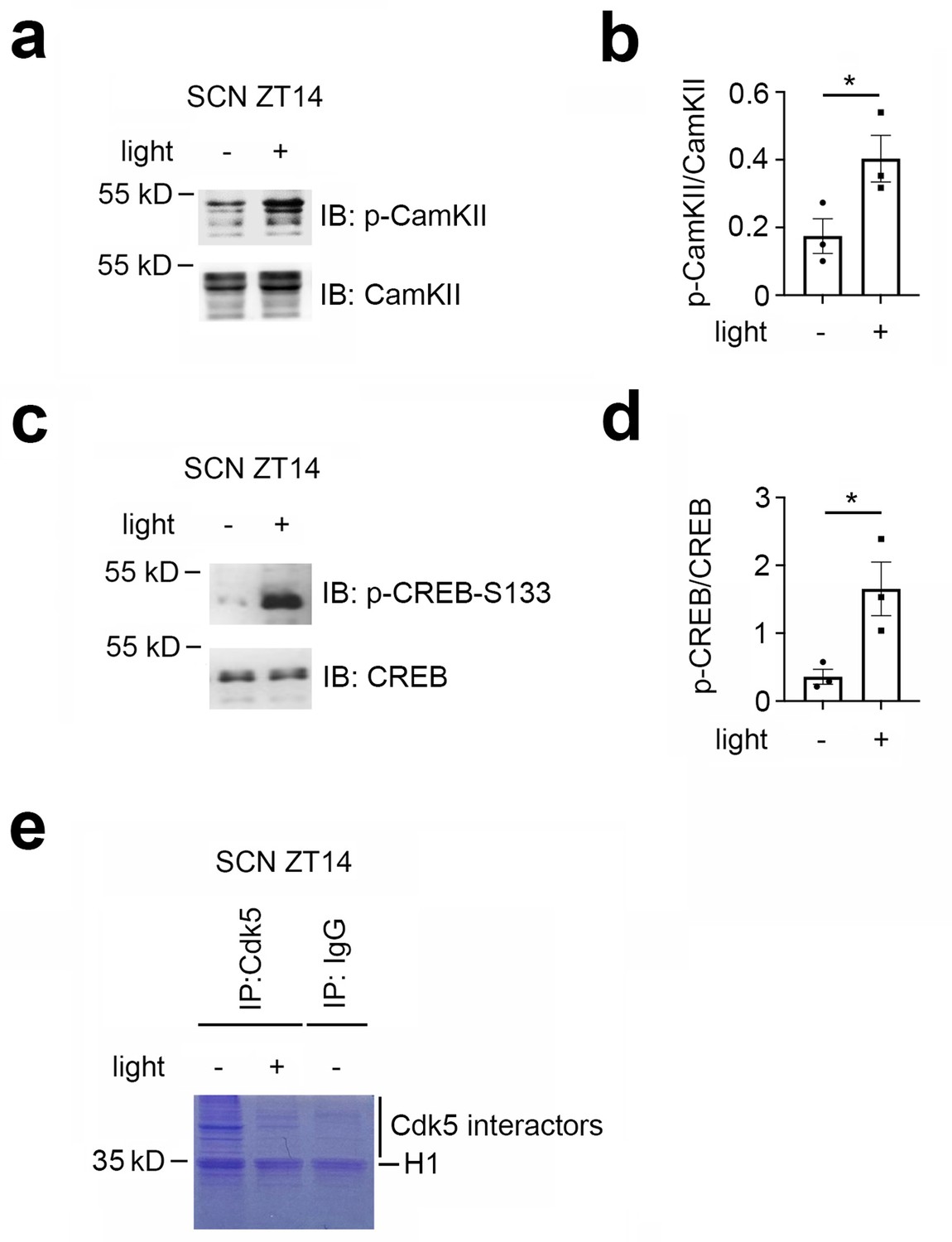
Light-induced phosphorylation of CaMKII and control immunoprecipitation.
(a) Western blot of suprachiasmatic nucleus (SCN) tissue extract at zeitgeber time (ZT) 14 with and without light pulse. The lower panel shows total CaMKII that was recognized by a polyclonal antibody. The upper panel shows increased levels of phosphorylated CaMKII (p-CaMKII) after light administration at ZT14. (b) Quantification of p-CaMKII relative to total CaMKII. Values are the mean ± SEM. Unpaired t-test, n=3, *p<0.05. (c) Western blot of suprachiasmatic nucleus (SCN) tissue extract at zeitgeber time (ZT) 14 with and without light pulse. The lower panel shows total CREB that was recognized by a polyclonal antibody. The upper panel shows increased levels of phosphorylated CREB (p-CREB-S133) after light administration at ZT14. (d) Quantification of p-CREB in relation to unphosphorylated CREB. Values are the mean ± SEM. Unpaired t-test, n=3, *p<0.05. (e) SDS-PAGE gel after immunoprecipitation of the SCN extracts (-/+ a light pulse) with anti-Cdk5 and anti-IgG, respectively.
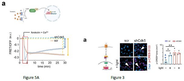
Figure 3 with 1 supplement
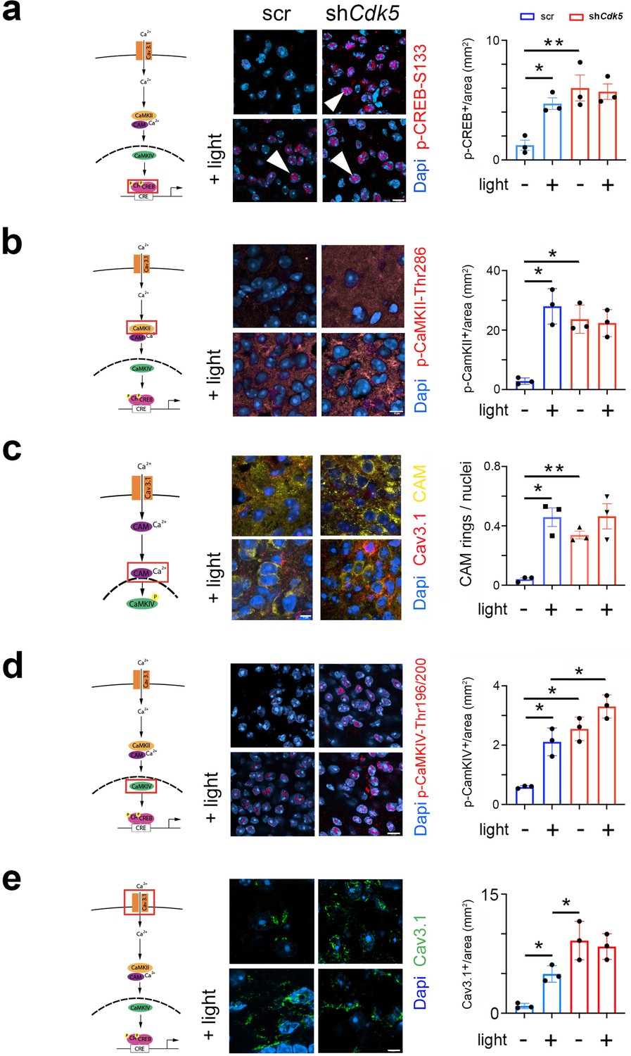
Cdk5 impacts on the cAMP response element-binding protein (CREB) signaling pathway via calcium/calmodulin-dependent kinases (CaMK).
The cartoons on the left of each figure depict the CaMK pathway with the red rectangle indicating the visualization of a particular component. (a) Immunohistochemistry on the suprachiasmatic nucleus (SCN) of control (scr) and shCdk5 mice using an antibody recognizing phospho-serine 133 of CREB (p-CREB-S133) before and after a light pulse at zeitgeber time (ZT) 14. The red color shows p-CREB-S133 and the blue color represents DAPI-stained nuclei of SCN cells. Scale bar: 8 µm. The right panel shows the quantification of the p-CREB-S133 signal. Values are the mean ± SEM. Unpaired t-test with Welch’s correction, n=3, *p<0.05. (b) Immunohistochemistry on the SCN of control (scr) and shCdk5 mice using an antibody recognizing phosphorylated Thr286 Cam kinase II (p-CaMKII) before and after a light pulse at ZT14. The red color shows p-CaMKII and the blue color represents DAPI-stained nuclei of SCN cells. Scale bar: 8 µm. The right panel shows the quantification of the p-CaMKII signal. Values are the mean ± SEM. Unpaired t-test with Welch’s correction, n=3, *p<0.05. (c) Translocation of calmodulin (CAM) in response to a light pulse at ZT14 in SCN neurons of control (scr) and Cdk5 knock-down (shCdk5) animals. CAM (yellow) accumulates around the nuclei in scr controls. In shCdk5 SCN neurons, this accumulation around the nuclei is already seen before the light pulse which is clearly different from scr controls. Scale bar: 7 µm The right panel shows the quantification of CAM rings. Values are the mean ± SEM. Unpaired t-test with Welch’s correction, n=3, *p<0.05, **p<0.01. (d) Immunohistochemistry on the SCN of control (scr) and shCdk5 mice using an antibody recognizing phosphorylated Thr196-200 Cam kinase IV (p-CaMKIV) before and after a light pulse at ZT14. The red color shows p-CaMKIV and the blue color represents DAPI-stained nuclei of SCN cells. Scale bar: 5 µm. The right panel shows the quantification of the p-CaMKIV signal. Values are the mean ± SEM. Unpaired t-test with Welch’s correction, n=3, *p<0.05. (e) Immunohistochemistry on the SCN of control (scr) and shCdk5 mice using an antibody recognizing the calcium channel Cav3.1 before and after a light pulse at ZT14. The green color shows Cav3.1 and the blue color represents DAPI-stained nuclei of SCN cells. Scale bar: 5 µm. The right panel shows the relative Cav3.1 signal. Values are the mean ± SEM. Unpaired t-test with Welch’s correction, n=3, *p<0.05.
Figure 3—figure supplement 1
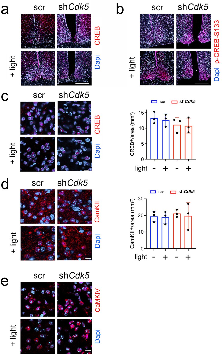
Lower magnification images of suprachiasmatic nucleus (SCN) sections and quantification of total cAMP response element-binding protein (CREB), CaMKII, and CaMKIV.
(a) Immunohistochemistry on the SCN of control (scr) and shCdk5 mice using an antibody recognizing total CREB before and after a light pulse at zeitgeber time (ZT) 14. The red color shows CREB and the blue color represents DAPI-stained nuclei of SCN cells. Scale bar: 250 µm. (b) Immunohistochemistry on the SCN of control (scr) and shCdk5 mice using an antibody recognizing phospho-serine 133 of CREB (p-CREB-S133) before and after a light pulse at ZT14. The red color shows p-CREB-S133 and the blue color represents DAPI-stained nuclei of SCN cells. Scale bar: 250 µm. (c) Left panel: Same as (a) but at higher magnification. Right panel: Quantification of total CREB in scr (blue) and shCdk5 (red) SCN. Values are the mean ± SEM. Unpaired t-test, n=3. Scale bar: 10 µm. (d) Left panel: Immunohistochemistry on the SCN depicting total CaMKII (red) and cell nuclei (blue). Right panel: Quantification of total CaMKII in scr (blue) and shCdk5 (red) SCN. Values are the mean ± SEM. Unpaired t-test, n=3. Scale bar: 10 µm. (e) Immunohistochemistry on the SCN depicting total CaMKIV (red) and cell nuclei (blue). Scale bar: 10 µm.
Figure 4 with 1 supplement
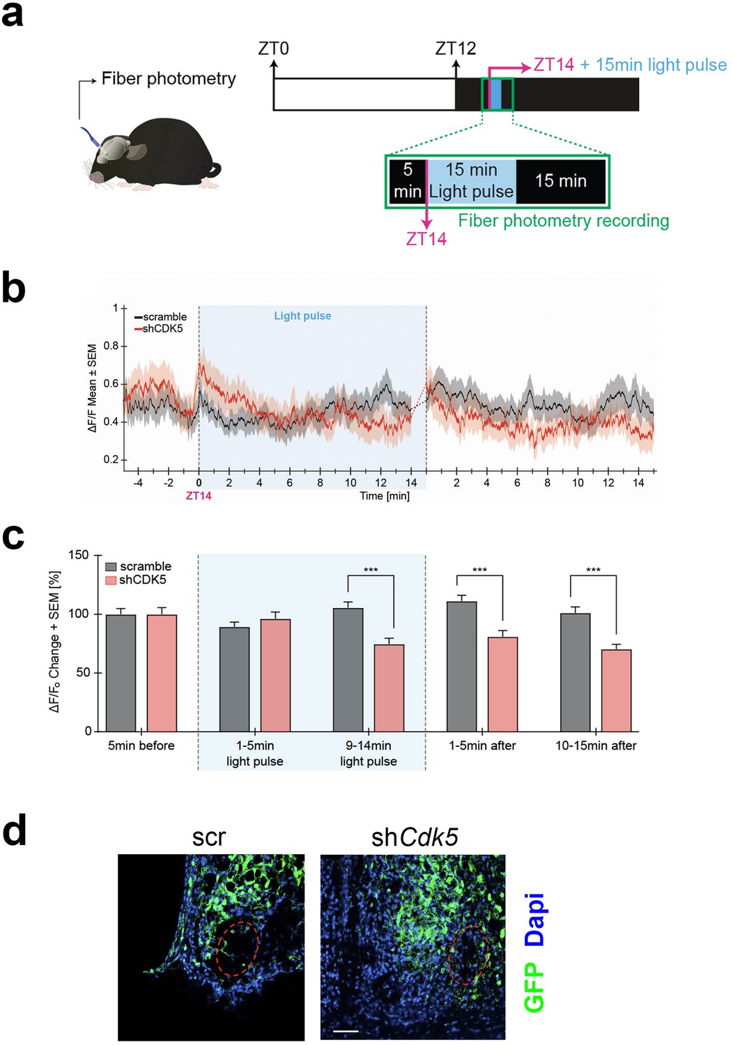
Neuronal activity in response to light at zeitgeber time (ZT) 14 is modulated by CDK5.
(a) Illustration of the chronic optic fiber implantation in the suprachiasmatic nucleus (SCN) for fiber photometry recording in freely moving mice (left). The animals were previously infected either with AAV9-hSyn1-chl[1x(shNS)]-jGCaMP7b-WPRE-SV40p(A)(scr) or AAV9-hSyn1-chl[mouse(shCdk5)]-jGCaMP7b-WPRE-SV40p(A)(shCdk5). The experimental timeline of one trial is shown on the right. White and dark boxes represent the light and dark phase, respectively. Fiber photometry recordings were done in the 5 min before the light pulse, during the light pulse, and 15 min after. The light pulse was 15 min long and was delivered at ZT14 (blue box; dashed lines between min 14–15 are not included in the analysis). (b) Mean traces ± SEM of cell activity (normalized ΔF/F0) of GCaMP7b-expressing SCN neurons (black: scr, red: shCdk5) 5 min before, 15 min during, and 15 min after the light pulse (±20 s) light pulse delivered at ZT14. N=15 trials, n=5 mice/red = shCdk5, N=12 trials, n=4 mice. (c) Bar plot showing the percentage of ΔF/F0 changes ± SEM 5 min before the light pulse, in the first and last 5 min during the light pulse and the first and last 5 min after the light pulse. 9–14 min of light pulse: scramble (black bar) 105.5±19.3 ΔF/F0 vs. shCdk5 (red bar) 74.7±17.2 ΔF/F0. 1–5 min after light pulse: scramble (black bar) 111.2±19.3 ΔF/F0 vs. shCdk5 (red bar) 81.0±17.8 ΔF/F0. Black = scr, N=15 trials, n=5 mice/red = shCdk5, N=12 trials, n=4 mice. Bar values represent the mean ± SEM. ***p<0.001; two-way ANOVA corrected with Bonferroni post hoc test. (d) Photomicrograph of the expression of GCaMP7b (green) in the SCN in both control (scr, left) and experimental (shCdk5, right) animals. The red hatched oval indicates the placement of the optic fiber. Blue: DAPI, green: GFP (produced by jGCaMPP7). Scale bar: 50 µm.
Figure 4—figure supplement 1
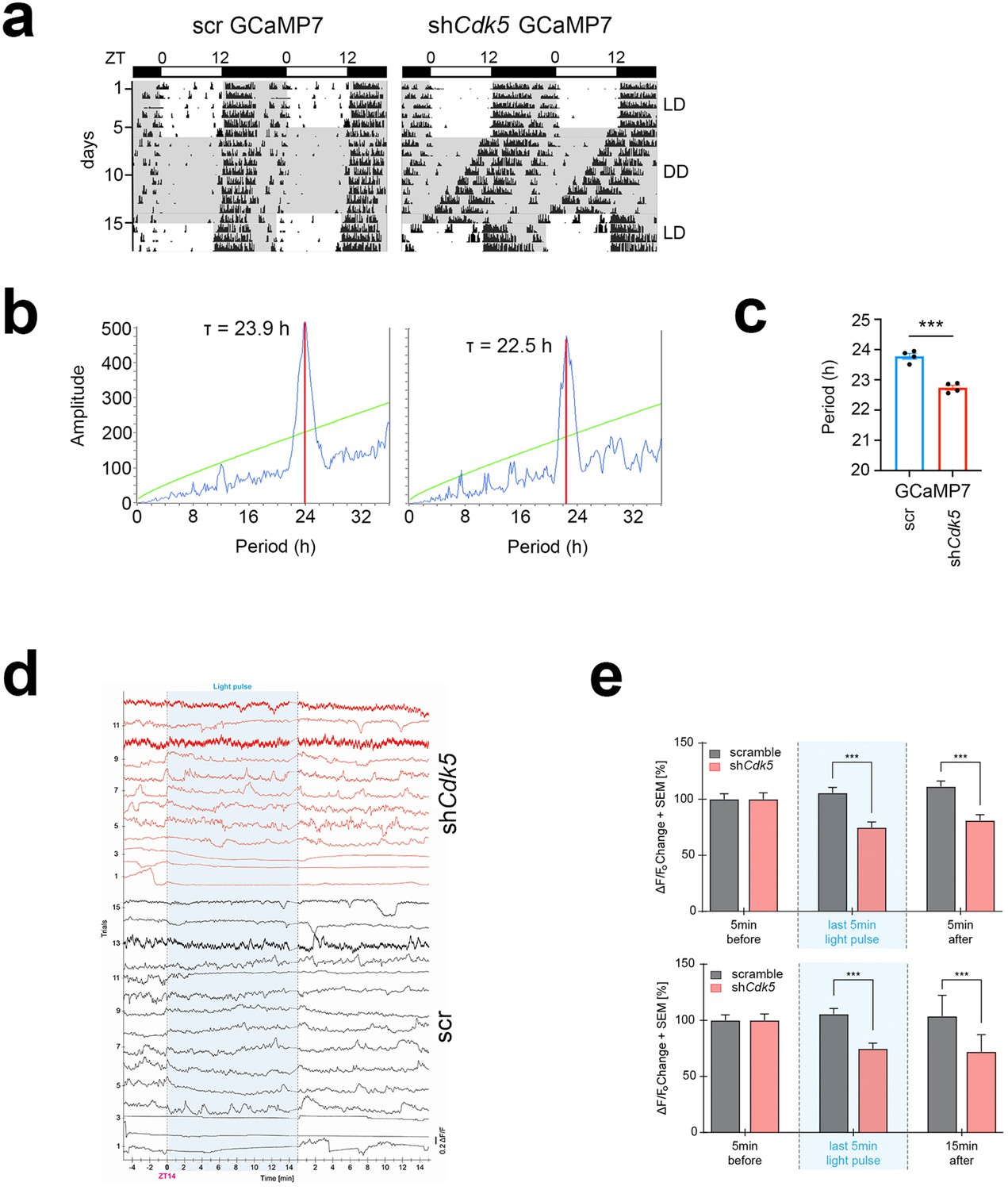
GCaMP7 reporter does not affect knock-down of Cdk5 in the suprachiasmatic nucleus (SCN).
(a) Examples of double-plotted wheel-running actograms of control (scr) and Cdk5 knock-down (shCdk5) mice containing the reporter GCaMP7. Animals were kept under a 12 hr light/12 hr dark cycle (white and gray areas, respectively) (LD). After 6 days they were released into constant darkness (DD). (b) χ2-periodogram analysis showing circadian period (τ) for control GCaMP7 and shCdk5 GCaMP7 mice, τ scr = 23.9 hr, τ shCdk5=22.5 hr. (c) Quantification of circadian period (τ). τ scr = 23.78±0.08 hr, τ shCdk5=22.74±0.09 hr. All values are mean ± SEM, unpaired t-test with Welch’s correction, n=5–6, ***p<0.001. (d) Activity of SCN neurons expressing GCamP7b (normalized ΔF/F) in individual trials in the dark phase, 5 min before and 15 min after the 15 min (±20 s) light pulse delivered at ZT14. The 15th minute of the light recording has not been analyzed to homogenize the lightning time precisely. Black = scramble, N=15 trials, n=5 mice/red = shCdk5, N=12 trials, n=4 mice. (e) Bar plots showing the percentage of ΔF/F0 changes ± SEM (normalized to the 5 min before the light pulse) in the dark phase, 5 min (top) or 15 min (bottom) after the light pulse delivered at ZT14 and in the last 5 min of the light pulse (min 10–14). Black: scramble, N=15 trials, n=5 mice; red: shCdk5, N=12, trials, n=4 mice. ***p<0.001; two-way ANOVA corrected with Bonferroni post hoc test.
Figure 5 with 1 supplement
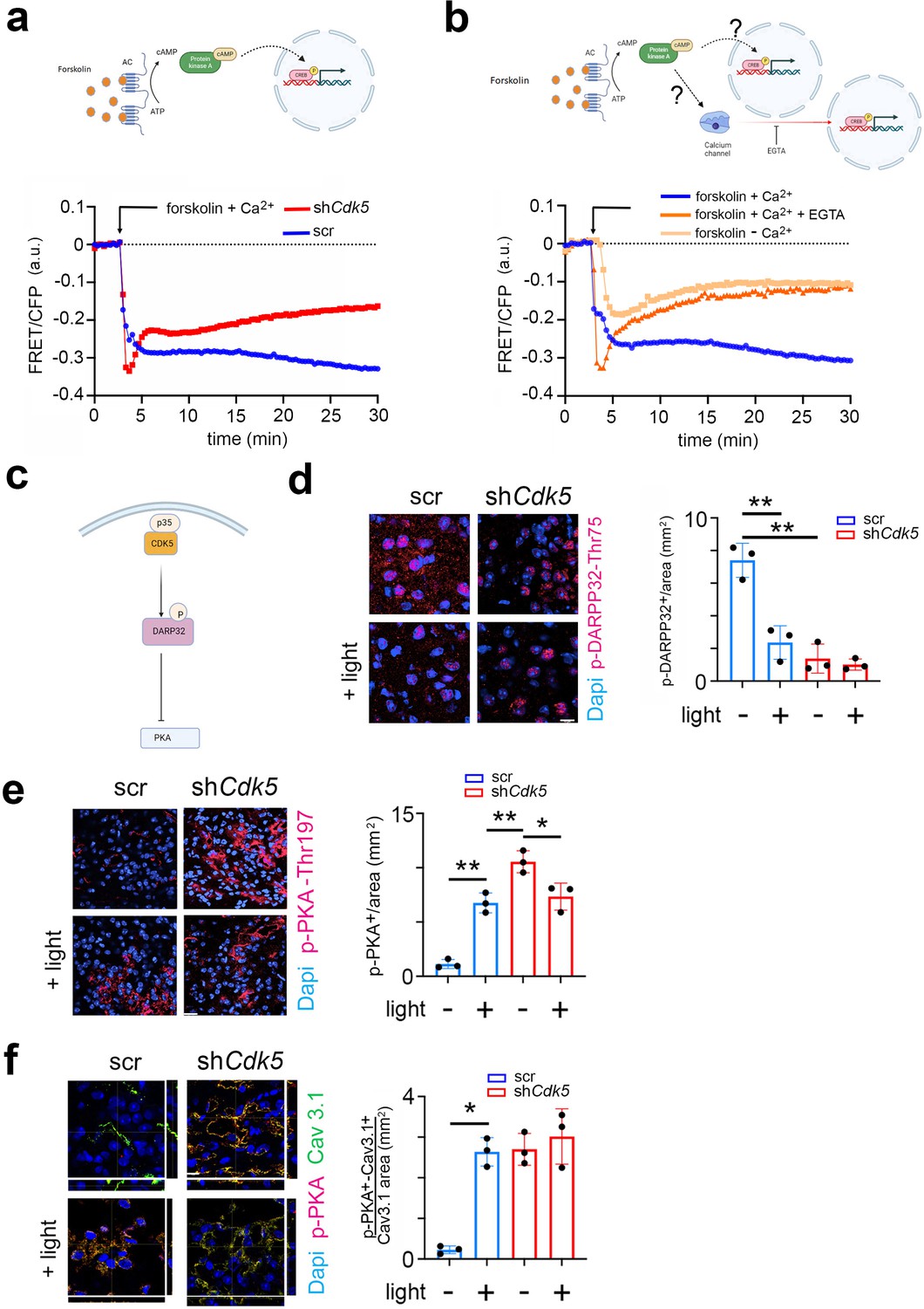
CDK5 regulates PKA phosphorylation via DARPP32 phosphorylation.
(a) Top: Scheme of the forskolin-PKA-CREB signaling pathway. Bottom: Förster resonance energy transfer (FRET)/CFP signal ratio changes in response to forskolin treatment in NIH 3T3 cells transfected with either a scr control (blue) or shCdk5 (red) expression construct. Values are the mean ± SD. Two-way ANOVA revealed a significant difference between the curves, n=3, ****p<0.0001. (b) Top: Scheme of the forskolin-PKA-CREB signaling pathway and calcium signaling. Bottom: FRET/CFP signal ratio changes in response to forskolin treatment in NIH 3T3 cells with the addition of Ca2+ (blue), without the addition of Ca2+ (salmon colored), and with the addition of Ca2+ and EGTA (orange). Values are the mean ± SD. Two-way ANOVA revealed a significant difference between the gray and blue/orange curves, n=3, ****p<0.0001. (c) Scheme of CDK5-DARPP32-PKA pathway. (d) Immunohistochemistry on the suprachiasmatic nucleus (SCN) of control (scr) and shCdk5 mice using an antibody recognizing phosphorylated Thr-75 of DARPP32 (p-DARPP32) before and after a light pulse at zeitgeber time (ZT) 14. The red color shows p-DARPP32 and the blue color represents DAPI-stained nuclei of SCN cells. Scale bar: 10 µm. The right panel shows the quantification of the p-DARPP32 signal. Values are the mean ± SEM. Unpaired t-test with Welch’s correction, n=3, **p<0.01. (e) Immunohistochemistry on the SCN of control (scr) and shCdk5 mice using an antibody recognizing phosphorylated Thr-197 of PKA (p-PKA) before and after a light pulse at ZT14. The red color shows p-PKA and the blue color represents DAPI-stained nuclei of SCN cells. Scale bar: 20 µm. The right panel shows the quantification of the p-PKA signal. Values are the mean ± SEM. Unpaired t-test with Welch’s correction, n=3, *p<0.05, **p<0.01. (f) Immunohistochemistry on the SCN of control (scr) and shCdk5 mice using an antibody recognizing phosphorylated Thr-197 of PKA (p-PKA) and Cav3.1 before and after a light pulse at ZT14. The red color shows p-PKA, the green color Cav3.1, and the blue color represents DAPI-stained nuclei of SCN cells. The yellow color signifies the co-localization of PKA and Cav3.1. The stripes on the left and bottom of each micrograph show the z-stacks to confirm co-localization. Scale bar: 10 µm. The right panel shows the quantification of relative p-PKA/Cav3.1. Values are the mean ± SEM. Unpaired t-test with Welch’s correction, n=3, *p<0.05.
Figure 5—figure supplement 1
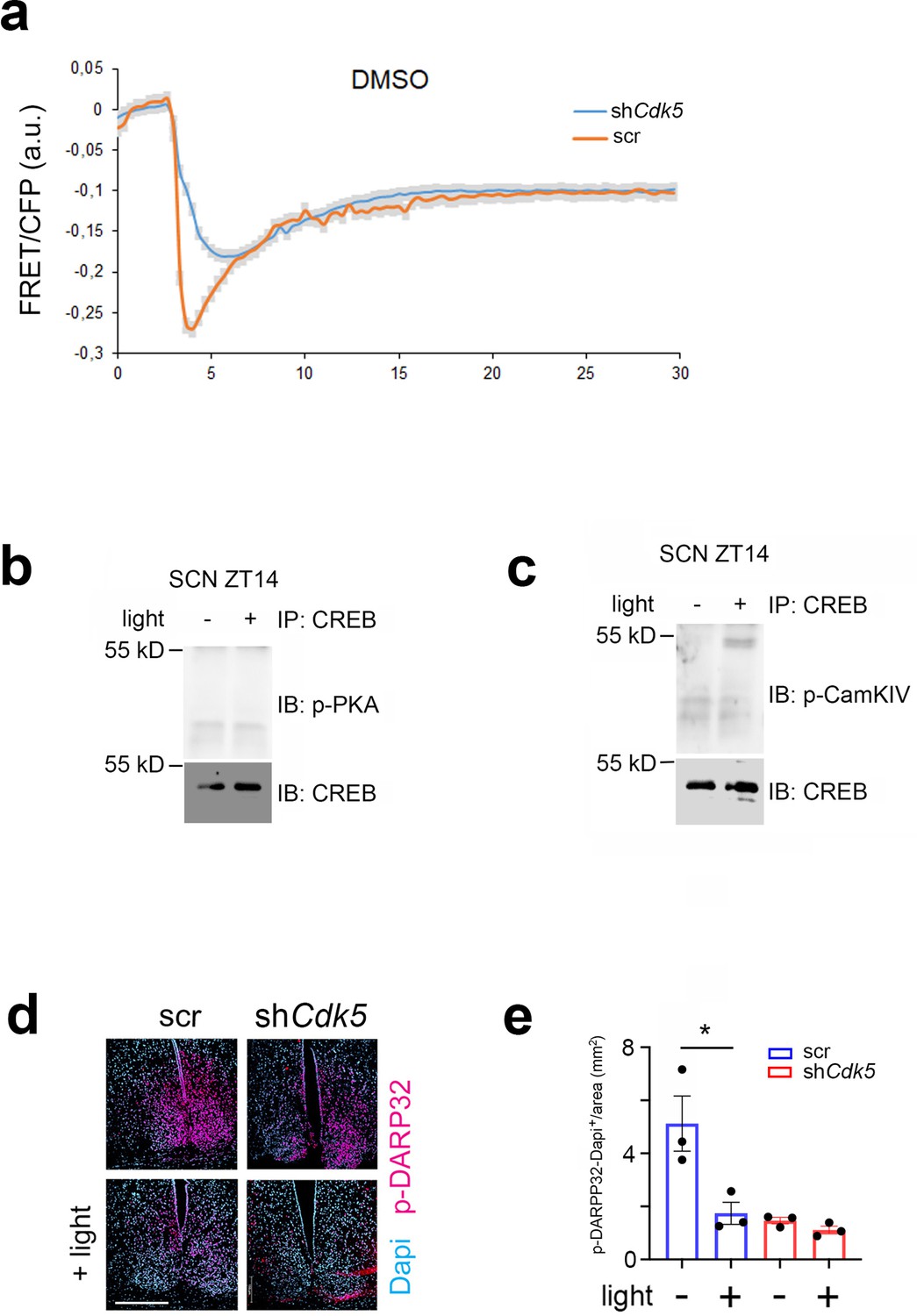
Solvent control for Förster resonance energy transfer (FRET) experiment.
Immunoprecipitation of cAMP response element-binding protein (CREB) and low magnification of suprachiasmatic nucleus (SCN) stained with p-DARPP32. (a) FRET/CFP signal ratio changes with DMSO treatment in NIH 3T3 cells transfected with either an scr control (red) or shCdk5 (blue) expression construct. Values are the mean ± SD. Two-way ANOVA revealed no difference between the curves, n=3. (b) Immunoprecipitation (IP) of CREB from SCN tissue at zeitgeber time (ZT) 14 before and after a light pulse. Immunoblot (IB) shows no interaction with p-PKA, IB: CREB loading control. (c) IP of CREB from SCN tissue at ZT14 before and after a light pulse. IB shows interaction with p-CaMKIV, IB: CREB loading control. (d) Immunohistochemistry on the SCN of control (scr) and shCdk5 mice using an antibody recognizing p-DARPP32 before and after a light pulse at ZT14. The red color shows p-DARPP32 and the blue color represents DAPI-stained nuclei of SCN cells. Scale bar: 250 µm. (e) Quantification of nuclear p-DARPP32 signal. Values are the mean ± SEM. Unpaired t-test with Welch’s correction, n=3, *p<0.05.
Figure 6 with 1 supplement
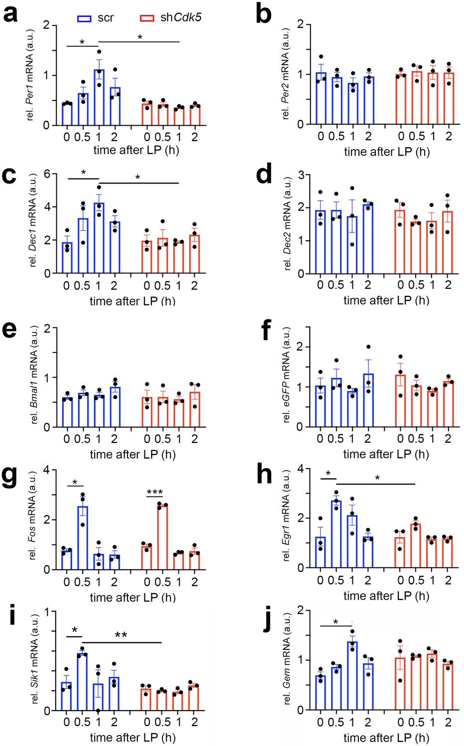
Cdk5 regulates light-induced gene expression in the suprachiasmatic nucleus (SCN) of some clock genes.
Relative mRNA values are represented as blue bars for scr control animals and as red bars for shCdk5 mice. The values were determined 0, 0.5, 1, and 2 hr after a light pulse (LP) given at zeitgeber time (ZT) 14. (a) Induction of Per1 mRNA expression by light with a maximum of 1 hr after light in scr control animals. In contrast, Per1 is not induced in shCdk5 SCN. Scr: 0 hr: 0.44±0.01, 0.5 hr: 0.65±0.12, 1 hr: 1.12±0.19, 2 hr: 0.44±0.01; shCdk5: 0 hr: 0.44±0.01, 0.5 hr: 0.44±0.01, 1 hr: 0.44±0.01, 2 hr: 0.77±0.18. Values are the mean ± SEM. Unpaired t-test, n=3, *p<0.05. (b) Per2 mRNA expression is not induced by light neither in scr controls nor in shCdk5 animals. Scr: 0 hr: 1.04±0.16, 0.5 hr: 0.95±0.10, 1 hr: 0.83±0.10, 2 hr: 0.96±0.07; shCdk5: 0 hr: 1.01±0.05, 0.5 hr: 1.07±0.10, 1 hr: 1.04±0.14, 2 hr: 1.04±0.12. Values are the mean ± SEM. Unpaired t-test, n=3. (c) Induction of Dec1 mRNA expression by light with a maximum at 1 hr after light in scr control animals. In contrast, Dec1 is not induced in shCdk5 SCN. Scr: 0 hr: 1.87±0.37, 0.5 hr: 3.32±0.75, 1 hr: 4.25±0.49, 2 hr: 3.13±0.34; shCdk5: 0 hr: 1.96±0.34, 0.5 hr: 2.13±0.51, 1 hr: 1.87±0.07, 2 hr: 2.32±0.40. Values are the mean ± SEM. Unpaired t-test, n=3, *p<0.05. (d) Dec2 mRNA expression is not induced by light neither in scr controls nor in shCdk5 animals. Scr: 0 hr: 1.93±0.29, 0.5 hr: 1.94±0.25, 1 hr: 1.75±0.49, 2 hr: 2.11±0.07; shCdk5: 0 hr: 1.94±0.23, 0.5 hr: 1.58±0.07, 1 hr: 1.61±0.25, 2 hr: 1.90±0.33. Values are the mean ± SEM. Unpaired t-test, n=3. (e) Bmal1 mRNA expression is not induced by light in the SCN of scr control and shCdk5 animals. Scr: 0 hr: 0.60±0.04, 0.5 hr: 0.69±0.06, 1 hr: 0.65±0.05, 2 hr: 0.81±0.12; shCdk5: 0 hr: 0.61±0.14, 0.5 hr: 0.61±0.12, 1 hr: 0.56±0.06, 2 hr: 0.71±0.15. Values are the mean ± SEM. Unpaired t-test, n=3. (f) eGFP mRNA expression is detected in the SCN of scr control and shCdk5 animals demonstrating proper injection of expression constructs (scr: ssAAV-9/2-hSyn1-chl[1x(shNS)]-EGFP-WPRE-SV40p(A), shCdk5: ssAAV-9/2-hSyn1-chl[mouse(shCdk5)]-EGFP-WPRE-SV40p(A)). Scr: 0 hr: 1.04±0.19, 0.5 hr: 1.23±0.22, 1 hr: 0.90±0.07, 2 hr: 1.34±0.34; shCdk5: 0 hr: 1.31±0.28, 0.5 hr: 1.04±0.13, 1 hr: 0.90±0.05, 2 hr: 1.15±0.06. Values are the mean ± SEM. Unpaired t-test, n=3. (g) Induction of Fos mRNA 0.5 hr after the light pulse in both scr controls and shCdk5 SCN. Scr: 0 hr: 0.77±0.07, 0.5 hr: 2.55±0.38, 1 hr: 0.64±0.25, 2 hr: 0.61±0.15; shCdk5: 0 hr: 0.95±0.08, 0.5 hr: 2.57±0.05, 1 hr: 0.68±0.04, 2 hr: 0.74±0.12. Values are the mean ± SEM. Unpaired t-test, n=3, *p<0.05, ***p<0.001. (h) Induction of Egr1 mRNA 0.5 hr after the light pulse in scr control but not shCdk5 SCN. Scr: 0 hr: 1.25±0.38, 0.5 hr: 2.71±0.19, 1 hr: 2.12±0.41, 2 hr: 1.26±0.13; shCdk5: 0 hr: 1.23±0.24, 0.5 hr: 1.77±0.13, 1 hr: 1.16±0.08, 2 hr: 1.18±0.06. Values are the mean ± SEM. Unpaired t-test, n=3, *p<0.05. (i) Sik1 mRNA expression is induced by light in the SCN of scr control but not shCdk5 animals. Scr: 0 hr: 0.29±0.07, 0.5 hr: 0.58±0.02, 1 hr: 0.27±0.14, 2 hr: 0.34±0.07; shCdk5: 0 hr: 0.22±0.03, 0.5 hr: 0.20±0.01, 1 hr: 0.19±0.02, 2 hr: 0.25±0.02. Values are the mean ± SEM. Unpaired t-test, n=3, *p<0.05, **p<0.01. (j) Gem mRNA expression is induced by light in the SCN of scr control but not shCdk5 animals. Scr: 0 hr: 0.70±0.07, 0.5 hr: 0.86±0.05, 1 hr: 1.38±0.11, 2 hr: 0.94±0.11; shCdk5: 0 hr: 1.05±0.24, 0.5 hr: 1.08±0.03, 1 hr: 1.13±0.07, 2 hr: 0.93±0.05. Values are the mean ± SEM. Unpaired t-test, n=3, *p<0.05.
Figure 6—figure supplement 1
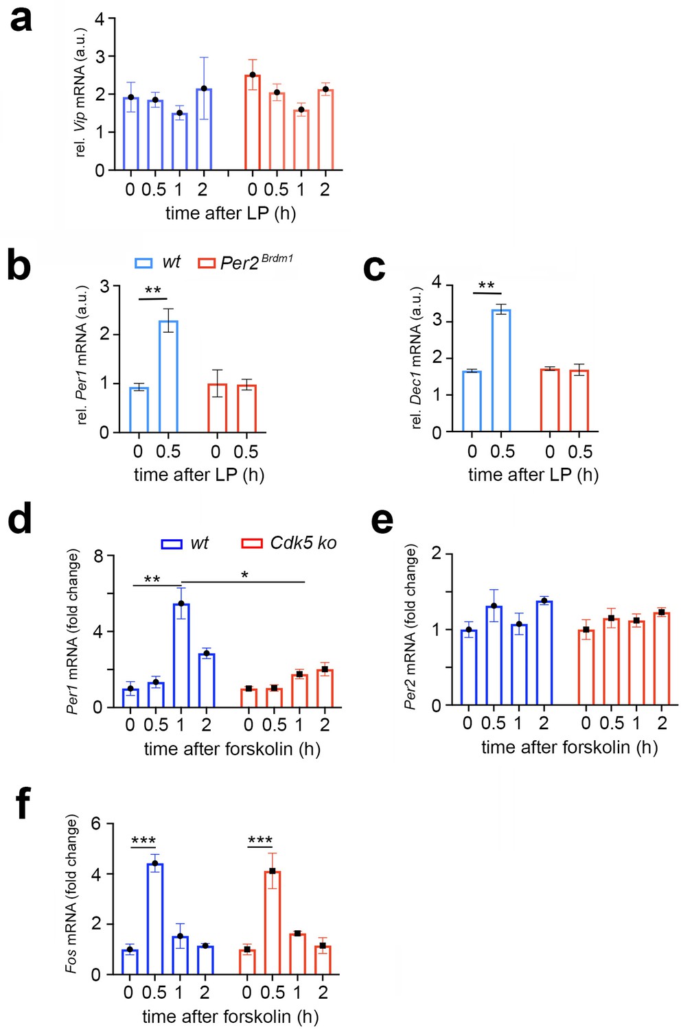
Vip, Per1, Dec1 levels in the suprachiasmatic nucleus (SCN) of shCdk5 and Per2Brdm1 mice and Per1, 2 and Fos gene expression in NIH 3T3 cells.
(a) Vip mRNA expression is not induced by light in the SCN of scr control and shCdk5 animals. Scr: 0 hr: 1.92±0.39, 0.5 hr: 1.85±0.20, 1 hr: 1.51±0.19, 2 hr: 2.15±0.82; shCdk5: 0 hr: 2.51±0.40, 0.5 hr: 2.05±0.22, 1 hr: 1.59±0.17, 2 hr: 2.13±0.17. Values are the mean ± SEM. Unpaired t-test, n=3. (b) Relative mRNA values are represented as blue bars for scr control animals and as red bars for Per2Brdm1 mice. The values were determined 0 and 0.5 hr after a light pulse (LP) was given at zeitgeber time (ZT) 14. Induction of Per1 mRNA expression by light 0.5 hr after light in wt control animals. In contrast, Per1 is not induced in Per2Brdm1 SCN. wt: 0 hr: 0.93±0.07, 0.5 hr: 2.29±0.24; Per2Brdm1: 0 hr: 1.00±0.28, 0.5 hr: 0.98±0.11. Values are the mean ± SEM. Unpaired t-test, n=3, **p<0.01. (c) Induction of Dec1 mRNA expression 0.5 hr after light in wt control animals. In contrast, Dec1 is not induced in the Per2Brdm1 SCN. wt: 0 hr: 1.66±0.04, 0.5 hr: 3.35±0.14; Per2Brdm1: 0 hr: 1.72±0.05, 0.5 hr: 1.69±0.16. Values are the mean ± SEM. Unpaired t-test, n=3, **p<0.01. (d) Relative mRNA values are represented as blue bars for control wild-type (wt) cells and as red bars for Cdk5 knock-out (Cdk5 ko) cells. The values were determined 0, 0.5, 1, and 2 hr after forskolin treatment. Induction of Per1 mRNA expression is maximal 1 hr after forskolin treatment in wt cells, but not in Cdk5 ko cells. Wt: 0 hr: 1±0.36, 0.5 hr: 1.34±0.30, 1 hr: 5.48±0.81, 2 hr: 2.85±0.28; Cdk5 ko: 0 hr: 1±0.10, 0.5 hr: 1.03±0.17, 1 hr: 1.76±0.25, 2 hr: 2.02±0.35. Values are mean ± SD. Unpaired t-test, *p<0.05, **p<0.01, n=3. (e) Per2 mRNA expression is not induced neither in wt cells nor in Cdk5 ko cells. Wt: 0 hr: 1±0.10, 0.5 hr: 1.32±0.21, 1 hr: 1.07±0.14, 2 hr: 1.38±0.05; Cdk5 ko: 0 hr: 1±0.13, 0.5 hr: 1.15±0.13, 1 hr: 1.12±0.09, 2 hr: 1.23±0.06. Values are mean ± SD. Unpaired t-test, n=3. (f) Induction of Fos mRNA expression is maximal 0.5 hr after forskolin treatment in wt cells, and in Cdk5 ko cells. Wt: 0 hr: 1±0.21, 0.5 hr: 4.42±0.35, 1 hr: 1.53±0.49, 2 hr: 1.15±0.08; Cdk5 ko: 0 hr: 1±0.21, 0.5 hr: 4.12±0.71, 1 hr: 1.64±0.09, 2 hr: 1.15±0.32. Values are mean ± SD. Unpaired t-test, n=3.
Figure 7
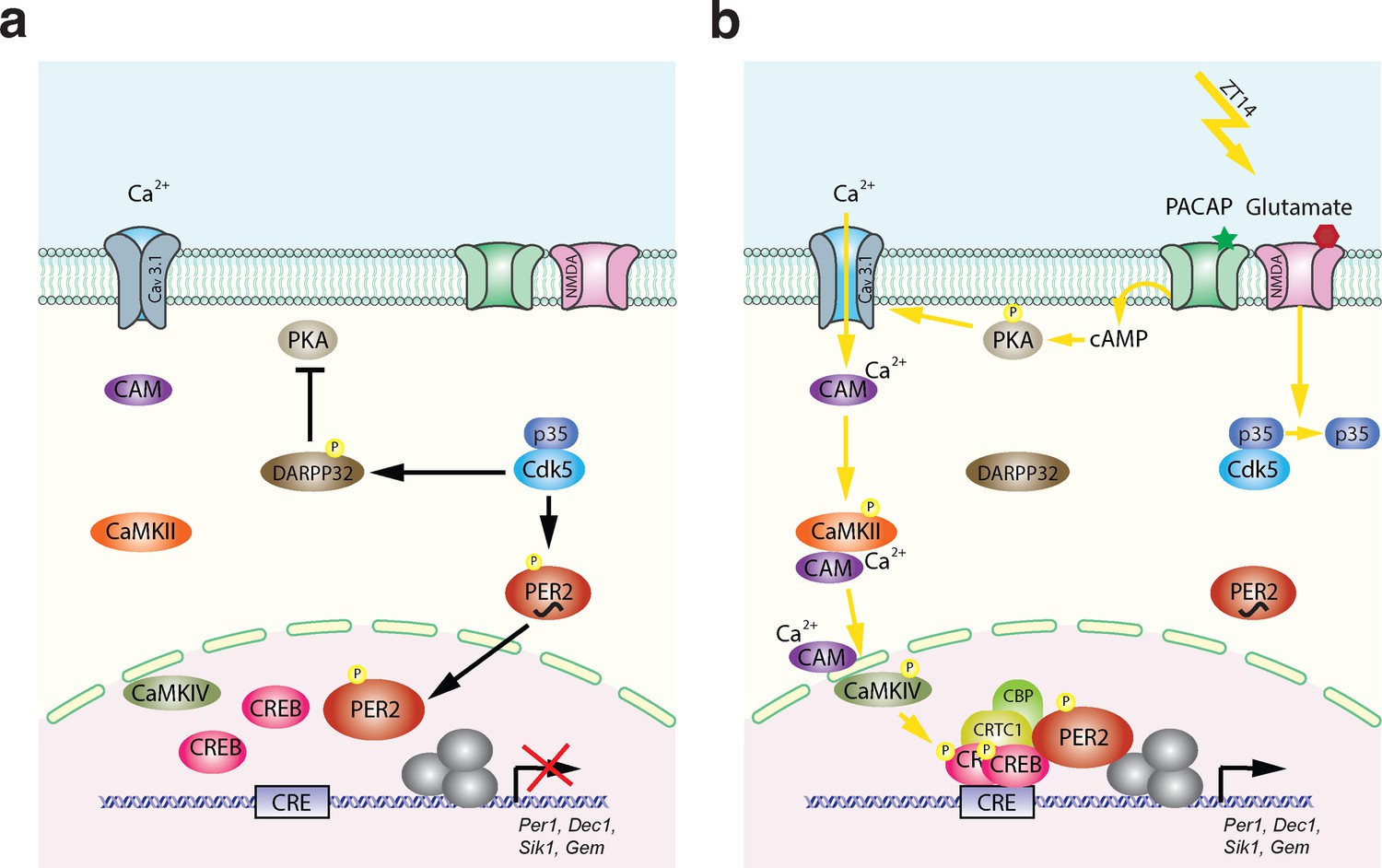
Model of Cdk5-gated light signal.
(a) Cdk5 is active during the dark portion of the day. Active Cdk5 with its co-activator p35 phosphorylates PER2, which leads to stabilization and nuclear translocation of this protein that is abundant at zeitgeber time (ZT) 12. At the same time CDK5 phosphorylates DARPP32, which inhibits the PKA signaling pathway. (b) Light perceived in the dark phase at ZT14 leads to detachment of p35 from Cdk5 stopping Cdk5 activity. DARPP32 is not phosphorylated and hence can’t inhibit PKA. PKA that is activated by the light signal is phosphorylated and can mediate cAMP response element-binding protein (CREB) phosphorylation via T-type calcium channels (Cav3.1) and the CaMK pathway leading to a transcriptionally active complex on the CRE element present in the promoters of many light-responsive genes such as Per1, Dec1, Gem, and Sik1. Overall, a light pulse at ZT14 will activate CREB phosphorylation and a protein complex will form. This complex needs phosphorylated PER2 that has accumulated in the nucleus between ZT12 and ZT14 to initiate the transcription of light-responsive genes. Both arms are necessary to build up a transcriptionally functional complex. Both arms depend on the presence and activity of CDK5, which therefore gates the light signal at ZT14. It is very likely that the amount of PER2 protein in the nucleus determines at least in part the magnitude of the phase delay, which depends on the timing of the light signal.
Author response image 1
Author response image 2
Author response image 3

FRET model.
On the left is a schematic representation of how ICAP works. On the right, an example of the quantified FRET decrease associated with increased KID: KIX interaction.
Author response image 4
Author response image 5

Tables
Table 1
Antibodies used for the immunostainings.
| Antibody | Species | Company | Catalog number | Dilution |
|---|---|---|---|---|
| Anti-Cdk5 clone 2H6 | Mouse | Origene | CF500397 | 1:100 |
| Anti-GFP | Rabbit | Abcam | ab6556 | 1:500 |
| Anti-Creb | Rabbit | Cell Signaling | D76D11 | 1:200 |
| Anti-Creb (pSer133) | Rabbit | Abcam | Ab32096 | 1:500 |
| Anti-CaMKII | Rabbit | Abcam | Ab52470 | 1:200 |
| Anti-CaMKII (pThr286) | Mouse | Invitrogen | MA1-047 | 1:100 |
| Anti-CaMKIV | Rabbit | Abcam | Ab3557 | 1:200 |
| Anti-CaMKIV (pThr196/200) | Rabbit | Invitrogen | PA5-105011 | 1:100 |
| Anti-CaV3.1 | Rabbit | Invitrogen | PA5-50635 | 1:100 |
| Anti-Calmodulin | Mouse | Invitrogen | MA3-917 | 1:100 |
| Anti-PKA (pT197) | Rabbit | Abcam | Ab75991 | 1:100 |
| Anti-Darpp32 (pThr75) | Rabbit | Invitrogen | PA5-105037 | 1:100 |
Table 2
Antibodies used for the western blots.
| Antibody | Species | Company | Catalog number | Dilution |
|---|---|---|---|---|
| Anti-Cdk5 | Rabbit | Cell Signaling | D1F7M | 1:3000 |
| Anti-Creb | Rabbit | Cell Signaling | D76D11 | 1:3000 |
| Anti-Creb (pSer133) | Rabbit | Abcam | Ab32096 | 1:1000 |
| Anti-PKA (pT197) | Rabbit | Abcam | Ab75991 | 1:1000 |
| Anti-p35 | Rabbit | Invitrogen | MA5-14834 | 1:1000 |
| Anti-CaMKIV (pThr196/200) | Rabbit | Invitrogen | PA5-105011 | 1:1000 |
Additional files
Download links
A two-part list of links to download the article, or parts of the article, in various formats.
Downloads (link to download the article as PDF)
Open citations (links to open the citations from this article in various online reference manager services)
Cite this article (links to download the citations from this article in formats compatible with various reference manager tools)
Cyclin-dependent kinase 5 (Cdk5) activity is modulated by light and gates rapid phase shifts of the circadian clock
eLife 13:RP97029.
https://doi.org/10.7554/eLife.97029.3


