Cingulate cortex shapes early postnatal development of social vocalizations
- Section on Behavioral Neuroscience, National Institutes of Health, United States
- NeuroImaging Facility, National Institutes of Health, United States
- Rodent Behavioral Core, National Institutes of Health, United States
Figures
Figure 1 with 1 supplement
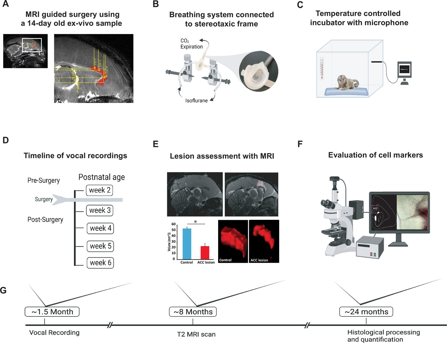
Experimental design and timeline.
(A) Reference MR scan was obtained using an ex vivo sample of a 14-day-old marmoset. Parasagittal view of the reference scan shows location of injection coordinates targeting the rostral portion of the dorsal anterior cingulate cortex (ACC) (24a and 24b), bilaterally (red). (B) Gas anesthesia was supplied through a custom-made breathing system comprising a facemask fitted with a palate bar with a 0.6 mm diameter hole. The palate bar was connected to a vital monitor to accurately detect small tidal end volumes during anesthesia while the animal was secured in the stereotaxic frame. (C) Five-minute vocalization recordings were obtained from infants placed in a softly padded temperature-controlled incubator. (D) Timeline of vocal recordings from postnatal week 2 to postnatal week 6. The ACC lesion was conducted at postnatal week 2 when animals were 14–16 days of age. (E) Representative sagittal view of postoperative T2-weighted MR images of a control (left panel) and lesioned (right panel) infant to reveal extent of white hypersignal, which reflects edema due to injections of the excitotoxin and therefore approximate site of the ACC lesion. There was a significant reduction in total ACC volume in the ACC group relative to controls (n=4/per group; F(1,6) = 82.78, p<0.0001). A representative three-dimensional view of area 24 is presented, showing the reduced volume of the ACC (right panel) relative to the normal volume in the control (left panel). (F) Schematic illustration highlights the end point of the experiment involving histological processing and evaluation of cell markers. (G) Longitudinal timeline shows approximate age of animals following vocal recordings, MRI lesion assessments, and histological processing.
Figure 1—figure supplement 1
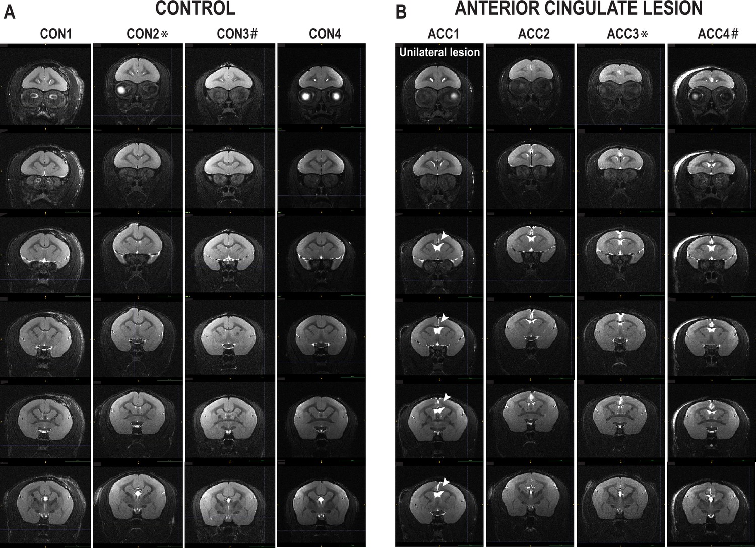
Bilateral MRI scans.
Images from MRI scans of each marmoset representing coronal planes ranging from pregeniculate region until retrosplenial area. Each slice is 0.25 mm apart. Anterior cingulate cortex (ACC)1 shows a unilateral lesion depicted by the arrow. CON2* and ACC3* are twins, and CON3# and ACC4# are twins.
Figure 2 with 1 supplement
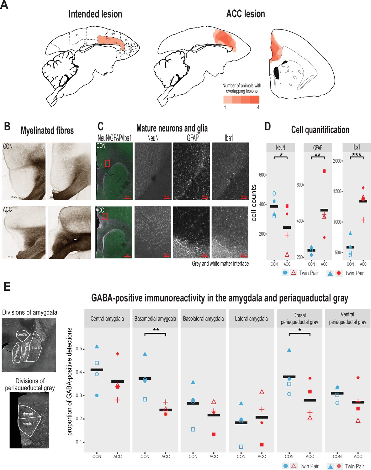
Lesion verification and impact of early life anterior cingulate cortex (ACC) lesion on vocal downstream structures.
(A) Left panel shows a sagittal section from the standard marmoset brain depicting the intended ACC lesion shaded in red. The right panel shows schematic lesion reconstructions superimposed on a sagittal and coronal marmoset brain section depicting the extent of the ACC lesion shaded in red. Regions that appear darker indicate greater overlap in the damage present among different animals. (B) Magnified images of area 24 stained to visualize myelinated fibers in a representative control (top panel) and ACC-lesioned (bottom panel) animal. The normal radial arrangement of the myelinated fibers is disrupted following the ACC lesion. (C) Histological visualization of mature neurons and glia in representative control (top row) and ACC-lesioned animal (bottom row). Leftmost image shows neurons (green), astrocytes (violet), and microglia/macrophages (white) in the same image. Red square represents the magnified grayscale sections showing cell loss (NeuN), high levels of astrocytes (GFAP), and microglia/macrophages (Iba1) surrounding the lesion site at the gray and white matter interface in the ACC-lesioned animal (bottom row) relative to the controls (top row). (D). Histological quantification of mature neurons and glia in cortical area 24 in the ACC-lesioned animals, or in the corresponding intact cortical tissue bordering white matter in the controls. There was a reduction in the number of mature neurons (NeuN) and an increase in glia (GFAP and Iba1) in the ACC group relative to controls. *p<0.05; **p<0.001; ***p<0.00001. (E) Left panel shows grayscale images with anti-NeuN staining depicting divisions of amygdala (AMY) and periaqueductal gray (PAG) where relative distribution of GABA-positive immunoreactive expression was quantified. Right graphs show the proportion of GABA expression in each division depicted in the AMY and PAG. Each symbol represents one animal (ACC-lesioned animal is red, control animal is blue). Mean expression is represented by a black bar. Data for two animals in the ACC group overlap for basomedial AMY quantification. GABA-positive immunoreactivity was significantly down in the basomedial AMY and dorsal PAG.
Figure 2—figure supplement 1
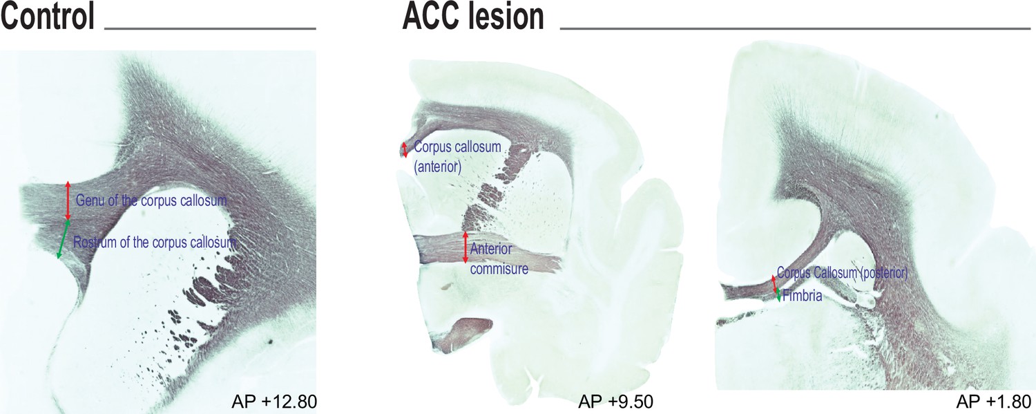
Representative images of white matter tract from a CON and anterior cingulate cortex (ACC)-lesioned subject.
Both the anterior and posterior corpus callosum are visibly narrowed in the area of the ACC lesion. The transverse widths of the white matter tracts were measured from sections stained for myelin at the following approximate rostrocaudal planes (in reference to the interaural axis): genu of the corpus callosum and rostrum of the corpus callosum (both +12.80 mm AP) were measured at the nadir of the overlaying cortex; anterior corpus callosum was measured at the midline and the anterior commissure was measured at the medial juncture with the internal capsule (both +9.50 mm AP); posterior corpus callosum and fimbriae were both measured at the junction with each other (both +1.80 mm AP).
Figure 3 with 1 supplement
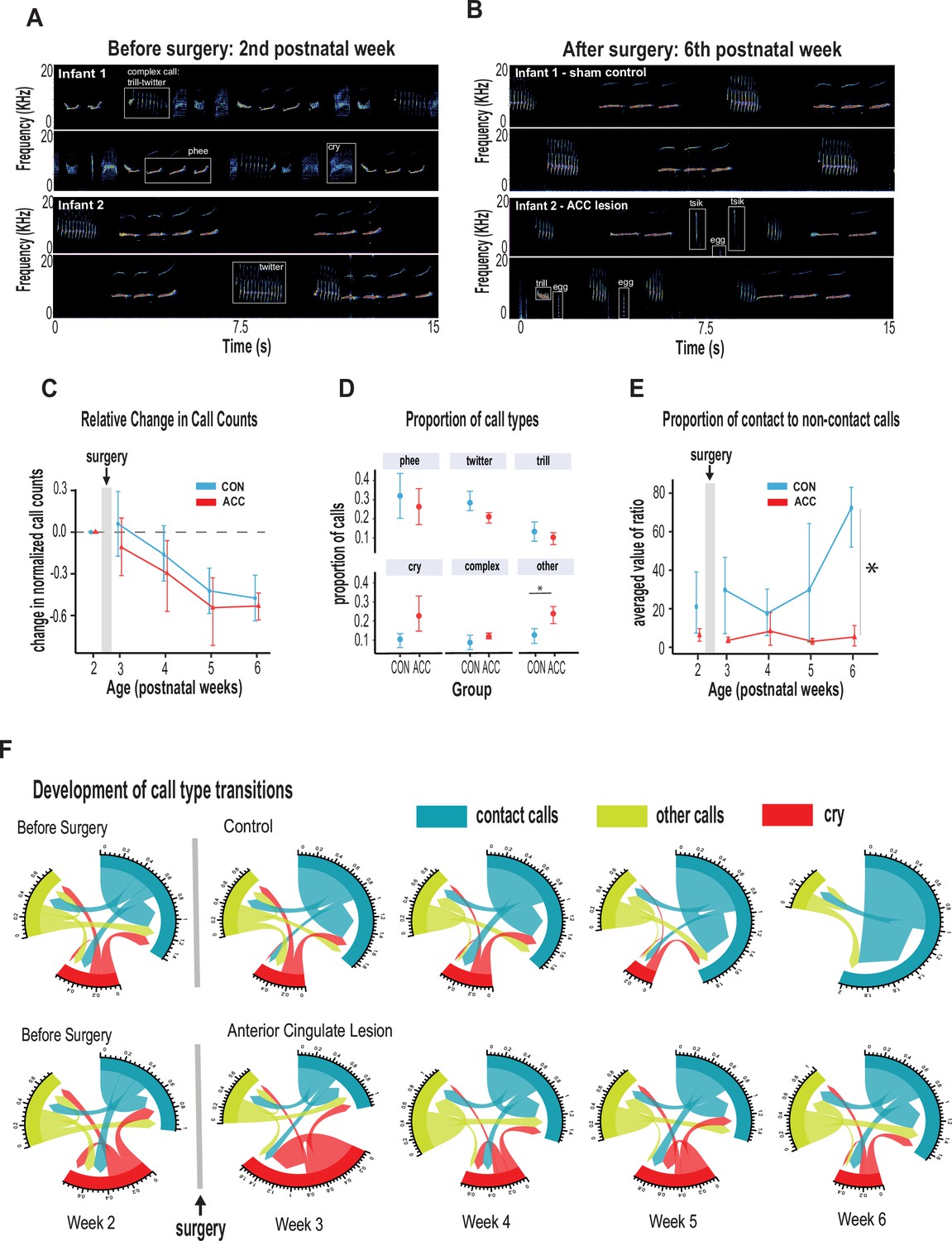
Anterior cingulate cortex (ACC) in early life is integral to postnatal development of social contact calls.
(A–B) Spectrograms show sample 30 s vocal recordings of a representative control and ACC-lesioned marmosets before (postnatal week 2) and after surgery (postnatal week 6). Before surgery, the infants ‘babbled’ by emitting a wide range of immature concatenated calls, each with its own spectrogram motif illustrated and labeled in boxes. After surgery, at postnatal week 6, calls show reduced variability separated by distinct gaps or inter-call intervals. (C) Both groups show a reduction in the relative call count with increasing age. (D) Average proportion of each call type pooled from week 3 to week 6 following surgery. Animals in both groups were able to emit calls of different call types. Those with ACC lesions made minor calls designated as ‘other’ more frequently than controls, but all major call types were produced at equivalent rates. (E) Proportion of mature contact calls relative to immature non-social contact calls. The y-axis represents the averaged value of the ratios of the number of social calls divided by the number of nonsocial calls: x̄ (# mature calls/# immature calls). Despite their ability to produce all call types, the proportion of mature contact calls comprising phee, twitter, and trills was substantially reduced in animals with early life ACC lesions at postnatal week 6. Due to factors beyond our control (COVID-19), the number of recordings varied between animals: week 3: CON n=5, ACC n=5; week 4: CON n=5, ACC n=4; week 5: CON n=4, ACC n=3; week 6: CON n=4, ACC n=3. (F) Chord diagrams illustrate the likelihood of transitioning between call types. At 6 weeks of age, animals with ACC lesions showed a higher likelihood of transitioning between all call types, but less frequent transitions between social contact calls relative to the sham group. The chord diagrams visualize the weighted probabilities and directionality of these transitions between different call types. Weighted probabilities were used to account for variations in call counts. The thickness of the arrows or links indicates the probability of a call transition, and the numbers surrounding each chord diagram represent the relative probability value for each specific transition.
Figure 3—figure supplement 1
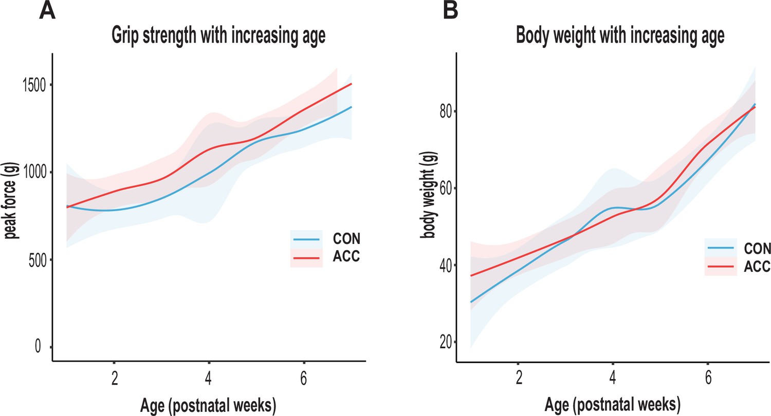
Physical factors in developing marmosets.
(A) Muscular strength was quantified using the Bioseb (BIO-GS4) grip strength monitor. The infant was allowed to grip onto a bar while being gently pulled backward in a horizontal plane to determine the maximal peak force (g). The anterior cingulate cortex (ACC) lesion did not cause any changes to forelimb muscular strength. (B) Average body weight (g) with increasing age. Animals in both groups showed comparable body weights with increasing age. CON (n=5), ACC (n=5).
Figure 4
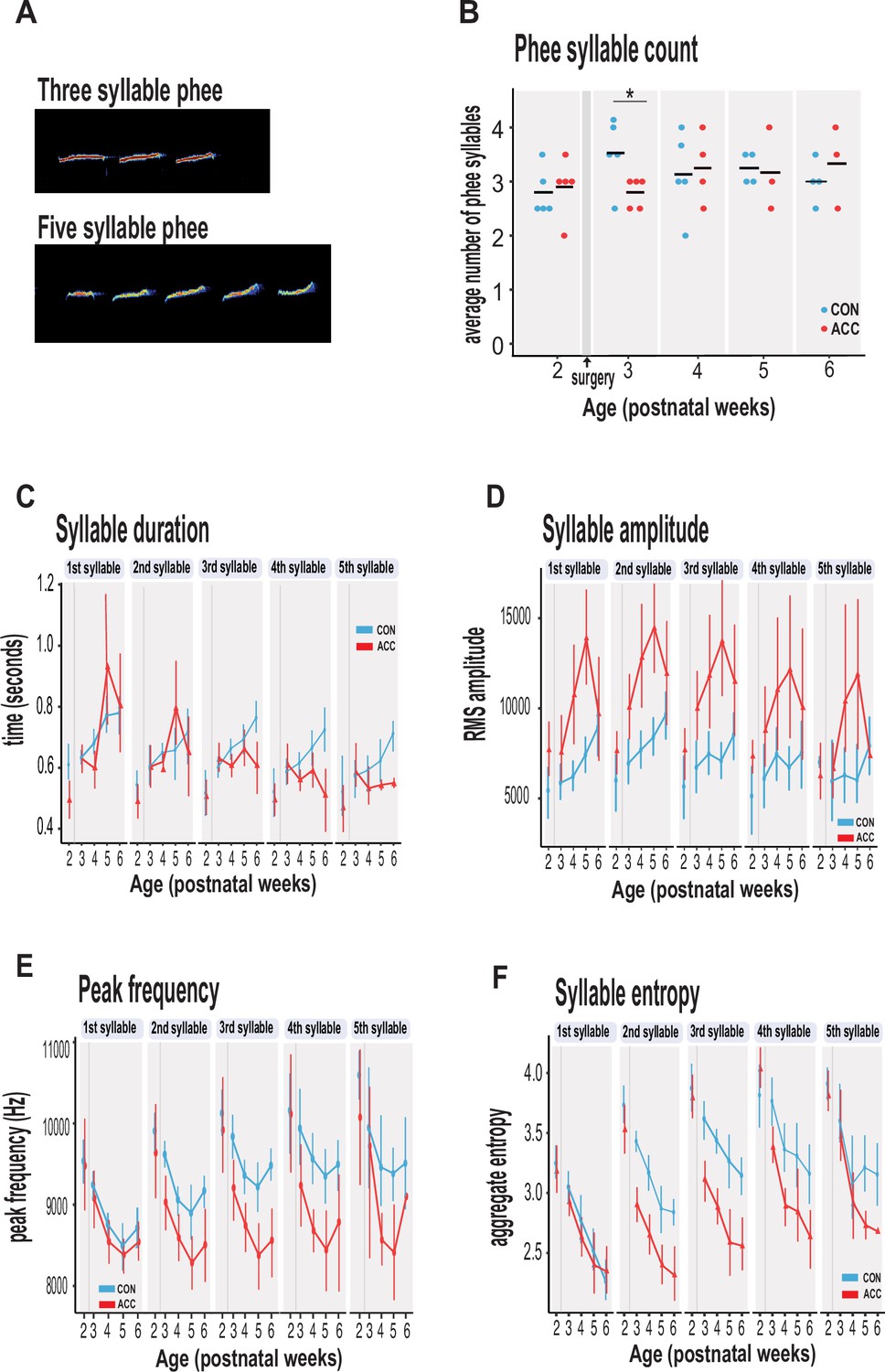
Anterior cingulate cortex (ACC) lesion alters structural characteristics of long-distance social phee calls.
(A) Sample spectrogram with examples of three and five syllable phee calls. (B) The ACC lesion caused a reduction in average phee syllable counts immediately after the ACC lesion (red dots) at postnatal week 3, but then normalized to 3–4 syllables thereafter. (C) Phee syllable duration in the ACC-lesioned group became shorter for multisyllabic phees ≥3, especially with increasing age. (D) The effective amplitude for each phee syllable increased for the ACC group until postnatal week 6. (E) Animals with ACC lesions emitted low entropy phees for calls as low as 2 syllables and continued until postnatal week 6. (F) Decrease in peak frequency of phee calls immediately following ACC lesion. Data in D–F represents average of specific syllable in a phee sequence irrespective of the number of syllables in a phee (e.g. 1 syllable phee, the 1st syllable in a 2-syllable phee, in a 3-syllable phee, and in a 4-syllable phee). Due to factors beyond our control (COVID-19), the number of recordings varied between animals: week 3: CON n=5, ACC n=5; week 4: CON n=5, ACC n=4; week 5: CON n=4, ACC n=3; week 6: CON n=4, ACC n=3. Error bars are confidence intervals. Gray shaded lines or bars represent time of surgery.
Additional files
Download links
A two-part list of links to download the article, or parts of the article, in various formats.
Downloads (link to download the article as PDF)
Open citations (links to open the citations from this article in various online reference manager services)
Cite this article (links to download the citations from this article in formats compatible with various reference manager tools)
Cingulate cortex shapes early postnatal development of social vocalizations
eLife 13:RP97125.
https://doi.org/10.7554/eLife.97125.3


