Single-cell dissection of prognostic architecture and immunotherap response in Helicobacter pylori infection-associated gastric cancer
- State Key Laboratory of Pathogen and Biosecurity, Institute of Biotechnology, Academy of Military Medical Sciences, China
- Department of Pharmacy, Medical Supplies Center, Chinese PLA General Hospital, China
- Center for Immune Ageing and Rejuvenation, Department of Rheumatology and Immunology, The First Affiliated Hospital of Chongqing Medical University, China
- Department of General Surgery, First Medical Center, Chinese PLA General Hospital, China
- Department of Gastroenterology and Hepatology, First Medical Center, Chinese PLA General Hospital, China
- Department of Pathology, Fourth Medical Center, Chinese PLA General Hospital, China
- Department of Nephrology, First Medical Center of Chinese PLA General Hospital, China
Figures
Figure 1 with 1 supplement
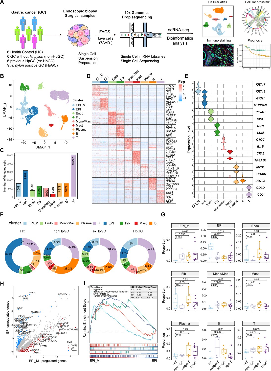
Global analysis of the tumor microenvironment and malignant features of H. pylori infection-associated gastric cancer (GC).
(A) A general workflow of GC sample preparation and processing of single-cell suspensions for scRNA-seq analysis. In total, 27 gastric samples, including gastric tissues of healthy control (HC, n=6), GC without H. pylori infection (non-HpGC, n=6), GC with previous H. pylori infection (ex-HpGC, n=6), and GC with current H. pylori infection (HpGC, n=9), were collected to perform scRNA-seq. (B) Uniform Manifold Approximation and Projection (UMAP) plot for unbiased clustering and cell type annotation of 86,637 high-quality cells. EPI_M: malignant epithelium; EPI: non-malignant epithelium; Endo: endothelium; Fib: fibroblast; Mono/Mac: monocyte/macrophage; Plasma: plasma cells; Mast: mast cells; B: B cells; T: T cells. (C) The absolute quantities of nine different cell types. (D) Heatmap showing the top eight differentially expressed genes (DEGs) of nine main cell types. (E) Violin plots showing the expression of signature genes of nine cell types. (F) Pie plot revealing the proportions of nine cell types in HC, non-HpGC, ex-HpGC and HpGC. (G) Box plot showing statistical analysis of proportion of nine cell types in HC, non-HpGC, ex-HpGC, and HpGC. The p-value of Student’s t-test is shown. (H) Volcano plot displaying the differentially upregulated genes in EPI and EPI_M. (I) Gene set enrichment analysis (GSEA) showing the pathway activity (scored per cell by GSEA) in malignant epithelium (EPI_M) and non-malignant epithelium (EPI).
Figure 1—figure supplement 1
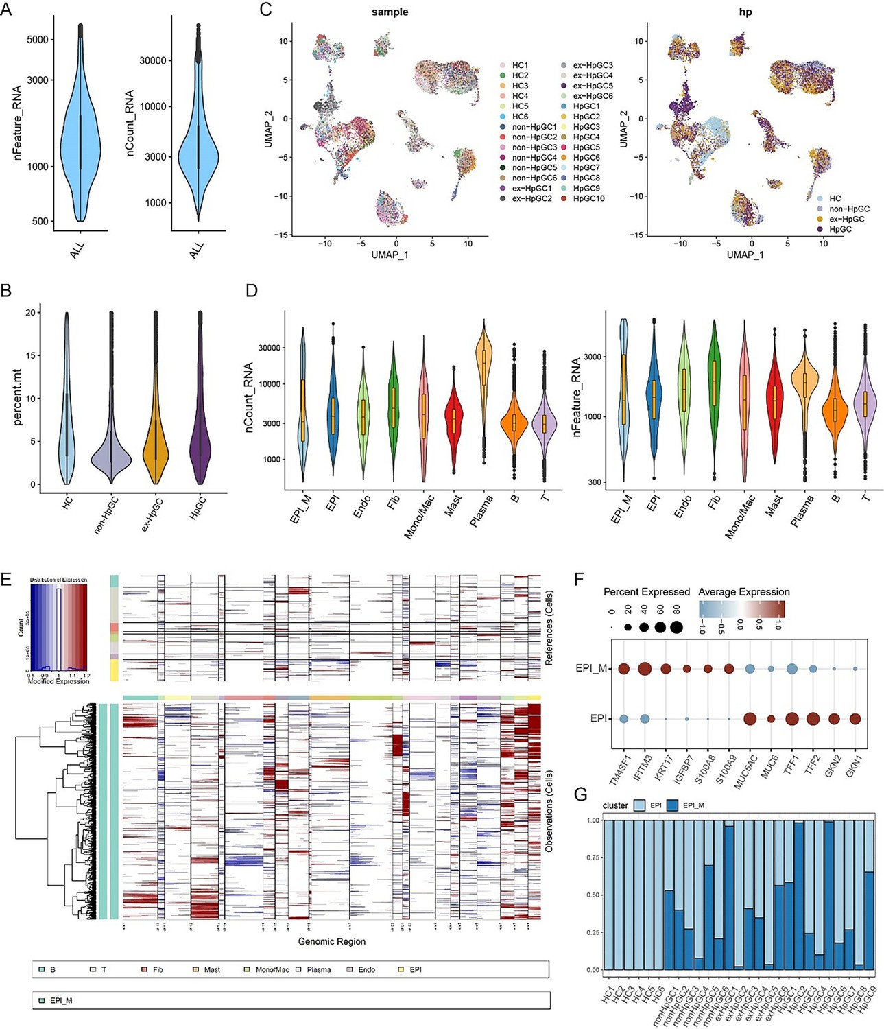
Data quality control and filtering criteria of gastric cancer (GC) scRNA-seq.
(A) The number of detected genes and UMI counts in all the cells. (B) The percentage of mitochondria genome in gastric mucosal samples of HC, non-HpGC, ex-HpGC, and HpGC. (C) Uniform Manifold Approximation and Projection (UMAP) plot showing the sample (right) and pathological (left) distributions of 83,637 high-quality cells. (E) The copy number variation (CNV) signal across cell types, estimated using inferCNV. (F) The expression of differentially expressed genes (DEGs) in assumed non-malignant epithelium (EPI) and malignant epithelium (EPI_M). (G) Bar plot showing the percentage of EPI and EPI_M in each gastric mucosal sample of HC, non-HpGC, ex-HpGC, and HpGC.
Figure 2 with 1 supplement
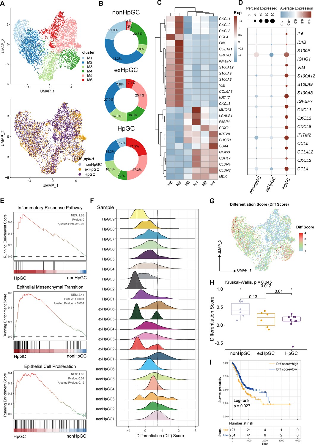
Characterization of the malignant epithelium within gastric cancer (GC) with different H. pylori infection status.
(A) Uniform Manifold Approximation and Projection (UMAP) plot showing six subpopulations of malignant epithelium, colored by different cell types (upper) and different H. pylori infection status (lower). (B) Pie plot showing the proportion of six subsets of malignant epithelium in non-HpGC, ex-HpGC, and HpGC. (C) Heatmap displaying the differentially expressed genes (DEGs) among the six subsets of malignant epithelium. (D) Bubble plot showing the difference in representative molecular among the non-HpGC, ex-HpGC, and HpGC. (E) Gene set enrichment analysis (GSEA) showing the pathway activity (scored per cell by GSEA) in malignant epithelium of HpGC compared to that of non-HpGC. NES, normalized enrichment score. (F) The ridge plot showing the differentiation score (Diff Score) of malignant epithelium within each sample. (G) Heatmap showing the Diff Score of six subpopulations of malignant epithelium. (H) Box plot showing differentiation score among non-HpGC, ex-HpGC, and HpGC, p-values were assessed by Wilcoxon test, two-way ANOVA test is used for comparison of multiple groups. (I) Kaplan–Meier survival analysis of The Cancer Genome Atlas (TCGA) stomach adenocarcinoma (STAD) patients stratified by tumor sample differentiation scores, which was used to group samples into high and low groups based on 33rd and 67th percentile. The p-value of two-sided log-rank test is shown.
Figure 2—figure supplement 1
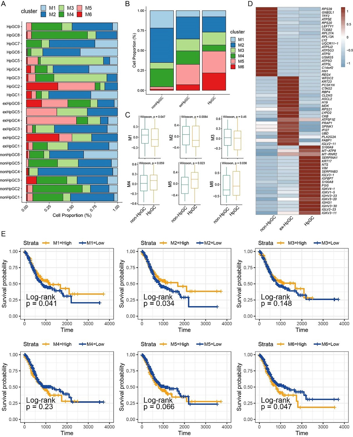
Malignant epithelium characteristic in gastric cancer (GC) with different H. pylori infection status.
(A, B) Proportion of six malignant epithelium subtypes in each GC sample (A) and non-HpGC, ex-HpGC, and HpGC(B). (C) The malignant epithelium subtypes relative abundance in H. pylori infection-associated GC using The Cancer Genome Atlas (TCGA) stomach adenocarcinoma (STAD) samples (estimated by gene set variation analysis [GSVA]). p-Values were assessed by Wilcoxon test. (D) Heatmap showing expression of top 20 differentially expressed genes (DEGs) in distinct malignant epithelium subtypes. (E) Kaplan–Meier survival analysis of TCGA STAD patients stratified by the relative abundance of six malignant epithelium subclusters, which was used to group samples into high and low groups based on 33rd and 67th percentile. The p-value of two-sided log-rank test is shown.
Figure 3
Characterization of the non-malignant epithelium within gastric cancer (GC) with different H. pylori infection status.
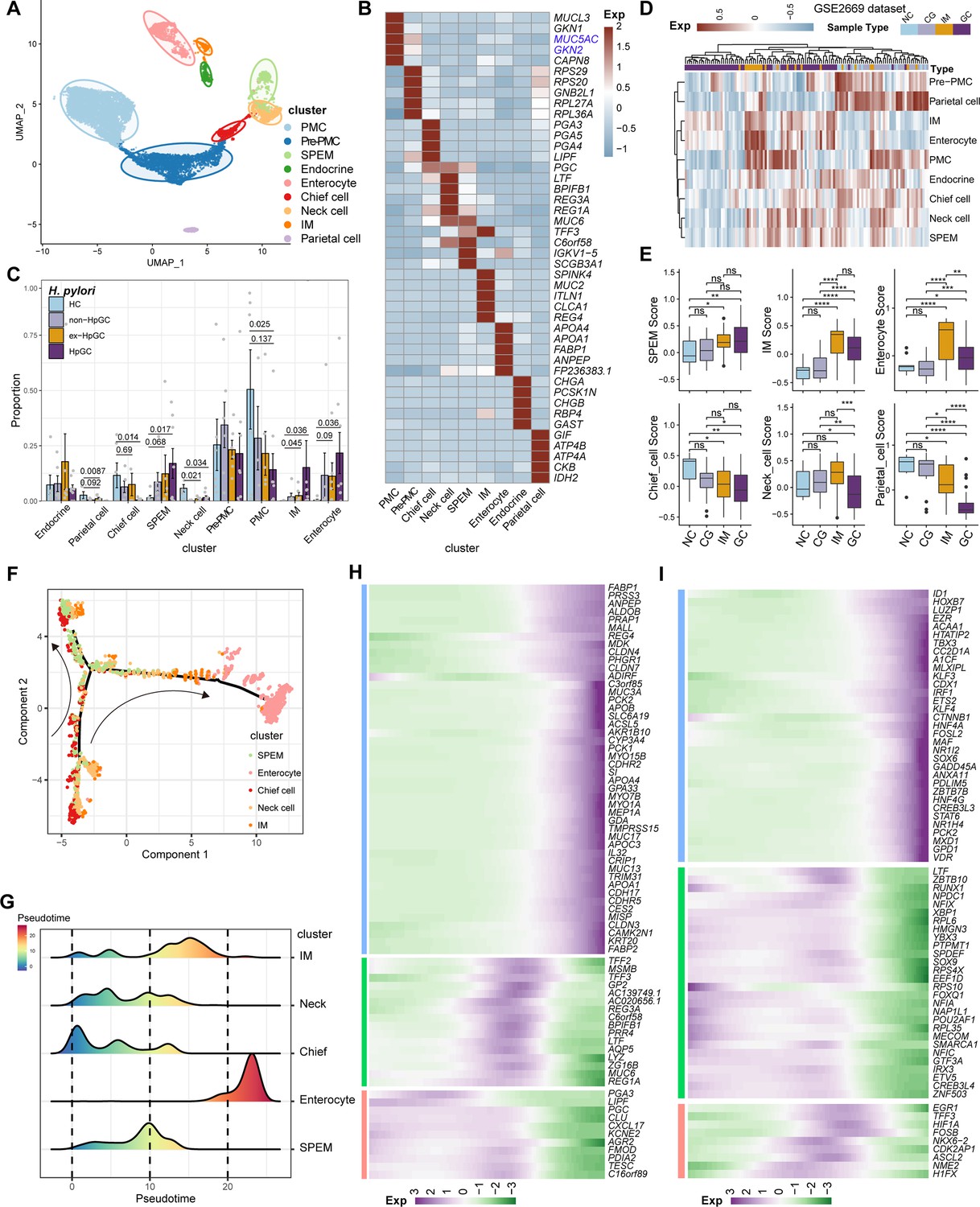
(A) Unbiased clustering of non-malignant epithelium generated nine subtypes. (B) Heatmap showing the molecular feature of non-malignant epithelium according to the top five differentially expressed genes (DEGs). (C) Box plot showing the dynamic proportion of different cell types in non-malignant epithelium with different H. pylori infection status. (D) Heatmap showing the relative abundance (estimated by gene set variation analysis [GSVA]) of non-malignant epithelium subtypes in normal control (NC), chronic gastritis (CG), intestinal metaplasia (IM), GC samples (GSE2669). (E) Boxplot showing the relative abundance (estimated by GSVA) of non-malignant epithelium subtypes SPEM, IM, enterocytes, chief cells, neck cells, and parietal cells within NC, CG, IM, and GC samples (GSE2669). The p-value of Student’s t-test is shown. (F) The trajectory analysis shows potential differentiation and transition trajectories in non-malignant epithelium clusters. (G) The ridge plot showing the pseudotime of non-malignant epithelium revealed the gastric pre-lesion process. (H, I) Heatmap showing scaled expression of dynamic genes (G) and TFs (H) along cell pseudotime.
Figure 4 with 1 supplement
Characterization of tumor-infiltrating T and natural killer (NK) cells in H. pylori infection-associated gastric cancer (GC).
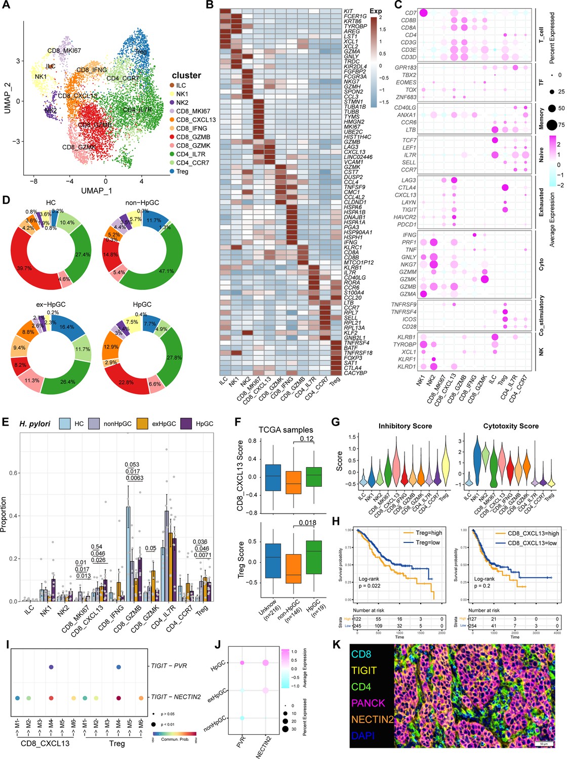
(A) Unbiased clustering of T and NK cells generated 11 clusters. (B, C) Molecular features annotations according to the top five differentially expressed genes (DEGs) (B), and representative genes (C). (D) Pie plot showing the T/NK cell subtype abundance distribution in HC, non-HpGC, ex-HpGC, and HpGC. (E) The percentage contribution of T/NK cell subtype in HC, non-HpGC, ex-HpGC, and HpGC samples. p-Values were assessed by Student’s t-test. (F) The deconvolution analysis showing the relative abundance of CD8_CXCL13 and Tregs in GC with different H. pylori infection status with The Cancer Genome Atlas (TCGA) stomach adenocarcinoma (STAD) dataset, p-values were assessed by Wilcoxon test. (G) The cytotoxic and inhibitory expression scores in T and NK clusters. (H) Kaplan–Meier survival analysis of TCGA STAD patients stratified by CD8_CXCL13 and Tregs relative abundance, which was used to group samples into high and low groups based on 33rd and 67th percentile. The p-value of two-sided log-rank test is shown. (I) Bubble plot showing intercellular interactions between suppressive T cells and malignant cells. (J) Dot plot showing the expression of NECTIN2 and PVR in malignant non-HpGC, ex-HpGC, and HpGC cells. (K) Immunostaining showing the ligand TIGIT expressed in suppressive T cells and the receptor NECTIN2 expressed on the malignant epithelium, respectively, in one HpGC sample.
Figure 4—figure supplement 1
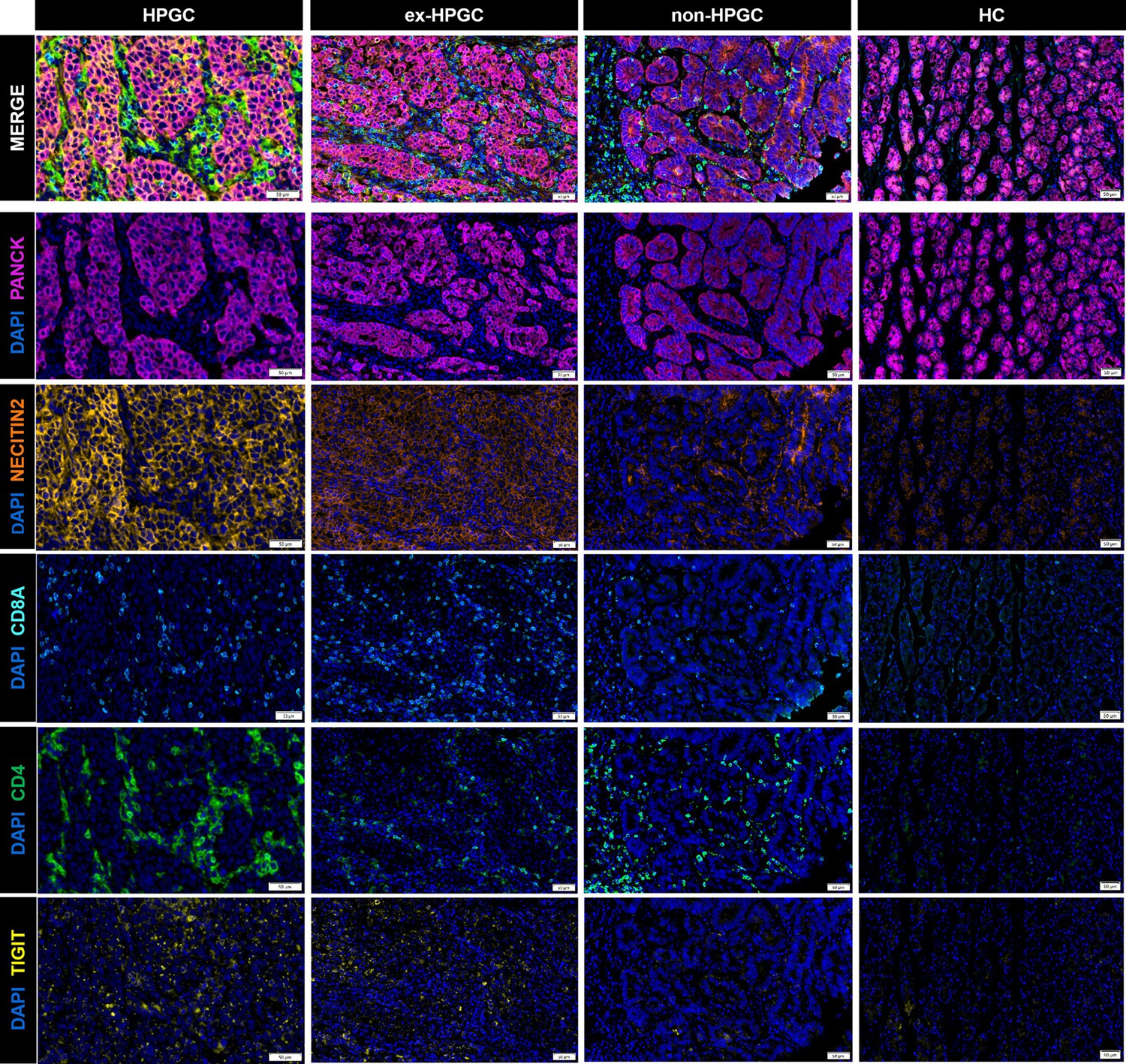
Immunostaining of the ligand TIGIT and the receptor NECTIN 2 on suppressive T cells and the malignant epithelium, respectively, in HpGC, ex-HpGC, non-HpGC, and HC.
Figure 5 with 2 supplements
Characterization of tumor-infiltrating myeloid cells by scRNA-seq in H. pylori infection-associated gastric cancer (GC).
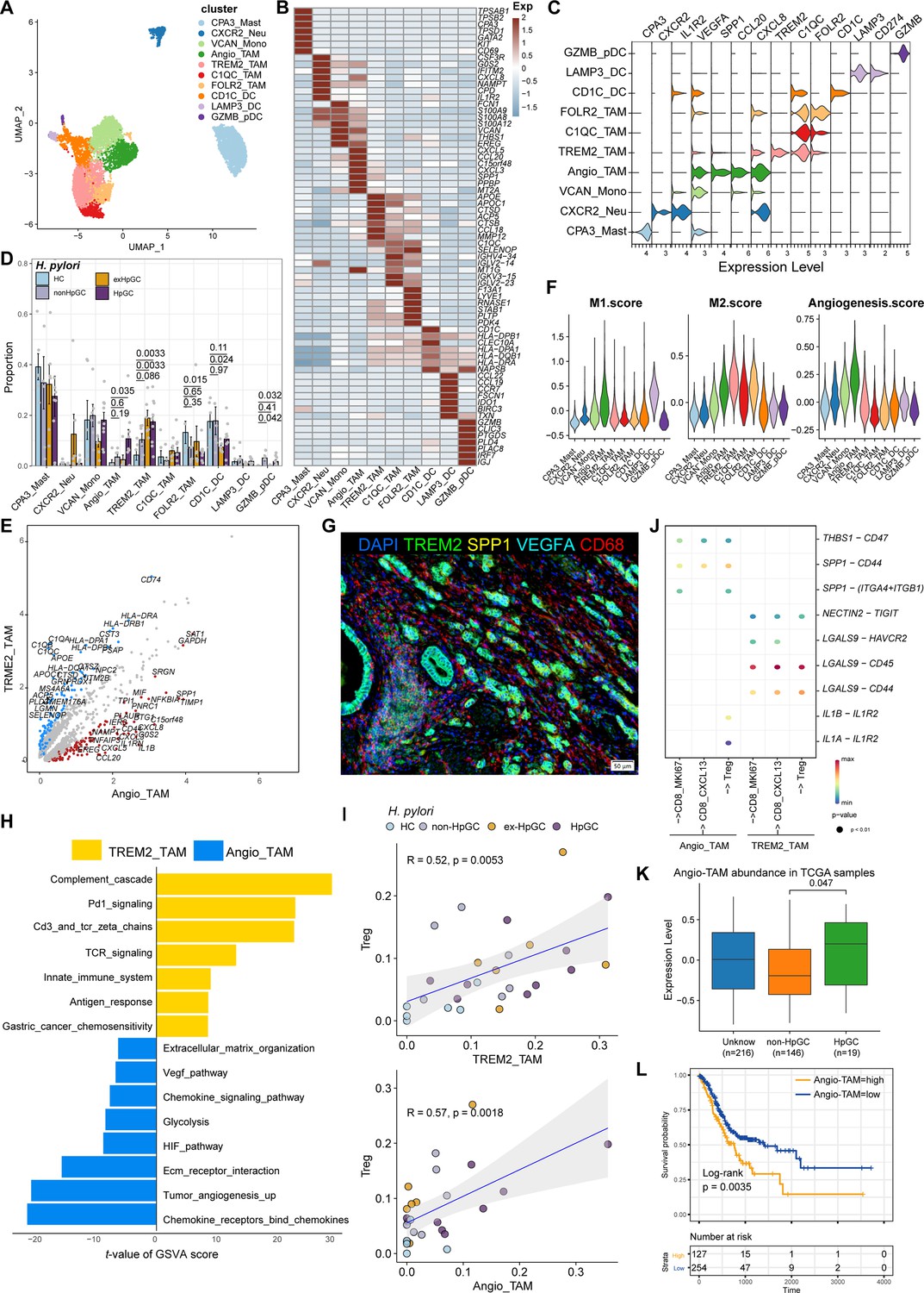
(A–C) Unbiased clustering of myeloid cells generated nine clusters (A), and molecular features were annotated according to the top seven differentially expressed genes (DEGs) (B) and representative genes (C). (D) The percentage contribution of each myeloid cell cluster in HC, non-HpGC, ex-HpGC, and HpGC. p-Values were assessed by Student’s t-test. (E) Volcano plot showing the DEGs between Angio-TAM and TREM2+ TAM. (F) Violin plot showing the expression of functional gene sets in myeloid clusters. (G) Immunostaining showing the distribution of Angio-TAM and TREM2+ TAM in one HpGC sample. (H) Bar plot showing the enriched signaling pathway between Angio-TAM and TREM2+ TAM. (I) The correlation of cell type (percentage) between Tregs and Angio-TAM and TREM2+ TAM. (J) Dotplot showing intercellular interactions among suppressive T cells and Angio-TAM and TREM2+ TAM. (K) The relative abundance of Angio-TAM in HpGC and non-HpGC in the The Cancer Genome Atlas (TCGA) stomach adenocarcinoma (STAD) dataset, p-values were assessed by the Wilcoxon test. (L) Kaplan–Meier survival analysis of TCGA STAD patients stratified by Angio-TAM relative abundance, which was used to group samples into high and low groups based on the 33rd and 67th percentiles. The p-value of the two-sided log-rank test is shown.
Figure 5—figure supplement 1
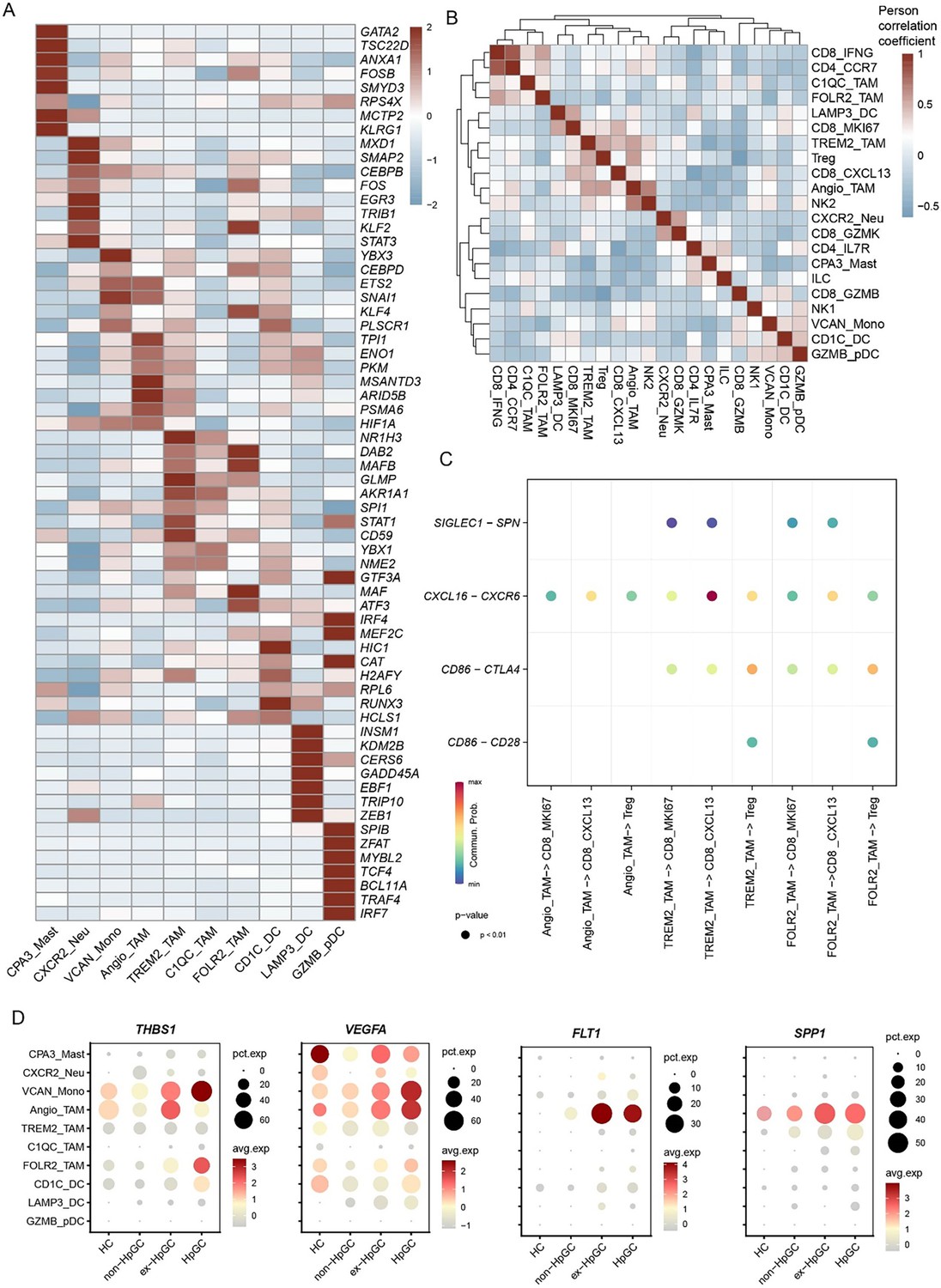
Myeloid cell characteristics in gastric cancer (GC) with different H. pylori infection status.
(A) Heatmap showing the top seven TFs in different myeloid cell subtypes. (B) Heatmap showing the Pearson correlation coefficient of myeloid cell types and T/NK cell subtype abundance. (C) Bubble plot showing the expression of ligand–receptor gene pairs associated with immune signaling pathway networks: CD86, SN, and CXCL signaling pathways involved in the interactions between TAM and T cell subtypes. (D) Dot plot showing the expression of THBS1, VEGFA, FLT1 (VEGFR1), and SPP1 in TAM derived from HC and GC with distinct H. pylori infection status.
Figure 5—figure supplement 2
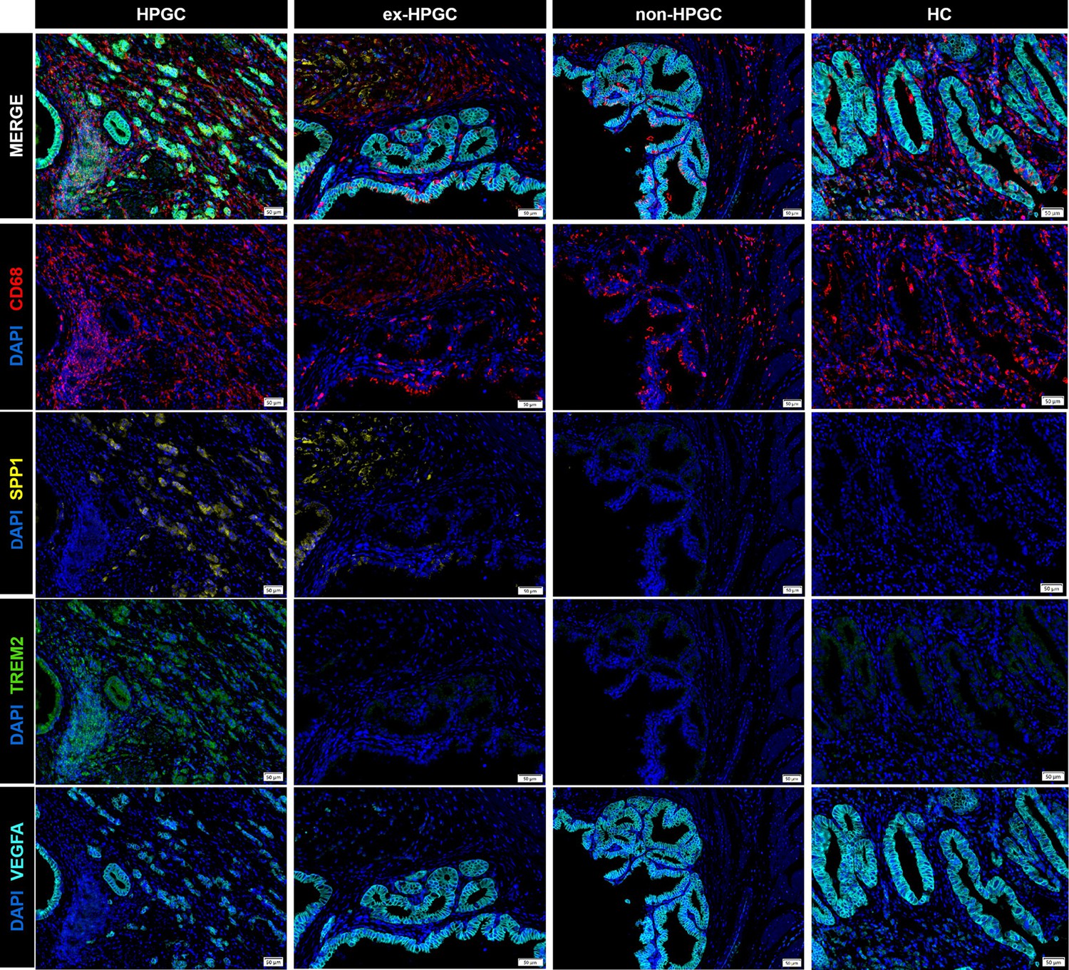
Immunostaining showing the expression of Angio-TAM and TREM2+ TAM, respectively, in HpGC, ex-HpHC, non-HpGC, and HC.
Figure 6 with 1 supplement
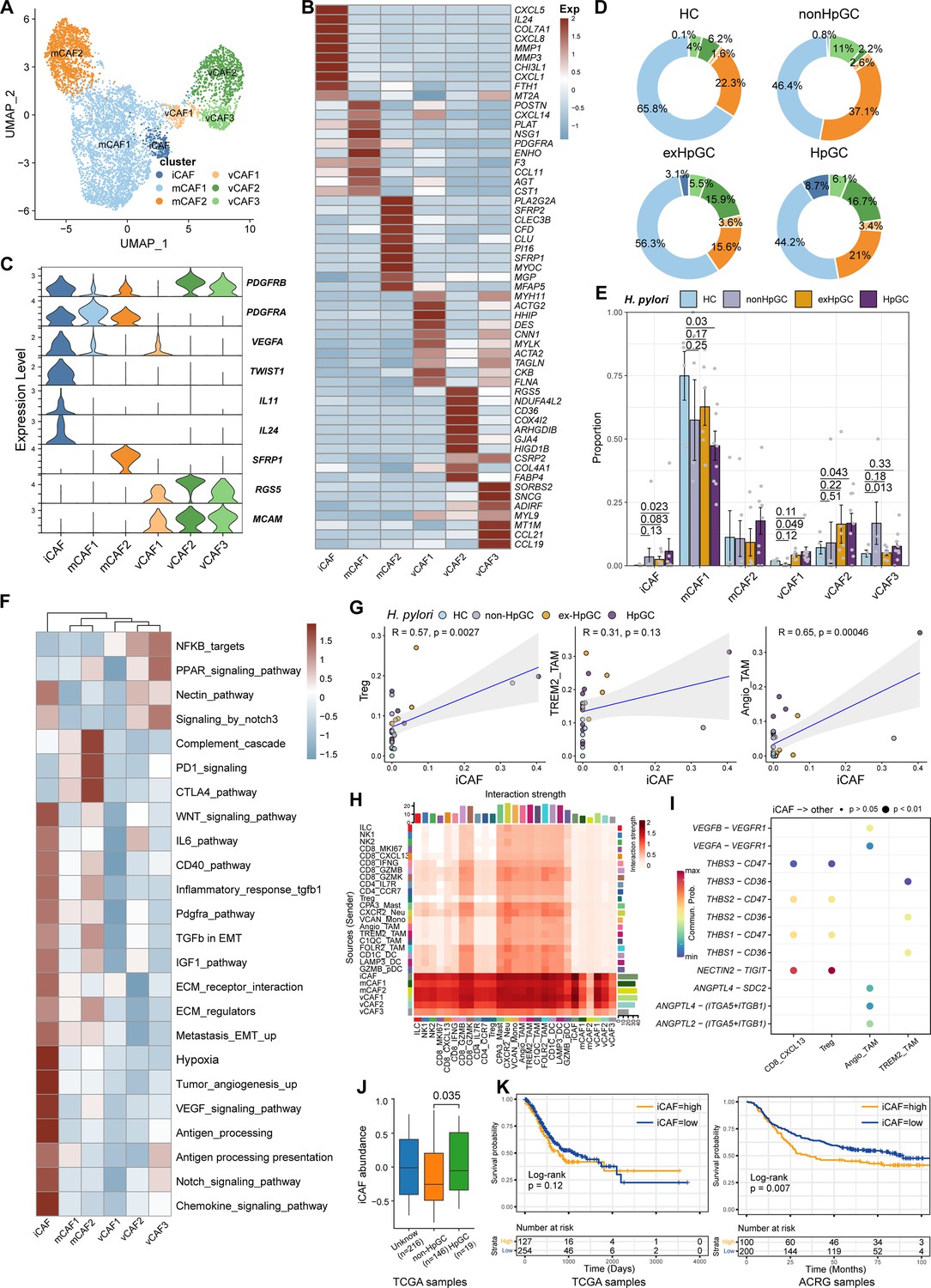
Characterization of cancer-associated fibroblasts (CAFs) by scRNA-seq in H. pylori infection-associated gastric cancer (GC).
(A) Unbiased clustering of CAF generated six clusters. (B, C) Molecular features annotations according to the top ten differentially expressed genes (DEGs) (B) and representative genes (C). (D) The pie plot showing the abundance distribution of six cancer-associated fibroblast (CAF) subset in HC, non-HpGC, ex-HpGC, and HpGC. (E) The percentage contribution of each CAF cluster in HC, non-HpGC, ex-HpGC, and HpGC. p-Values were assessed by Student’s t-test. (F) Heatmap showing the enriched signaling pathway among six CAF clusters. (G) The cell type percentage correlation between iCAF and Treg, Angio-TAM, and TREM2+ TAM. (H) Heatmap showing intercellular interaction strength among immune cells and different subsets of CAF. (I) Enriched signaling pathway among suppressive T cell, Angio-TAM, and TREM2+ TAM with iCAF. (J) The relative abundance of iCAF in HpGC and non-HpGC in TCGA STAD dataset. p-Values were assessed by Wilcoxon test. (K) Kaplan–Meier plot shows that the abundance of iCAF predicts poor prognosis of GC using two public bulk RNA sequencing dataset (left, TCGA; right, ACRG). The abundance of iCAF was used to group samples into high and low groups based on 33rd and 67th percentile. The p-value of two-sided log-rank test is shown. ACRG: Asian Cancer Research Group.
Figure 6—figure supplement 1
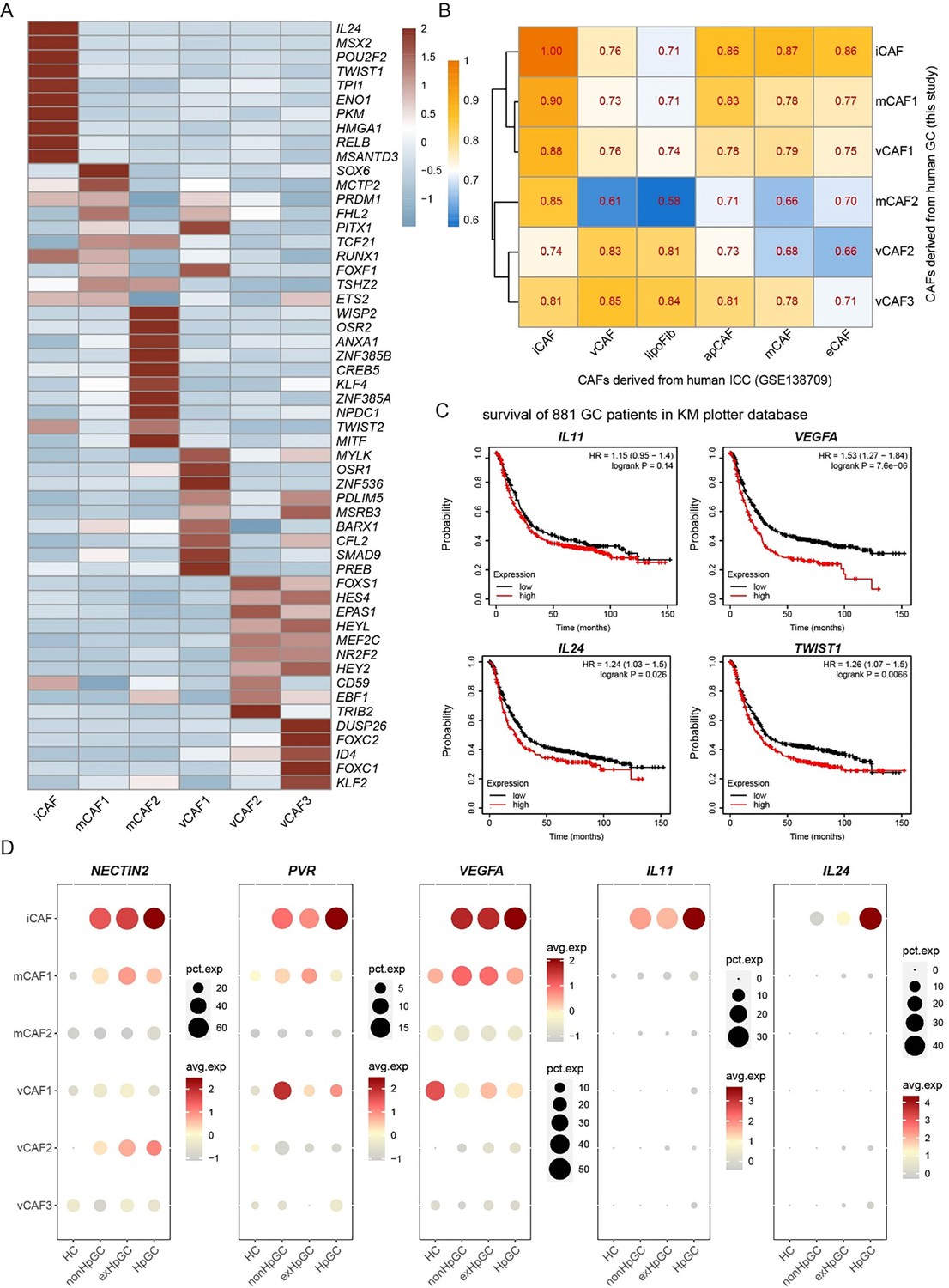
Cancer-associated fibroblast (CAF) subtypes characteristic in gastric cancer (GC) with different H. pylori infection status.
(A) Heatmap showing the top five TFs in different CAF subtypes. (B) Heatmap showing the Pearson correlation coefficient of CAF subtypes in human GC and ICC (GSE138709). (C) Kaplan–Meier survival analysis of 881 GC patients (km plotter database) stratified by high and low expression of iCAF associated genes IL11, IL23, VEGFA, TWIST1. The p-value of two-sided log-rank test is shown. (D) Dot plot showing the expression of IL11, IL24, PVR, VEGFA, and NECTIN2 in CAF subtypes derived from HC and GC with distinct H. pylori infection status.
Figure 7 with 1 supplement
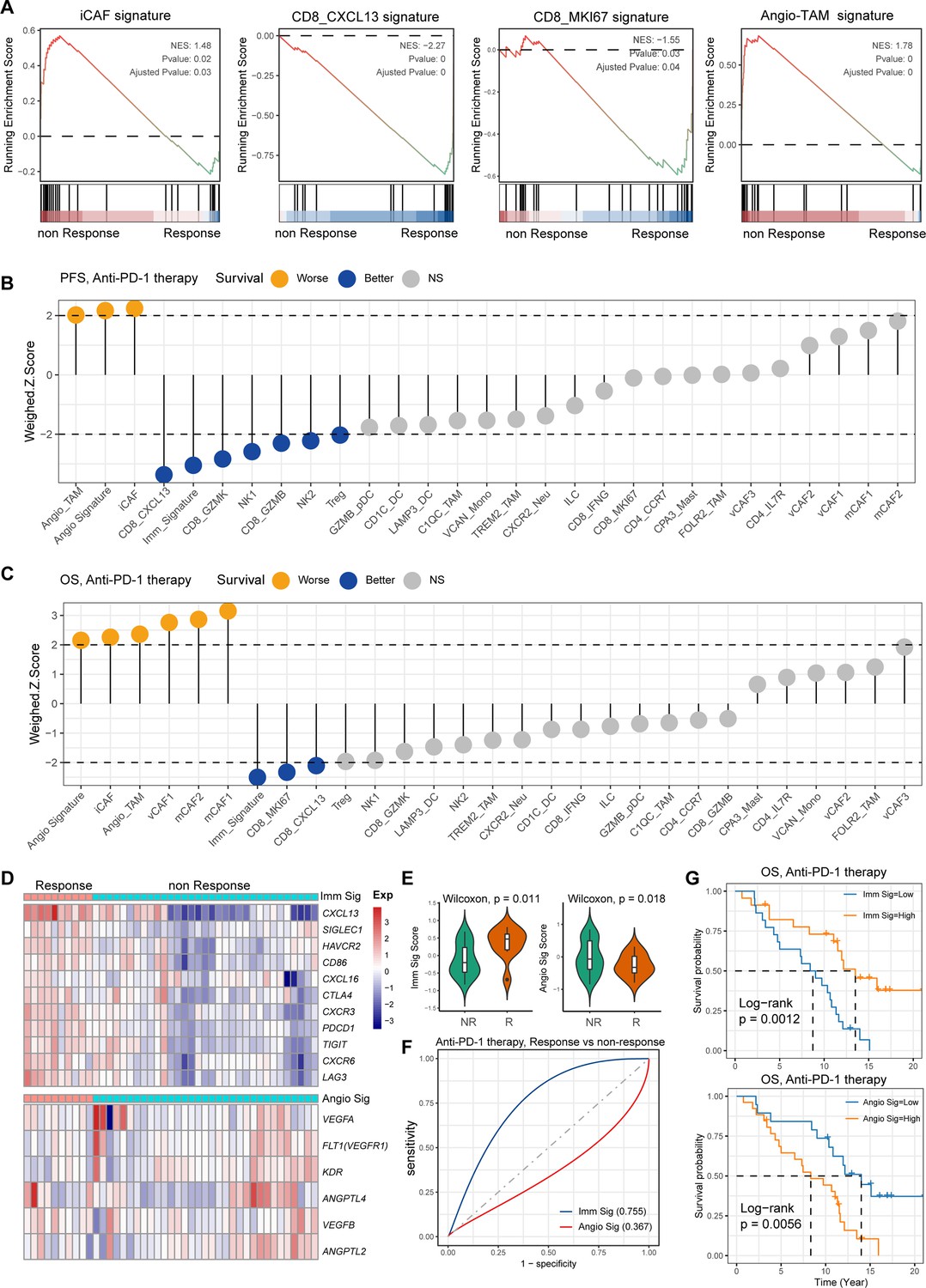
Single-cell tumor microenvironment (TME) composition associated with gastric cancer (GC) immunotherapy outcome.
(A) Gene set enrichment analysis (GSEA) plot showing the enrichment of iCAF, CD8_MKI67, Angio-TAM, and CD8_CXCL13 in anti-PD-1-responsive or non-responsive GC. NES, normalized enrichment score. (B, C) Bar chart showing the cell subtypes relative abundance, the immune signature, angiogenesis signature derived from scRNA-seq predicted GC immunotherapy outcome, progression-free survival (B), and overall survival (C). (D) Heatmap showing the expression of immune and angiogenesis signature derived from scRNA-seq in immunotherapy responsive and non-responsive GC. (E) Violin plot showing the expression of immune signature and angiogenesis signature in immunotherapy responsive and non-responsive GC. The p-value of Wilcoxon test is shown. (F) A model for evaluating the GC immunotherapy sensitivity and specificity using the immune signature and angiogenesis signature derived from scRNA-seq. (G) The Kaplan–Meier plot showing the immune signature and angiogenesis signature could efficiently predict prognosis of GC anti-PD-1 therapy. The p-value of two-sided log-rank test is shown.
Figure 7—figure supplement 1
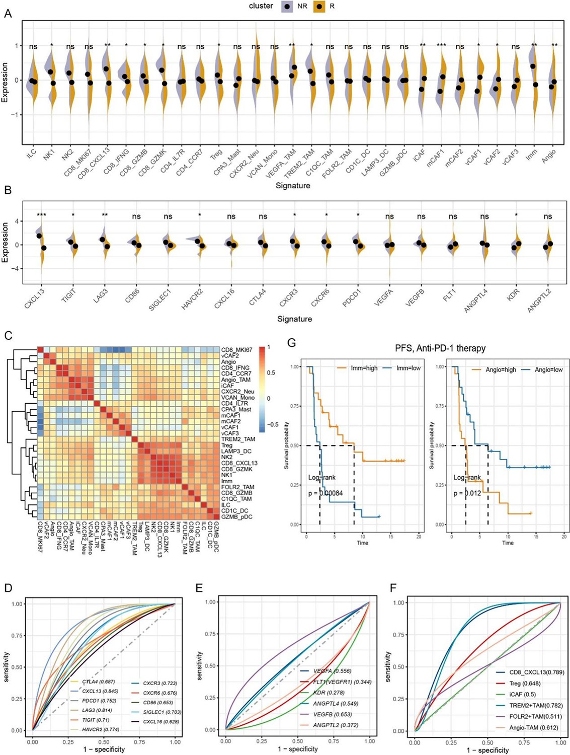
Gastric cancer (GC) tumor microenvironment (TME) characteristics related to GC immunotherapy response.
(A) Relative abundances of cell types identified using scRNA-seq data predicts GC immunotherapy efficacy, R: responsive, NR: non-responsive. p-Values were assessed by Wilcoxon test. (B) The immune and angiogenesis signature identified using scRNA-seq data predicts GC immunotherapy efficacy, R: responsive, NR: non-responsive. p-Values were assessed by Wilcoxon test. (C) Heatmap showing the Pearson correlations between cell subtypes and immune and angiogenesis signature identified in the GC TME. (D−F) Evaluation of the sensitivity of GC to immunotherapy based on immune signature, angiogenesis signature, and cell component. (G) Kaplan–Meier plot shows that the expression of Angio and Immune signature predicts anti-PD1 immunotherapy response (PFS) of GC.
Author response image 1

A.
The dot plot illustrates the enrichment of the TIGIT-PVR/NECTIN axis in the interaction between malignant epithelial cells and immunosuppressive T cells. B. T Dotplot showing the expression of NECTIN2 and PVR in non-HpGC, ex-HpGC, and HpGC cells. C. The bubble plot showing the expression of NECTIN, PVR, VEGF, IL11 and IL24 in the CAF within non-HpGC, ex-HpGC, and HpGC sample. D. The correlation of cell type (percentage) between Tregs, Angio-TAM, TREM2+ TAM and iCAF.
Tables
Table 1
Patient characteristics of each sample in gastric cancer (GC) scRNA-seq.
| Sample | Stage | Age | Sex | Histopathological diagnosis | Site of origin | Lauren’s classification | H. pylori serum antibody | H. pylori DNA | H. pylori cagA | H. pylori on H&E slide |
|---|---|---|---|---|---|---|---|---|---|---|
| exHpGC1 | III | 62 | F | Moderately differentiated adenocarcinoma | Gastric antrum and corpus | Intestinal | + | - | - | - |
| exHpGC2 | III | 61 | M | Moderately differentiated adenocarcinoma | Gastric antrum | Mixed | + | - | - | - |
| exHpGC3 | I | 75 | M | Poorly differentiated adenocarcinoma | Gastric antrum | Diffuse | + | - | - | - |
| exHpGC4 | II | 67 | F | Signet ring cell carcinoma | Gastric body | Diffuse | + | - | - | - |
| exHpGC5 | III | 64 | M | Moderately poorly differentiated adenocarcinoma | Cardia | Intestinal | + | - | - | - |
| exHpGC6 | I | 65 | M | Moderately differentiated adenocarcinoma | Gastric antrum | Intestinal | + | - | - | - |
| HpGC1 | II | 73 | M | Moderately poorly differentiated adenocarcinoma | Stomach angle | Intestinal | + | + | - | + |
| HpGC2 | IV | 55 | M | Poorly differentiated adenocarcinoma | Gastric antrum and corpus | Diffuse | + | + | - | + |
| HpGC3 | I | 49 | F | Moderately-well differentiated adenocarcinoma | Gastric antrum and gastric angle | Intestinal | + | + | - | + |
| HpGC4 | I | 47 | M | Poorly differentiated adenocarcinoma, mostly signet ring cell carcinoma | Gastric antrum | Diffuse | + | + | - | + |
| HpGC5 | II | 67 | F | Signet ring cell carcinoma | Gastric antrum | Diffuse | + | + | - | + |
| HpGC6 | I | 58 | M | Moderately poorly differentiated adenocarcinoma | Greater curvature | Mixed | + | + | + | + |
| HpGC7 | III | 67 | F | Moderately poorly differentiated adenocarcinoma | Angular incisure | Intestinal | + | + | N/A | + |
| HpGC8 | III | 74 | M | Moderately poorly differentiated adenocarcinoma | Greater curvature | Intestinal | + | + | N/A | + |
| HpGC9 | II | 62 | M | Poorly differentiated adenocarcinoma, partial signet ring cell carcinoma | Angular incisure | Mixed | + | + | N/A | + |
| nonHpGC1 | I | 54 | F | Poorly differentiated adenocarcinoma | Cardia | Diffuse | - | - | N/A | - |
| nonHpGC2 | III | 65 | M | Moderately differentiated adenocarcinoma | Cardia | Intestinal | - | - | N/A | - |
| nonHpGC3 | III | 56 | M | Moderately differentiated adenocarcinoma | Cardia | Intestinal | - | - | N/A | - |
| nonHpGC4 | III | 72 | M | Moderately poorly differentiated adenocarcinoma | Angular incisure | Intestinal | - | - | N/A | - |
| nonHpGC5 | I | 56 | M | Poorly differentiated adenocarcinoma | Antrum | Mixed | - | - | N/A | - |
| nonHpGC6 | II | 63 | M | Poorly differentiated adenocarcinoma | Angular incisure | Mixed | - | - | N/A | - |
| HC1 | N/A | 38 | M | Normal | Greater curvature | N/A | - | - | N/A | - |
| HC2 | N/A | 43 | F | Normal | Greater curvature | N/A | - | - | N/A | - |
| HC3 | N/A | 62 | F | Chronic gastritis | Gastric body | N/A | - | - | - | - |
| HC4 | N/A | 61 | F | Normal | Gastric antrum | N/A | - | - | - | - |
| HC5 | N/A | 25 | M | Normal | Gastric antrum | N/A | - | - | - | - |
| HC6 | N/A | 32 | F | Normal | Gastric antrum | N/A | - | - | - | - |
Additional files
-
Supplementary file 1
Top 30 DEGs of nine main cell types.
- https://cdn.elifesciences.org/articles/99337/elife-99337-supp1-v1.xlsx
-
Supplementary file 2
Top 30 DEGs of six malignant epithelium subclusters.
- https://cdn.elifesciences.org/articles/99337/elife-99337-supp2-v1.xlsx
-
Supplementary file 3
Top 30 DEGs of nine non-malignant epithelium subclusters.
- https://cdn.elifesciences.org/articles/99337/elife-99337-supp3-v1.xlsx
-
Supplementary file 4
Top 30 DEGs of T/NK subclusters.
- https://cdn.elifesciences.org/articles/99337/elife-99337-supp4-v1.xlsx
-
Supplementary file 5
Top 30 DEGs of myeloid cells subclusters.
- https://cdn.elifesciences.org/articles/99337/elife-99337-supp5-v1.xlsx
-
Supplementary file 6
Top 30 DEGs of cancer-associated fibroblasts subclusters.
- https://cdn.elifesciences.org/articles/99337/elife-99337-supp6-v1.xlsx
-
Supplementary file 7
Multilabel immunofluorescence staining antibody.
- https://cdn.elifesciences.org/articles/99337/elife-99337-supp7-v1.xlsx
-
MDAR checklist
- https://cdn.elifesciences.org/articles/99337/elife-99337-mdarchecklist1-v1.pdf
Download links
A two-part list of links to download the article, or parts of the article, in various formats.
Downloads (link to download the article as PDF)
Open citations (links to open the citations from this article in various online reference manager services)
Cite this article (links to download the citations from this article in formats compatible with various reference manager tools)
Single-cell dissection of prognostic architecture and immunotherap response in Helicobacter pylori infection-associated gastric cancer
eLife 13:RP99337.
https://doi.org/10.7554/eLife.99337.3


