Genetic inactivation of the β1 adrenergic receptor prevents cerebral cavernous malformations in zebrafish
- Department of Medicine, University of California, San Diego, United States
Figures
Figure 1 with 2 supplements
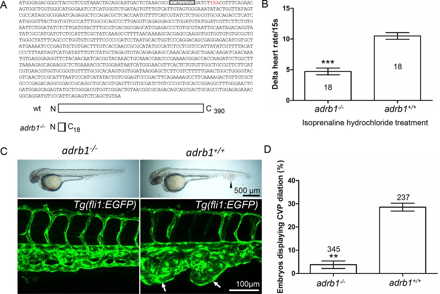
Adrb1 signaling is essential for CVP dilation.
(A) The targeted adrb1 allele shows an 8-nucleotide deletion producing a pre-stop codon. Adrb1 null cDNA is predicted to encode truncated adrb1 protein. The wild type adrb1 protein contains 390 amino acids, while the predicted adrb1 null protein would contain 2 missense amino acids (gray bar) and would terminate after amino acid 18. (B) Isoprenaline hydrochloride (50µM) treatment at 72hpf lead to a heart rate increase in zebrafish, while the delta heart rate in adrb1-/- is significantly smaller than that of wild type. Heartbeat was counted in 18 embryos of each group before and immediately after adding the chemical. Paired two-tailed t test, p<0.0001. (C) After ccm2 CRISPR injection, representative bright field and confocal images of 2dpf Tg(fli1:EGFP) embryos show that wild type embryos display CVP dilation, while adrb1-/- embryos were resistant to this defect. Arrowhead and arrows indicate the dilation in CVP. Scale bar: 500 µm (bright field), 100 µm (confocal). (D) Paired two-tailed t test shows that percentage of embryos displaying CVP dilation is significantly smaller on adrb1-/- background than that of control embryos. p=0.0012. 345 adrb1-/- embryos and 237 control embryos from four experiments were examined for CVP cavernoma.
Figure 1—figure supplement 1
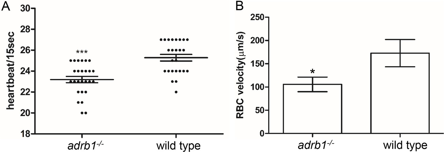
Adrb1-/- zebrafish embryos displayed a decrease of heart rate and blood flow in CVP.
(A) adrb1-/- embryos displayed a decrease of heart rate compared to wild type embryos at 28hpf. The heartbeats of 26 adrb1-/- and 25 wild type embryos were counted over a 15-second period. Unpaired two-tailed t test was performed and p<0.0001. (B) adrb1-/- embryos showed significant decrease of RBC velocity compared to wild type embryos. Time-lapses on a single z-plane was performed at the frequency of 160.97ms/frame (372 frames/minute) on Fast Airyscanning. 10 embryos from each group were scanned, and 3 red blood cells were traced from each embryo. The measurement was performed using ImageJ. Unpaired two-tailed t-test, p=0.0346.
Figure 1—figure supplement 2
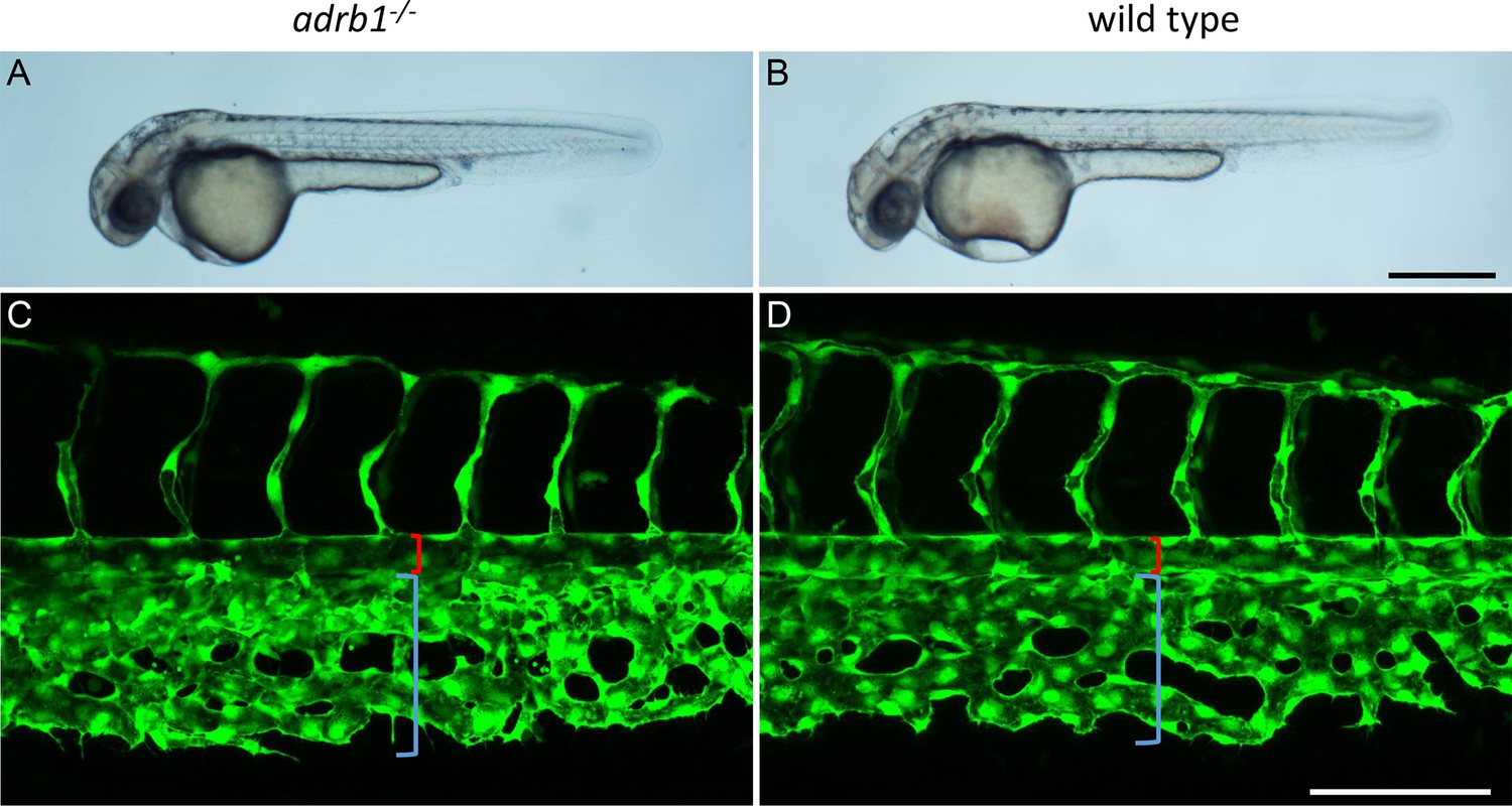
No significant difference of CVP development was observed between adrb1-/- and wild type embryos.
(A and B) Representative bright field pictures of adrb1-/- (A) and wild type (B) embryos at 36hpf. Scale bar: 500 µm. (C and D) Representative confocal pictures of CVP in adrb1-/- (C) and wild type (D) embryos at 36hpf. Red and blue brackets indicate the aorta and CVP respectively. Scale bar:100 µm.
Figure 2
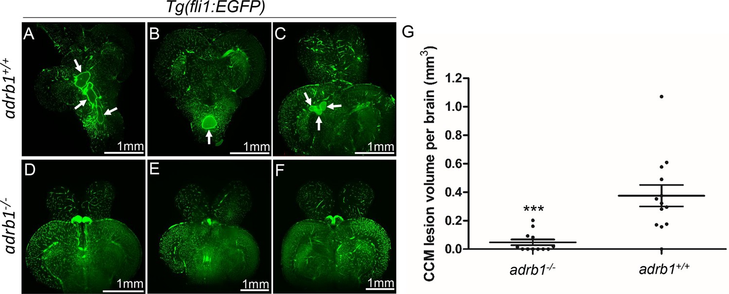
Genetic inhibition of adrb1 signaling could rescue CCM in ccm2 CRISPR zebrafish.
(A through F) Representative light sheet microscopy scanning pictures of brains from ccm2 CRISPR adult zebrafish of adrb1+/+ (A through C) and of adrb1-/- (D through F) onTg(fli1:EGFP) background. Brains from ccm2 CRISPR on wild type background show lesions indicated by arrows (A through C), while brains from ccm2 CRISPR on adrb1-/- do not show lesions (D through F). Scale bar: 1 mm. (G) Statistical analysis of total lesion volume by unpaired two-tailed t test. p=0.0005. 12 adrb1-/- brains and 13 adrb1+/+ brains were analyzed.
Figure 3
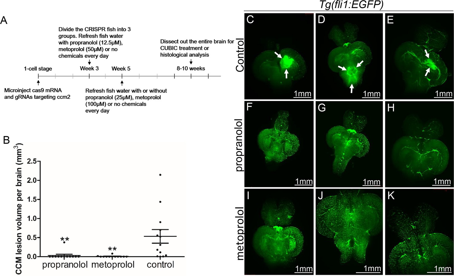
Both propranolol and metoprolol could rescue CCM in ccm2 CRISPR zebrafish.
(A) A diagram outlines the drug treatment experiment, CUBIC treatment and following recording of CCMs in adult zebrafish brain. The chemical treatment was started from week 3 with 12.5 µM propranolol or 50 µM metoprolol, and increased to 25 µM propranolol and 100 µM metoprolol, respectively from week 5. The fish water with chemicals or vehicle control are refreshed on a daily basis. (B) Statistical analysis of lesion volume by one-way ANOVA followed by Tukey’s multiple comparison test. p<0.01. 12 propranolol treated, 12 metoprolol treated, and 13 vehicle brains were analyzed. (C through K) Representative light sheet microscopy scanning pictures of brains from ccm2 CRISPR adult zebrafish onTg(fli1:EGFP) background. In controls without chemical treatment (C, D, and E) there were vascular anomalies indicated by arrows. Neither propranolol (F, G, and H) nor metoprolol (I, J, and K) treated fish showed vascular lesions in the brain. Scale bar: 1 mm.
Figure 4 with 1 supplement
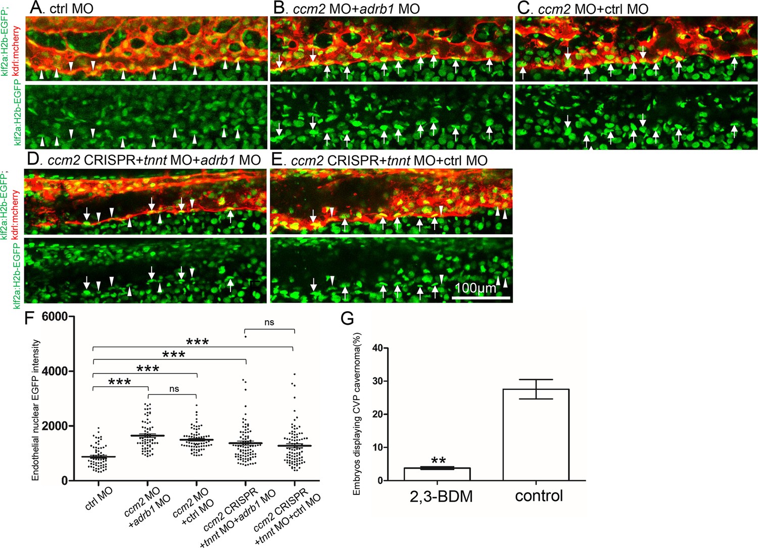
Adrb1 signaling does not alter klf2a expression in ccm2 CRISPR embryos.
Tg(klf2a:H2b-EGFP; kdrl:mcherry) embryos were injected and nuclear EGFP signal in mcherry labeled vascular endothelial cells is recorded by confocal. Representative images from each group are shown. (A) Control MO alone injected embryos were used as control. (B and C) Ccm2 morphant embryos co-injected with adrb1 MO (B) or control MO (C) both displayed significant increase of endothelial nuclear EGFP intensity (p<0.0001) compared to that of control (A), and there is no significant difference between them. (D and E) All the ccm2 CRISPR embryos were co-injected with tnnt MO, which are absent of blood flow. Compared to that of control (A), ccm2 CRISPR embryos co-injected with adrb1 MO (D) or control MO (E) both displayed a mosaic increase of nuclear EGFP intensity of vascular endothelial cells compared to control (A) (<0.0001), and there is no significant difference between them. Arrows indicated the endothelial nuclei with significant higher EGFP intensity than those indicated by arrowheads. Scale bar:100 µm. (F) EGFP intensity of endothelial nuclei were quantified with Image J. The number of analyzed nuclei were: 63 from 10 embryos (control MO), 70 from 10 embryos (ccm2 MO + adrb1 MO), 77 from 10 embryos (ccm2 MO + control MO), 93 from 13 embryos (ccm2 CRISPR +adrb1 MO), and 94 from 13 embryos (ccm2 CRISPR +control MO). Statistical analysis is performed by one-way ANOVA followed by Tukey’s multiple comparison test. (G) At 2dpf, 2,3-BDM prevented the CVP cavernoma dramatically. 164 embryos in 2,3-BDM treated group and 177 in control group were used for Two-tailed paired t-test. p=0.0013.
Figure 4—figure supplement 1
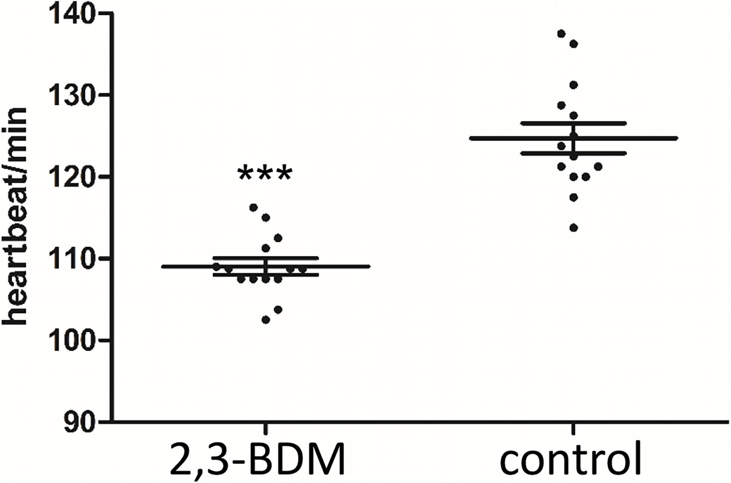
2,3-BDM decreases the heart rate in 30hpf zebrafish embryos.
The heartbeats of 14 embryos treated with 2,3-BDM and 14 vehicle-treated embryos were counted over a 1-minute period. Two-tailed paired t-test was used for statistical analysis. p<0.0001.
Author response image 1

42 endothelial nuclei from 7 embryos were scored as described in the Experimental Procedures of the manuscript.
Two tailed t test were performed. P=0.4529
Videos
Video 1
The cardiac pumping in adrb1-/- embryos at 28hpf.
Video 2
The cardiac pumping in wild type embryos at 28hpf.
Video 3
The blood flow in CVP in adrb1-/- embryos at 28hpf.
Video 4
The blood flow in CVP in wild type embryos at 28hpf.
Additional files
-
Supplementary file 1
Comparison of the two-phase zebrafish CCM model with mouse CCM model and human CCM.
“?” means it is yet to be determined. “-” means it is not applicable. “1”. Perilesional red blood cell leakage was seen.
- https://cdn.elifesciences.org/articles/99455/elife-99455-supp1-v1.xlsx
-
Supplementary file 2
The predicted off-targets genomic sites produced by adrb1 CRISPR.
These genomic sites were sequenced and found no mutations. Primer sequence used for amplifying these sites were listed.
- https://cdn.elifesciences.org/articles/99455/elife-99455-supp2-v1.xlsx
-
MDAR checklist
- https://cdn.elifesciences.org/articles/99455/elife-99455-mdarchecklist1-v1.docx
Download links
A two-part list of links to download the article, or parts of the article, in various formats.
Downloads (link to download the article as PDF)
Open citations (links to open the citations from this article in various online reference manager services)
Cite this article (links to download the citations from this article in formats compatible with various reference manager tools)
Genetic inactivation of the β1 adrenergic receptor prevents cerebral cavernous malformations in zebrafish
eLife 13:RP99455.
https://doi.org/10.7554/eLife.99455.3


