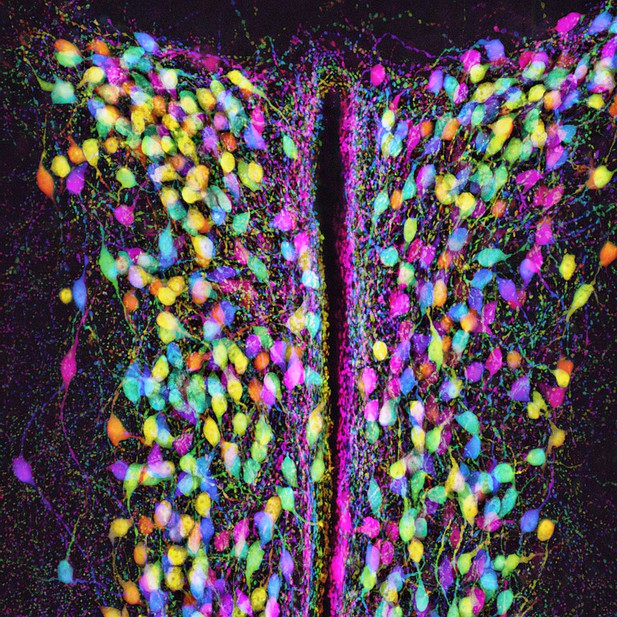
Oxytocin-synthesizing neurons in the hypothalamus. Image credit: Wanhui Sheng, Scott W. Harden, and Charles J. Frazier (CC BY 4.0)
Oxytocin is often referred to as a ‘love hormone’ because it can be released during activities such as hugging, snuggling, or sex. Reality, of course, can be a bit more complicated. In the brain, oxytocin can have powerful and diverse effects on mood, stress, anxiety, and social interactions. In the body it helps regulate fluid balance, promotes contractions during childbirth, and stimulates the letdown of milk during breastfeeding.
Much of the oxytocin produced in both humans and rodents comes from oxytocin-synthetizing magnocellular neurons located in an area of the brain called the hypothalamus. These very specialized neurons have separate, but overlapping, mechanisms for releasing oxytocin into the brain and into the rest of the body. This means that while certain signals cause the neurons to release oxytocin into the body and the brain at the same time, others can cause them to release the hormone preferentially into the body or the brain.
Sheng et al. wanted to better understand how these different release mechanisms work, and, in particular, to learn more about how release of oxytocin into the brain is regulated. This is important, because when oxytocin is given as a medicine, much of it fails to reach the brain.
A lot of the oxytocin that acts in the brain is released from a specific part of the oxytocin-synthesizing magnocellular neurons called the dendrites. When these neurons are stimulated, calcium enters the dendrites, triggering the release of oxytocin directly into the brain. Sheng et al. used electrical and optical tools on brain tissue extracted from mice to measure how different signals change the amount of calcium that enters the dendrites of oxytocin-synthesizing magnocellular neurons in response to a consistent stimulus.
The results showed that increasing the osmolarity, the amount of water-soluble particles that cannot spontaneously cross the cell membrane, in the liquid surrounding the neurons reduced the amount of calcium that flowed into the dendrites during stimulation. Meanwhile, decreasing osmolarity had the opposite effect. Sheng et al. also found that the influx of calcium induced by stimulating the neurons can be strongly regulated by activating receptors in the dendrites that detect a common molecule in the brain called GABA. This occurs even absent a change in osmolarity.
These results shed light on some of the physiological processes that control the release of oxytocin into the brain. Understanding these processes is a necessary step towards developing new drugs intended to regulate levels of oxytocin in the brain. Such drugs could be useful in the treatment of several types of mental health disorders.


