Scientists have characterised the folding of the cerebellar cortex using data from a sample of 56 mammalian species, allowing them to study the diversity and evolution of cerebellar folding and its relationship with the anatomy of the cerebrum.
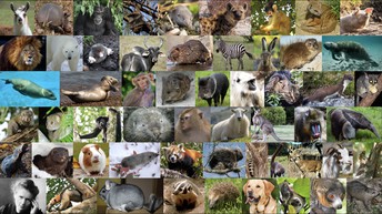
The study compared data from a sample of 56 mammalian species, pictured above. Image credit: Katja Heuer (CC BY 4.0)
The study from the Institut Pasteur, published today in eLife, suggests the size and folding of the cerebellum relate closely with the size and folding of the cerebrum across species, and demonstrates that large cerebellums are disproportionately more folded than smaller ones. Furthermore, the size of each individual fold appears to be constant across species, despite the large differences in brain size.
The findings provide new insights into the diversity and evolution of cerebellar folding, the mechanisms underlying brain folding, and its potential influence on the organisation of the brain across species.
The cerebrum is the largest part of the mammalian brain and handles a wide range of responsibilities, including vision, hearing, and the initiation and coordination of movement. The cerebellum is smaller, but contains a much larger number of neurons, and plays an important role in cognition and motion, including muscle control, balance and movement. While the cerebrum is unique to mammals, the cerebellum is present in all vertebrates. Brain folding, the process by which the brain’s surface forms ridges and grooves, greatly increases the surface-to-volume ratio allowing more neurons to be in a smaller volume than if it were unfolded. This process is thought to play an important role in the development, connectivity and organisation of both the cerebrum and cerebellum.
“Studying cerebellar folding is challenging due to the small size and abundance of its folia – little leaf-like structures of the highly folded cerebellar surface,” explains lead author Katja Heuer, a postdoctoral researcher at the Institut Pasteur, Paris, France. “As a consequence, little is known about its diversity and evolution across species.”
To remedy this, Heuer and colleagues examined data on the brain structure of 56 mammalian species, including humans, to explore the evolution of the cerebellum and its relationship with the cerebrum and body size. The data was obtained from several open sources, including the BigBrain Project for the human data, and the Comparative Mammalian Brain Collection for many other species. To visualise and segment the data, the team used a web tool they had previously developed, called MicroDraw.
They developed methods to measure the geometry of cerebellar folia across species and estimate the thickness of the molecular layer of the cerebellar cortex. They also used phylogenetic tree data from the TimeTree website, which depicts the lines of evolutionary descent of a given species from a common ancestor, for all the species included in their study. Since species are non-independent data points due to their evolutionary relatedness, it was crucial for the team to take phylogenetic tree structure into account. For example, variation in cerebellar structure could be due to different evolutionary processes, natural drift or selection. The researchers therefore used phylogenetic comparative methods to ensure their analyses considered the evolutionary relationships and history of each species.
Their results showed two groups of phenotypes, or observable characteristics. The first was a group of “diverse” characteristics that varied widely across the species, including body weight, brain weight, and cerebellar and cerebral section area and length. These all varied over several orders of magnitude together with body size. The second group of “stable” characteristics showed much less variation in comparison and included folial width and thickness of the molecular layer. These characteristics changed only slightly with changes in brain size.
By tracing the evolutionary pathway of the species, the team observed a strong pattern demonstrating a concerted change in brain size and body size. Primates, such as humans, were an outlier to this trend, with large brains relative to their body size.
In addition, the results confirmed that cerebellar size is strongly correlated with cerebral size, and revealed that large cerebella are disproportionately more folded than smaller ones. Similarly, the width of cerebellar folds was related to the thickness of the molecular layer. These patterns were highly conserved across all of the species studied, suggesting the presence of a common mechanism underlying the folding of the cerebrum and the cerebellum across mammals.
The authors note that there are a few limitations to the study. Due to the reduced number of species, only global neuroanatomical measurements could be analysed. A larger number of species would make it possible to study local variation, even full 3D reconstructions for each species. This presents challenges from both a technical standpoint in terms of computational neuroanatomy and phylogenetic comparative methods, as well as the need for collaboration and open science.
“Our results provide a closer look at the nature of macroscopic cerebellar anatomy, its relationship with cerebral anatomy, its diversity across mammalian species, and its evolution,” concludes senior author, Roberto Toro, Director of Research at the Institut Pasteur, and head of the Applied and Theoretical Neuroanatomy Unit. “The findings suggest to us that the process leading to cerebellar and cerebral folding is the same. In both structures, folding should lead to the formation of strongly conserved, mechanically canalised, neuroanatomical modules, which could have an important role in their functional organisation.”
Media contacts
Emily Packer
eLife
e.packer@elifesciences.org
+441223855373George Litchfield
eLife
g.litchfield@elifesciences.orgAurelie Perthuison
Institut Pasteur
presse@pasteur.fr
About
About eLife
eLife transforms research communication to create a future where a diverse, global community of scientists and researchers produces open and trusted results for the benefit of all. Independent, not-for-profit and supported by funders, we improve the way science is practised and shared. In support of our goal, we’ve launched a new publishing model that ends the accept/reject decision after peer review. Instead, papers invited for review will be published as a Reviewed Preprint that contains public peer reviews and an eLife assessment. We also continue to publish research that was accepted after peer review as part of our traditional process. eLife receives financial support and strategic guidance from the Howard Hughes Medical Institute, Knut and Alice Wallenberg Foundation, the Max Planck Society and Wellcome. Learn more at https://elifesciences.org/about.
To read the latest Evolutionary Biology research in eLife, visit https://elifesciences.org/subjects/evolutionary-biology.
And for the latest in Neuroscience see https://elifesciences.org/subjects/neuroscience.
About the Institut Pasteur
The Institut Pasteur, a non-profit foundation with recognized charitable status set up by Louis Pasteur in 1887, is today an internationally renowned center for biomedical research. In the pursuit of its mission to tackle diseases in France and throughout the world, the Institut Pasteur operates in four main areas: research, public health, training, and development of research applications. The Institut Pasteur is a globally recognized leader in infectious diseases, microbiology, and immunology, with research focusing on the biology of living systems. Among its areas of investigation are emerging infectious diseases, antimicrobial resistance, certain cancers, neurodegenerative diseases, and brain connectivity disorders. The Institut Pasteur's outstanding research is facilitated by the development of a technological environment of the highest standard, with core facilities for nanoimaging, computational biology and artificial intelligence. Since its inception, 10 Institut Pasteur scientists have been awarded the Nobel Prize for Medicine, including two in 2008 for the 1983 discovery of the human immunodeficiency virus (HIV) that causes AIDS. The Institut Pasteur is part of the Pasteur Network a worldwide network of more than 30 members on five continents, united by Pasteurian values, that contribute to global health.Since July 1, 2021, the Institut Pasteur is a research partner organization of Université Paris Cité. More information: www.pasteur.fr


