
Peer review process
Revised: This Reviewed Preprint has been revised by the authors in response to the previous round of peer review; the eLife assessment and the public reviews have been updated where necessary by the editors and peer reviewers.
Read more about eLife’s peer review process.Editors
- Reviewing EditorEsteban BeckwithInstituto de Investigación en Biomedicina de Buenos Aires (IBioBA) - CONICET, Buenos Aires, Argentina
- Senior EditorK VijayRaghavanNational Centre for Biological Sciences, Tata Institute of Fundamental Research, Bangalore, India
Reviewer #1 (Public review):
Summary:
The authors aim to use state-of-the art behaviour, imaging and connectome techniques to identify the neural interaction between sleep and long-term memory consolidation in the PAM-DPM circuits, a well-known dopaminergic pathway within Drosophila Mushroom Body.
Strengths:
The investigation follows a logical strategy to collect huge dataset of sleep, appetitive memory and live imaging. The authors identified and showed that activation of a PAM subset: alpha-1 reduces sleep quality and memory consolidation in a starvation dependant manner. The author also convincingly demonstrated the corresponding neuronal responses of DPM neurons following PAM alpha-1 activation, and the positive role of DPM neural activity in sleep and memory consolidation. Moreover, the new data provide TRIC-LUC provided better temporal resolution of neural activity correlates for PAMalpha1-DPM inhibition. Importantly, the author demonstrated that memory loss derived from PAM alpha 1 activation can be partly restored by ectopic sleep enhancement via feeding THIP at the memory consolidation period after training.
Weaknesses:
Although the revised version carries arguments to satisfy the reviewers' concern, the writing is now less cohesive. Crucially an explanation however remains required for the following experimental contradiction: the central observation of the study indicates that PAM alpha1 activation cause DPM inhibition which disrupt sleep and memory consolidation. Therefore, one would expect a reduced PAMalpha1 and increased DPM activities after memory training, but the authors found the opposite is true from now enhanced TRIC-LUC dataset. The authors indicate this data reinforce the inhibitory nature of PAM-alph1-DPM, but it does not explain why such a reduced DPM activity is observed after training.
Reviewer #2 (Public review):
Summary:
Sleep plays a critical role in memory consolidation, but the neural mechanisms underlying this relationship remain incompletely understood. The authors examined a specific subset of PAM dopaminergic neurons, PAM-α1, and DPM neurons in Drosophila. These neurons have previously been implicated in memory, and DPM neurons have also been linked to sleep. The study explores whether this circuit provides a mechanistic link between sleep and memory consolidation.
Strengths:
The authors report several novel findings. Brief activation or inhibition of PAM-α1 neurons, or brief inhibition of DPM neurons during the first few hours after training, impairs 24-hour LTM. Notably, these brief manipulations disrupt sleep for many hours afterward, particularly during the night. The authors further show that perturbation of PAM-α1 and DPM neurons impairs sleep and appetitive memory consolidation under starvation conditions, and that pharmacological sleep induction during the night rescues the LTM defects. Together, these findings suggest that PAM-α1 and DPM neurons are involved in sleep regulation and LTM consolidation under starvation. These are important observations that advance our understanding of the circuits regulating sleep and memory consolidation.
Weaknesses:
Some claims require additional evidence or clarification.
(1) Previous studies linking impaired memory to reduced sleep have primarily examined conditions involving severe sleep deprivation. In contrast, this manuscript argues that relatively modest decreases in total sleep, accompanied by sleep fragmentation, are sufficient to impair memory consolidation. It remains unclear whether sleep fragmentation of this magnitude is itself critical for LTM consolidation. An independent method for inducing comparably mild sleep loss and fragmentation would be needed to directly test this interpretation.
(2) It is unclear why both activation and inactivation of PAM-α1 neurons produce similar effects on sleep and memory. In addition, MB299B-labeled neurons exert stronger effects on memory than MB043B-labeled neurons, whereas MB043B-labeled neurons have stronger effects on sleep. If sleep disruption is the primary driver of impaired memory consolidation, a stronger correspondence between the sleep and memory phenotypes might be expected. The authors speculate that MB043B may affect sleep through non-PAM neurons, but without identifying the relevant neurons, this remains speculative.
(3) The complex schematic model (Fig. 12), with parallel circuits and unidentified neuronal groups, underscores the difficulty of interpreting the current data. In the "less activity" arm of the model, distinct circuits are proposed to regulate sleep and LTM, respectively, and DPM neurons are not included. This makes it difficult to reconcile the model with the central claim that the PAM-α1-to-DPM microcircuit links sleep and LTM consolidation.
(4) The TRIC-LUC reporter system is not ideal for resolving dynamic changes in neuronal activity. Activity-dependent Ca²⁺ signaling must first reconstitute the TRIC transcriptional system, which then drives luciferase transcription, translation, and accumulation. The original characterization of TRIC indicates that TRIC signals accumulate and decay over several hours. Thus, the kinetics of the TRIC-LUC reporter should be interpreted cautiously, particularly when inferring transient or precisely timed changes in neuronal activity.
(5) Including data from training under fed conditions would provide a more complete understanding of state-dependent neural activity and would help distinguish starvation-specific effects from more general circuit mechanisms.
Reviewer #3 (Public review):
Summary:
Understanding the neural circuits that link sleep and memory remains a fundamental challenge in neuroscience. In this study, Lin Yan and colleagues investigate how dopamine signaling in Drosophila regulates long-term memory (LTM) formation in the context of sleep. They identify a specific microcircuit between protocerebral anterior medial dopamine neurons (PAM-DANs) and dorsal paired medial (GABAergic DPM) neurons that modulates memory consolidation. Their findings suggest that disrupting the basal activity of PAM-α1 neurons during early consolidation impairs LTM, with particularly pronounced effects under starvation conditions. Notably, sleep fragmentation caused by this disruption can be pharmacologically rescued, restoring LTM. These results provide compelling evidence how dopamine signaling plays a crucial role in linking sleep and memory, offering new insights into the underlying mechanisms.
Strength:
This study presents a well-executed investigation into sleep-memory interactions, utilizing a combination of connectomics, behavioral assays, functional imaging, and pharmacological manipulations. The authors convincingly demonstrate that the PAM-α1 and DPM circuit interact, highlighting a potential mechanism by which sleep influences memory consolidation. The anatomical and functional dissection of this circuit is of high interest to the field, and the study's integration of sleep and memory processes contributes significantly to our understanding of the role of dopamine in cognitive functions. Additional experiments investigating the contribution of MBON-α1 to the circuit, connectomic analysis together with a dissection of dopamine receptor function further strengthen the proposed circuit motif and its biological relevance.
Weaknesses:
While the study is well designed, presents compelling findings and has been further strengthened by additional experiments, some aspects remain unclear. The role of DPM neurons in memory consolidation seems not yet fully resolved, as different genetic approaches yield variable results. Furthermore, some manipulations impair memory without affecting sleep fragmentation - or vice versa, suggesting that the observed memory deficits cannot be explained solely by impaired sleep-dependent consolidation. It would also have been interesting to discuss potential mechanisms by which dopamine receptor-mediated cAMP signaling could lead to a reduction in Ca²⁺ signals. I am confident that these questions can be addressed in future studies.
Conclusion:
Overall, this study provides valuable new insights into how sleep and dopaminergic circuits interact to regulate memory consolidation in Drosophila and may reveal general principles underlying the neural regulation of memory.
Author response:
The following is the authors’ response to the original reviews
eLife Assessment
This study approaches an important topic providing insight into the neuronal circuitry that interconnects memory consolidation and sleep. The data were collected and analysed using a solid methodology, contributing new findings for neurobiologists working on how memories are stored and the roles of sleep. However, the data is incomplete to support the proposed role of the PAM-DPM circuits as the link between sleep state and long-term memory consolidation.
We sincerely appreciate the editor and reviewers’ thoughtful and constructive comments on our study. Your insightful feedback has not only affirmed the significance of our work on the interplay between memory consolidation and sleep, but also provided valuable inputs for improving the clarity, rigour, and impact of our study.
We have carefully addressed all the comments raised by the reviewers and revised the manuscript accordingly. We have also streamlined the paper with the goal of making it more accessible to readers. We feel this revised version strengthens our conclusion that the PAM-DPM circuits as the link between sleep and memory consolidation.
The main improvements in terms of data addition are three complementary sets of circuit-specific experiments:
(1) To better characterize the dynamics of the PAM-DPM circuit following associative memory training, we performed 3-hour continuous neural activity recording in freely behaving flies. This experiment addresses the activity of the microcircuit in a much more relevant time frame than the CRTC data in the previous version of the paper which looked only at the first hour after training. Specifically, we expressed the Tric-LUC reporter gene, a calcium-responsive tool that harnesses the interaction between calmodulin and its cognate binding peptides to drive rapid luciferase transcription in a calcium-dependent manner (Gao et al., 2015; Guo et al., 2017), in PAM-α1 and DPM neurons, respectively. Flies were then subjected to either associative memory training or a no-training control condition, with real-time luciferase levels monitored throughout the recording window.
In the absence of training, both PAM-α1 and DPM neurons displayed similar neural activity over the 3-hour recording period. The first hour was characterized by a synchronous decrease in activity for both neuron types, with hours 2 and 3 achieving a stable baseline. Since the decrease in the first hour is also seen in the trained condition, we think it is likely a reflection of the animals becoming acclimated to the recording tubes.
Notably, associative memory training profoundly reshaped the activity profile of the PAM-DPM circuit in the LTM consolidation time window. Training induced a mild yet statistically significant elevation in PAM-α1 neural activity specifically during the third hour of recording, while concurrently eliciting a robust reduction in DPM neuron activity over the last two hours (revised Figure 8C-F). These findings not only support the hypothesized role of the inhibitory PAM-α1-DPM circuit in sleep and memory consolidation, but also advance our mechanistic understanding of underlying neural dynamics.
(2) To further support the functional connectivity of the PAM-DPM microcircuit, we conducted in vivo experiments to complement the dissected brain prep P2X2 data. Optogenetic activation of PAM neurons in intact flies via the red light-gated cation channel CsChrimson (Klapoetke NC et al., 2014) resulted in a significant reduction in GCaMP signals within DPM neurons (revised Figure 2B). These findings strongly confirm that PAM neurons exert direct inhibitory control over DPM neurons in the intact brain.
(3) Further, we investigated how dopamine signaling to the DPM inhibits its activity, and issue which has not been investigated previously. We conducted a series of experiments:
Firstly, we verified which dopamine receptors (Dop1R1, Dop1R2, DopEcR, and Dop2R) express on the DPM neurons via double-labeling with gene-embedded GAL4 lines. We found that DPM neurons have expression of both Dop1R1 and Dop1R2 (revised Figure 10A).
Secondly, to clarify which receptors on DPM neurons respond to dopamine and how they signal, in addition to EPAC experiments in the first submission, we recorded neural activity changes when we knocked down Dop1R1 and Dop1R2 in DPM neurons. DPM neurons exhibited a significantly reduced GCaMP level with DA application, regardless of whether Dop1R1 or Dop1R2 was intact or knocked down knockdown in comparison to the no-DA control condition (revised Supplemental Figure 3C-E). These data suggest that either residual Dop1R1 and Dop1R2 remaining in the RNAi condition is sufficient or that the two receptors may coordinate to mediate the inhibition of neural activity.
Finally, we investigated the behavioral contributions of Dop1R1 and Dop1R2 in DPM neurons to sleep and memory processes (revised Figure 10C-H). Dop1R1 knockdown resulted in a marked reduction in daytime sleep and a significant impairment of 24 h memory expression. In contrast, Dop1R2 knockdown selectively compromised 24 h memory without affecting sleep.
When integrated with our EPAC assay findings from the initial submission, which demonstrated, that Dop1R1 is the primary receptor mediating dopamine-induced cAMP elevation, these new data collectively delineate a more complex mechanistic framework: dopamine signaling in DPM neurons coordinates the dual regulation of sleep and memory predominantly via Dop1R1. Meanwhile, Dop1R2 are engaged in the selective modulation of memory.
All newly generated experimental datasets, comprehensive statistical analyses, and their corresponding figure panels (revised Figures 2B, 10, 11 and Supplemental Figure 3) have been fully incorporated into the revised manuscript.
In addition to adding the experiments described above, we have reorganized and streamlined the paper. First, the CRTC data have been replaced by the Tric-luc data. The CRTC data were taken in the first hour after training and do not shed light on the bulk of the consolidation window. Since the behavioral and sleep effects we see with manipulation of the PAM/DPM microcircuit all occur with a time delay, examining later times in consolidation is more relevant. Additionally, the first hour post-training is quite complex since there are sensory changes and STM processes overlaid on the processes we want to study. Second, we have moved the data in Figure 8 to supplemental (revised Supplemental Figure 2) since they are basically a control for the experiments in Figure 7 validating known requirements for appetitive LTM.
We have also substantially expanded the Discussion section to contextualize the PAM-DPM circuit within the broader framework of well-characterized memory-regulatory pathways, such as the intrinsic circuits of the mushroom body, and to explicitly delineate the hierarchical interplay between sleep-dependent synaptic plasticity and LTM consolidation.
We contend that these complementary experimental assays and targeted revisions markedly strengthen the causal evidence underscoring the role of the PAM-DPM circuit as a pivotal regulatory node bridging sleep states and LTM consolidation. We are confident that these revisions essentially address the concerns raised by the reviewers.
Public Reviews:
Reviewer #1 (Public review):
Summary:
The authors aim to use state-of-the art behavior, imaging, and connectome techniques to identify the neural interaction between sleep and long-term memory consolidation in the PAM-DPM circuits, a well-known dopaminergic pathway within Drosophila Mushroom Body.
Strengths:
From a Drosophila sleep researcher's perspective, the investigation follows a clear and logical strategy to collect a huge dataset of sleep, appetitive memory, and live imaging. The authors clearly identified and showed that activation of a PAM subset: alpha-1 reduces sleep quality and memory consolidation in a starvation-dependent manner. The authors also convincingly demonstrated the corresponding neuronal responses of DPM neurons following PAM alpha-1 activation, and the positive role of DPM neural activity in sleep and memory consolidation. Moreover, the authors applied a new way of sleep statistics to demonstrate hour-by-hour changes between treatment and genotypes. Importantly, the authors demonstrated that memory loss derived from PAM alpha 1 activation can be partly restored by ectopic sleep enhancement via feeding THIP during the memory consolidation period after training.
Weaknesses:
Two investigatory gaps relate to the misalignment between circuital activity and behaviors, due to the nature of large circuital functional analysis like this. Firstly, the central observation of the study indicates that PAM alpha1 activation causes DPM inhibition which disrupts sleep and memory consolidation. Therefore one would expect a reduced PAMalpha1 and increased DPM activities after memory training, but the authors found that the endogenous CRTC::GFP reported neuronal activity for PAMalpha1 and DPM are both increased after memory training (Figure 9). This can be due to the difficult functional demarcation among the 14 PAMalpha1 projections. Secondly, the authors acknowledged the contradicting finding that memory defect is detected in PAMalpha1 inactivation (Figure 7C), yet suggested a tight link between sleep and memory consolidation; it is clear loss of PAM subset activity can disrupt memory consolidation without affecting sleep (cf Figure 7C and 7I).
Thank you for your insightful analysis and the relevant possibilities you've raised. We agree that given that memory consolidation and sleep are time-dependent processes, the 1-hour window employed to capture neural activity changes via the CRTC::GFP reporter may not fully reflect the overall dynamics of neural activity in this microcircuit. To better characterize the dynamics of the PAM-DPM circuit in the consolidation window following associative memory training, we performed 3-hour continuous neural activity recording in freely behaving flies. Specifically, we expressed the Tric-LUC reporter gene, a calcium-responsive tool that harnesses the interaction between calmodulin and its cognate binding peptides to drive rapid luciferase transcription in a calcium-dependent manner (Gao et al., 2015; Guo et al., 2017), in PAM-α1 and DPM neurons, respectively. Flies were then subjected to either associative memory training or a no-training control condition, with real-time luciferase levels monitored throughout the recording window.
In the absence of training, PAM-α1 neurons displayed stable neural activity over the entire 3-hour recording period. However, associative memory training profoundly reshaped the activity profile of the PAM-DPM circuit. Training induced a mild yet statistically significant elevation in PAM-α1 neural activity specifically during the third hour of recording, while concurrently eliciting a robust reduction in DPM neuron activity over the last two hours (revised Figure 8C-F). These findings not only support to the hypothesized role of inhibitory PAM-α1-DPM circuit in sleep and memory consolidation, but also advance our mechanistic understanding of underlying neural dynamics.
Regarding the second question, the core finding underlying the link between sleep and memory elucidated in the present study lies in the whole PAM-α1-DPM microcircuit rather than the specific DANs alone. MB299B and MB043B, the two split-GAL4 drivers employed to target PAM-α1 neurons, were originally characterized previously (Aso et al., 2014). However, these drivers also exhibit non-specific labeling of additional cells, and we can not rule out the possibility that such off-target labeling may have masked the subtype-specific necessity in sleep or memory processes.
Reviewer #2 (Public review):
Summary:
Sleep plays a critical role in memory consolidation, but the neural mechanisms underlying this relationship remain poorly understood. The authors present novel findings implicating two small neuronal groups with inhibitory connections, PAM-a1 to DPM, in sleep regulation and LTM consolidation. However, whether the PAM-a1 to DPM microcircuit promotes LTM consolidation through sleep regulation requires further investigation.
Strengths:
The authors report several novel findings. Brief activation or inhibition of PAM-a1 neurons, or brief inhibition of DPM neurons during the first few hours after training, impairs 24-hour LTM. Notably, these brief manipulations disrupt sleep for many hours afterward, particularly at night. Interestingly, disruption of PAM-a1 and DPM neurons impairs sleep and appetitive memory consolidation only under starvation conditions, and pharmacological induction of sleep during the night rescues the LTM defects. These findings suggest that PAM-a1 and DPM neurons are involved in sleep regulation and LTM consolidation under starvation. These are important findings that advance our understanding of the link between sleep and memory consolidation.
Weaknesses
Some claims lack sufficient evidence or clarity:
(1) All sleep experiments are conducted under the "training" (temperature-change) condition. While genotypic controls are helpful, additional no-training controls are required to confirm that the observed differences are due to training rather than unknown genotype-related factors. The fact that experimental genotypes exhibit significantly altered sleep even before "training" (e.g., Figs. 7H, J, K, 8A, B, D) highlights the necessity of these controls.
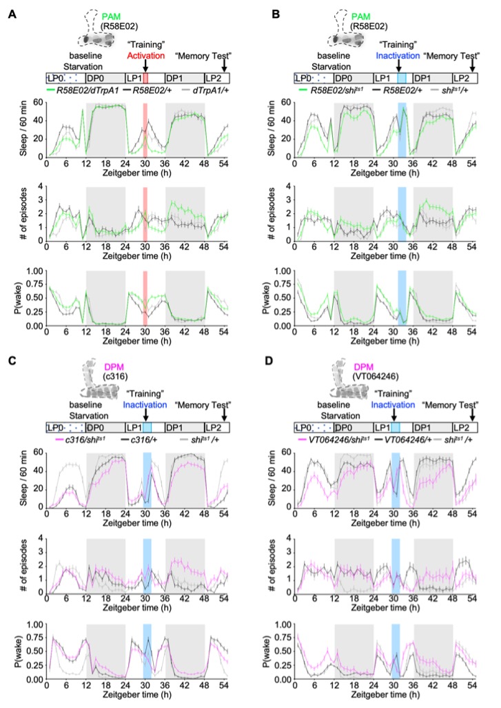
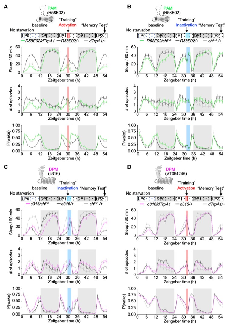
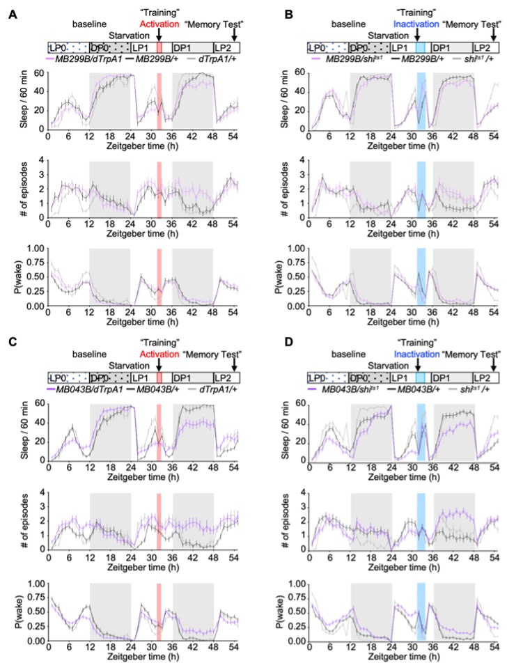
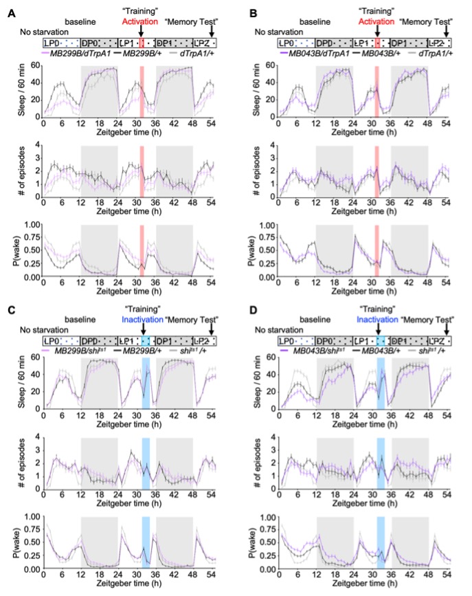
Thank you for raising this important question. We have re-examined the sleep profiles recorded over two acclimation days and one day of baseline sleep, which preceded the implementation of the “training” paradigm (temperature manipulation) and thus served as a valid no-training control. As shown in Author response images 1-4, subtle yet discernible genotype-dependent differences were indeed observed under baseline conditions. However, when animals were subjected to starvation, the experimental manipulations (activation or inactivation of the target cells) elicited marked, statistically significant alterations in sleep patterns that cannot be accounted for by the baseline genotype differences. Collectively, these data confirm that the observed sleep phenotypes are attributable to the “training” intervention, rather than to confounding, pre-existing genotype-related factors.
Author response image 1.
Baseline and manipulation day sleep profiles following PAM activation and PAM/DPM inactivation under starvation conditions.
Author response image 2.
Baseline and manipulation day sleep profiles following PAM activation and PAM/DPM inactivation under non-starvation conditions.
Author response image 3.
Baseline and manipulation day sleep profiles following PAM- α1 activation and inactivation under starvation conditions.
Author response image 4.
Baseline and manipulation day sleep profiles following PAM- α1 activation and inactivation under non-starvation conditions.
(2) Previous studies on disrupted memory due to sleep reduction have primarily examined conditions with severe sleep deprivation. In contrast, this report claims that relatively small decreases in total sleep accompanied by sleep fragmentation are responsible for impaired memory consolidation. It remains unclear whether sleep fragmentation at this level is truly critical for memory consolidation. The authors should cause sleep loss and fragmentation of similar magnitude through other means and determine whether it can impair LTM.
We appreciate the reviewer’s insightful suggestion. While alternative assays for inducing sleep loss or sleep fragmentation are indeed available, this line of investigation lies beyond the core scope of the present study. We will certainly take this valuable suggestion into consideration for the future studies.
(3) The authors employed a neural activity reporter to show that starvation increases the basal activity of PAM-a1 but not DPM neurons in untrained flies (Figures 9C-E). They observed small increases in the activity of both neuron groups immediately after training but not one hour later. Given the inhibitory connection from PAM-a1 to DPM, it is unclear why both neuron groups show increased activity after training. Additionally, as the authors acknowledge, it is puzzling how the inactivation of PAM-a1 produces similar effects on sleep and memory as DPM inhibition and PAM-a1 activation. Further experiments are needed to clarify these findings, such as manipulating PAM-a1 activity during the one-hour post-training period and evaluating the effect on DPM activity. Including data from training under fed conditions would provide a more comprehensive understanding of state-dependent neural activity. Even if certain experiments are not feasible, these issues warrant further discussion. It is also important to clarify that the term "synchronized" does not imply single-spike-level synchrony.
Thank you for raising these critical questions. To deepen our understanding of these issues, we have conducted additional experiments and have incorporated them into the revised manuscript. Below are our specific responses to each of your points:
(1) Regarding the contradiction between "PAM-α1 inhibition of DPM" and a transient increase in the activity of both neurons immediately after training:
PAM/PAM-α1 neurons are well-documented to respond to reward signals (Liu et al., 2012, Ichinose et al., 2015), while DPM neurons have been shown to respond to both olfactory stimuli and electric shocks, and to form delayed olfactory memory traces (Yu et al., 2005). Thus, the concurrent increase in the activity of PAM-α1 and DPM neurons immediately following training is likely a response to the olfactory and/or sucrose stimuli in the assay. Given that memory consolidation and sleep are time-dependent processes, the 1-hour window employed to capture neural activity changes via the CRTC::GFP reporter likely does not fully reflect the overall dynamics of neural activity in this microcircuit. Additionally, this time window overlaps with the period in which the animals are adapting to the new tubes and is likely contaminated with other sensory information.
To better characterize the dynamics of the PAM-DPM circuit following associative memory training, we performed 3-hour continuous neural activity recording in freely behaving flies. Specifically, we expressed the Tric-LUC reporter gene, a calcium-responsive tool that harnesses the interaction between calmodulin and its cognate binding peptides to drive rapid luciferase transcription in a calcium-dependent manner (Gao et al., 2015; Guo et al., 2017), in PAM-α1 and DPM neurons, respectively. Flies were then subjected to either associative memory training or a no-training control condition, with real-time luciferase levels monitored throughout the recording window.
In the absence of training, PAM-α1 neurons displayed stable neural activity over the entire 3-hour recording period. Notably, associative memory training profoundly reshaped the activity profile of the PAM-DPM circuit. Training induced a mild yet statistically significant elevation in PAM-α1 neural activity specifically during the third hour of recording, while concurrently eliciting a robust reduction in DPM neuron activity over the last two hours (revised Figure 9F-I). These findings not only support to the hypothesized role of inhibitory PAM-α1-DPM circuit in sleep and memory consolidation, but also advance our mechanistic understanding of underlying neural dynamics post-training. We have replaced the CRTC data with this more relevant data set.
(2) Regarding the state-dependent neural activity:
We agree that investigating state-dependent neural activity would be an interesting extension of our study. However, this falls beyond the scope of the current study and will be considered in future research. Our primary findings, including sleep disruptions and the associated memory impairments, were specifically observed under starvation conditions, which align with the appetitive memory paradigm employed here. Delving into neural activity changes under non-starvation state would not yield direct evidence to support the core conclusions of the present work, as the study’s focus is on the starvation-dependent interplay between sleep, neural circuitry, and appetitive memory consolidation.
(3) Regarding the terminology of “synchronization”:
We believe that the use of the term “synchronization” in our study is appropriate. In the context of neural circuitry, synchronization refers to the process by which distinct neurons or neural populations achieve temporal alignment of their activity, a phenomenon that supports neural communication and information integration. In the present work, this specifically describes how PAM-α1 and DPM neurons exhibit phase-related temporal coordination of their activity to regulate the interplay between sleep and memory consolidation.
(4) The authors considered that PAM-a1 and DPM might function in parallel, independent pathways for sleep and LTM. They rejected this possibility based on the lack of additive effects when both neuronal groups were simultaneously inactivated. However, they found that MB299B-labelled neurons exert stronger memory effects than MB043B-labelled neurons, while MB043B neurons have stronger sleep effects. If sleep is a primary driver of memory consolidation, a stronger correlation between memory and sleep effects would be expected. This observation merits further discussion.
We appreciate the reviewer’s constructive suggestions. We have performed additional experiments to explore a well-characterized memory-related PAM-α1 recurrent loop in sleep regulation. The new data, along with further discussion, have been incorporated into the revised manuscript.
The two split-GAL4 drivers (MB299B and MB043B) used to target PAM-α1 neurons were originally characterized previously (Aso et al., 2014). However, these drivers exhibit non-specific labeling of additional neuronal populations, a technical limitation that may have masked the subtype-specific functional requirements of PAM-α1 in sleep and memory processes.
In addition, we assessed sleep and LTM following the thermoactivation of DPM neurons (revised Supplemental Figure 1), and no significant changes were observed in either phenotype.
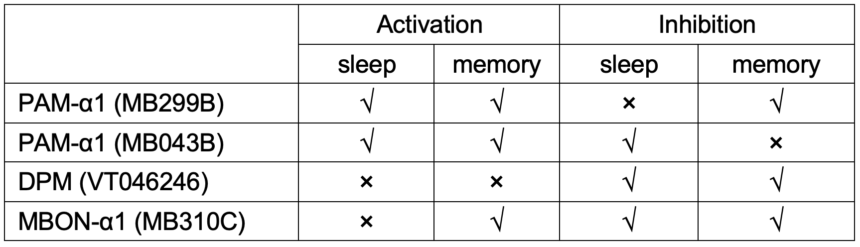
PAM-α1 has previously been demonstrated to drive appetitive LTM formation and consolidation via a recurrent loop with MBON-α1 (Ichinose et al., 2015). To investigate whether MBON-α1 also participates in sleep regulation, we activated or inactivated MBON-α1 neurons under both starvation and non-starvation conditions. Our results revealed that inhibition of MBON-α1 under both starvation and non-starvation conditions resulted in a significant reduction in sleep and a reduced arousal threshold (revised Figure 11B, D), suggesting that MBON-α1 participates in regulating sleep in a state-independent manner. However, no significant changes were observed upon activation of MBON-α1 neurons (revised Figure 11A, C). Combined with our observation that inhibition of MBON-α1 during the memory consolidation phase also impaired 24 h LTM, these new data indicate that MBON-α1-mediated sleep is necessary for effective memory consolidation. Notably, while activation of MBON-α1 during consolidation phase similarly impaired LTM, this manipulation did not alter the sleep profile, suggesting a dissociation between MBON-α1’s mechanistic roles in sleep regulation and LTM processing.
Taken together (see Author response table 1 and the new schematic diagram of revised Figure 12), these findings reveal a dedicated hierarchical, modular regulatory network that mediates sleep-LTM coupling via an activity-dependent mechanism. Within this network, activation of PAM-α1 acts as an upstream modulator to inhibit the activity of DPM, a downstream integrative hub that coordinates the execution of sleep and memory processes via recruiting different signaling cascades mediated by distinct dopamine receptors. MBON-α1, which is likely inhibited by PAM-α1, serves as parallel pathway to suppress sleep and impair LTM. Conversely, inactivation of PAM-α1 relieves its inhibitory control over MBON-α1, leading to MBON-α1 activation; MBON-α1 then functions as a signal amplifier that further exacerbates the reduced activity of PAM-α1, ultimately resulting in LTM impairment. Inactivation of PAM-α1, together with non-PAM-α1 neurons labeled by MB043B, contributes to the regulation of sleep. Sleep and memory are highly intertwined within this circuit, where distinct neuronal populations exhibit specialized yet interdependent functional roles, with overlapping and divergent regulatory contributions to sleep and LTM. The inherent complexity of this regulatory network thus merits further dedicated investigation in future studies.
Author response table 1.
(5) Given prior knowledge that PAM neurons are heterogeneous and that the R58E02 driver is broadly expressed, data in Figures 1-5 concerning PAM are outdated. The use of more restricted PAM-a1 drivers from the outset would make the manuscript easier to read and interpret.
We sincerely appreciate the reviewer’s point of view regarding the selection of PAM drivers. While we acknowledge the well-characterized heterogeneity of PAM neurons and the broad expression profile of the R58E02 driver, and fully agree that employing subtype-restricted drivers enhances the precision of functional interpretation, this set of experiments serves as an essential foundational step and logical basis for subsequent subtype-specific investigations and thus merits retention in the manuscript. As detailed above, the more specific drivers also have some drawbacks in terms of additional expression, making the broad driver critical for setting the stage.
(6) Some figures lack relevant data, certain experiments are missing necessary controls, and anomalies are present in some data sets.
We sincerely appreciate the reviewer’s detailed suggestions, and we have revised the manuscript comprehensively in accordance with them.
Reviewer #3 (Public review):
Summary:
Understanding the neural circuits that link sleep and memory remains a fundamental challenge in neuroscience. In this study, Lin Yan and colleagues investigate how dopamine signaling in Drosophila regulates long-term memory (LTM) formation in the context of sleep. They identify a specific microcircuit between protocerebral anterior medial dopamine neurons (PAM-DANs) and dorsal paired medial (GABAergic DPM) neurons that modulates memory consolidation. Their findings suggest that disrupting the basal activity of PAM-α1 neurons during early consolidation impairs LTM, with particularly pronounced effects under starvation conditions. Notably, sleep fragmentation caused by this disruption can be pharmacologically rescued, restoring LTM. These results provide compelling evidence that dopamine signaling plays a crucial role in linking sleep and memory, offering new insights into the underlying mechanisms.
Strengths:
This study presents a well-executed investigation into sleep-memory interactions, utilizing a combination of connectomics, behavioral assays, functional imaging, and pharmacological manipulations. The authors convincingly demonstrate that the PAM-α1 and DPM circuits interact, highlighting a potential mechanism by which sleep influences memory consolidation. The anatomical and functional dissection of this circuit is of high interest to the field, and the study's integration of sleep and memory processes contributes significantly to our understanding of dopamine's role in cognitive functions.
Weaknesses:
While the study is well-designed and presents compelling findings, some aspects require further clarification. The interpretation of dopamine receptor signaling remains incomplete, particularly regarding inhibitory pathways. The role of DPM in memory consolidation is not entirely conclusive, as different genetic approaches yield variable results. Additionally, some inconsistencies in neuronal activity patterns and experimental variability, especially regarding sleep patterns or pharmacological rescue, should be addressed to strengthen the mechanistic framework.
Conclusion:
Overall, this study provides valuable new insights into how sleep and dopamine circuits interact to regulate memory consolidation. While the findings are compelling, addressing the points above-particularly receptor signaling and the specific role of DPM and its activity patterns within the microcircuit would further solidify the study's conclusions.
We sincerely appreciate the reviewer’s constructive feedback and useful suggestions, which have been instrumental in enhancing the rigour and completeness of our study.
To address these points, we have performed a series of additional experiments that we believe strengthen the mechanistic framework of our work. The key new findings are summarized below:
(1) Regarding the dopamine receptor signaling
To define the dopamine receptor (DAR) signaling mechanisms underlying DPM neuron activity and its regulatory roles in sleep and memory, we first characterized DAR expression profile of the DPM neurons. Using double-labeling assay, we detected robust expression of Dop1R1 and Dop1R2 in DPM neurons, whereas no detectable colocalization was observed for DopEcR and Dop2R (revised Figure 10A). Accordingly, we refined our FRET-based EPAC data by removing the DopEcR knockdown group, and now present cAMP changes in DPM neurons following Dop1R1 and Dop1R2 knockdown, in direct comparison with the intact receptor control group (revised Figure 10B). These data conform that Gαs-coupled Dop1R1 is the primary receptor mediating DA-dependent cAMP elevation in DPM neurons.
To further identify the DARs responsible for transducing DA-induced inhibitory effect on DPM neural activity, we quantified GCaMP levels in DPM neurons with targeted knockdown of individual DARs. Knockdown of either Dop1R1 or Dop1R2 failed to abolish DA-induced Ca2+ decrease; only Dop1R2 knockdown exhibited a trend toward attenuating this Ca2+ decrease (revised Supplemental Figure 3C-E), suggesting that the two receptors cooperate to modulate DPM neural activity.
Finally, to dissect the specific contributions of DARs in DPM neurons to sleep and/or memory regulation, we performed sleep monitoring and memory assays in animals with DPM-specific knockdown of distinct DARs (revised Figure 10C-H). Knockdown Dop1R1 in DPM neurons resulted in statistically significant sleep reduction, decreased arousal threshold, and impaired 24 h LTM memory (revised Figure 10C-E). In contrast, knockdown Dop1R2 in DPM neuron selectively impaired 24 h LTM memory with no effect on sleep (revised Figure 10F-H). Collectively, these findings demonstrate that coupling sleep and LTM requires Dop1R1 in DPM neurons through the modulation of both cAMP signaling and neuronal activity, while Dop1R2 specifically mediates LTM regulation, likely through modulating DPM neural activity alone.
(2) We have additionally characterized the role of MBON-α1 in sleep, which has been previously shown as a PAM-α1-related recurrent feedback loop in the regulation of memory formation and consolidation (Ichinose et al., 2015).
To investigate whether MBON-α1 also participates in sleep regulation, we activated or inactivated MBON-α1 neurons under both starvation and non-starvation conditions (revised Figure 11A-D). Our results revealed that inhibition of MBON-α1 under both starvation and non-starvation conditions resulted in a significant reduction in sleep and a reduced arousal threshold (revised Figure 11B, D), suggesting that MBON-α1 participates in regulating sleep in a state-independent manner. However, no significant changes were observed upon activation of MBON-α1 neurons (revised Figure 11A, C). Moreover, inhibition of MBON-α1 during the memory consolidation phase significantly impaired 24 h LTM (revised Figure 11E-F). These results indicate that MBON-α1mediated sleep is necessary for effective memory consolidation. Notably, while activation of MBON-α1 during consolidation phase similarly impaired LTM, this manipulation did not alter the sleep profile, suggesting a dissociation between MBON-α1’s mechanistic roles in sleep regulation and LTM processing.
Taken together (see Author response table 1 and the new schematic diagram of revised Figure 12), these findings reveal a dedicated hierarchical, modular regulatory network that mediates sleep-LTM coupling via an activity-dependent mechanism. Within this network, activation of PAM-α1 acts as an upstream modulator to inhibit the activity of DPM, a downstream integrative hub that coordinates the execution of sleep and memory processes via recruiting different signaling cascades mediated by distinct dopamine receptors. MBON-α1, which is likely inhibited by PAM-α1, serves as parallel pathway to suppress sleep and impair LTM. Conversely, inactivation of PAM-α1 relieves its inhibitory control over MBON-α1, leading to MBON-α1 activation; MBON-α1 then functions as a signal amplifier that further exacerbates the reduced activity of PAM-α1, ultimately resulting in LTM impairment. Inactivation of PAM-α1, together with non-PAM-α1 neurons labeled by MB043B, contributes to the regulation of sleep. Sleep and memory are highly intertwined within this circuit, where distinct neuronal populations exhibit specialized yet interdependent functional roles, with overlapping and divergent regulatory contributions to sleep and LTM. The inherent complexity of this regulatory network thus merits further dedicated investigation in future studies (See Author response table 1).
We have modified the schematic diagram in the revised manuscript to illustrate the mechanistic framework (revised Figure 12).
Recommendations for the authors:
Reviewer #1 (Recommendations for the authors):
Here I listed details for potential clarification or further investigation related to the weaknesses:
(1) Line 145-147: I suspected the authors used previously verified RNAi lines, but it would be informative to include a citation or their own validation for the effectiveness of these RNAi lines.
We sincerely appreciate the reviewer’s suggestion. As our double-labeling assays confirmed that only Dop1R1 and Dop1R2 are colocalized with DPM neurons (see our responses to the public review from Reviewer #2 and #3), we refined the revised data to focus exclusively on these two DARs. Corresponding revisions have been made to the Materials and Methods, Results and Discussion sections. Additionally, we conducted qPCR analysis to verify the knockdown efficiency of these DARs, providing further support for our findings that Dop1R1 and Dop1R2 are functionally required in DPM neurons for the regulation of sleep and memory (revised Supplemental Figure 3).
(2) Line 169: Moving from describing Figure 3A/B to Figure 3C, it is not immediately clear from 3C-H, the authors follow the training paradigm of 3B?
To enhance clarity, we have added the referenced figure citations in the “Memory assay” section: “For all 24 h sucrose-odour memory, a single training session of sucrose paired with an odour for 2 min was employed (Figure 3A).”
(3) Line 179: before the PAM inactivation data are shown in Figure 7, the authors seem to be getting ahead of themselves by stating "suggesting that activity of PAM neurons is necessary for the consolidation window or that heterogeneity in the subsets of PAM neurons masks any phenotype." when the Figure 3 data collectively indicate that "suppression" of PAM is necessary.
This statement is based on our observation that inactivation of the majority of PAM neurons labeled by R58E02 results in sleep disruption but leaves memory intact, and we stand by this conclusion.
(4) Line 186-188: The labelling of DP1 is not entirely aligned between the figures and the text for a reader to follow which time period is described, as DP1 is embedded within the dark phase in the figures.
These experiments spanned two consecutive days. LP1 and DP1 denote the light and dark periods on the first day, respectively, whereas LP2 designates the light period on the second day. As only one full dark phase was monitored across the experimental interval, we characterized the relevant phenotype using the general terms dark phase or nighttime, rather than specifying DP1. We thank the reviewer for this thoughtful observation; nonetheless, we consider the original description correct and unambiguous, and thus appropriate for inclusion in the manuscript.
(5) Line 321: The statistics for Figure 9 CRTC::GFP measurement is crucial for interpretation but the referring and labelling for this on Figure 9 is poor: it is not apparent which comparisons are indicated. There is inconsistency between Table 1 Figure 9D and Table 3 Figure 9D entries: no significant between train and untrain indicated in Table 1 but it is described as significant in the text and Table 3?
Thank you for this observation. As described above, we have removed these data from the paper and replaced them with Tric-Luc data that capture the consolidation window more completely.
(6) Line 423: The starvation-mediated sleep suppression is not clear in this manuscript, can the author comment on this? The response to this may also alter the summary concept cartoon.
This is an important point. To directly address the reviewer’s question regarding starvation-mediated sleep suppression, we have generated a representative response figure comparing sleep duration under starvation versus non-starvation conditions (Author response image 5). This figure clearly demonstrates that sleep is suppressed under starvation, providing straightforward evidence to address this concern.
Author response image 5.
Examples of starvation-mediated sleep suppression.
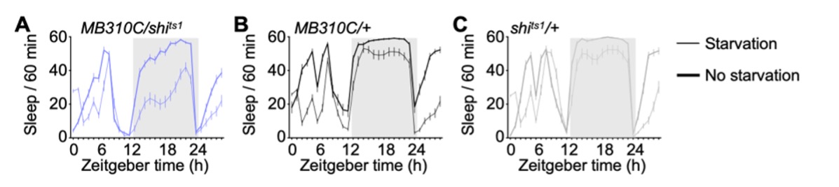
However, the key focus of our study is that changes in neuronal activity disrupt sleep under starvation conditions but not under non-starvation conditions. To emphasize this critical distinction, we have incorporated additional discussion focused specifically on this point.
“It is well established that starvation induces sleep suppression (MacFadyen, 1973; Thimgan et al., 2010; Melnattur and Shaw, 2019; Keene et al., 2010; He et al., 2020; Yangkyun et al., 2022), and our results are consistent with these previous findings: all genotypes exhibited less sleep under starvation than under fed conditions (i.e. Figures 4A-B, 5A-B and 11). Under normal appetitive memory training, starvation-induced sleep loss does not necessarily impair memory processing (Thimgan et al., 2010; Chouhan et al., 2021). PAM-α1 neuronal activity is higher in starved, trained flies than in fed or untrained flies (data not shown), suggesting that these neurons act as a critical node for integrating internal motivational and arousal states, as well as conveying positive valence for the normal appetitive memory process, independently of starvation-induced sleep loss. While DPM neurons are less sensitive to starvation, they still exhibit training-induced elevated activity (data not shown), indicating coherent responsiveness to upstream signaling. In the present study, we found that under fed conditions, sleep remained intact even when excessive changes in neural activity occurred within the PAM(-α1)-DPM circuit; in contrast, under starvation conditions, significant sleep reduction and fragmentation were observed. These observations indicate that starvation may trigger a transition from a physiologically normal brain state to an unstable, abnormally active state, which consequently elicits negative behavioral outputs.”
(7) Line 1121: The data points for Figure 9 D-E are surprisingly low considering there are 14 PAMalpha1 labelled, the data presented here indicated potentially only 1-2 neurons were counted per fly brain. Can this contribute to the large variation and the contradiction of PAM's memory-suppressing role?
We sincerely appreciate the reviewer’s critical comments regarding the sample size of labeled PAM-α1 neurons in Figure 9D–E. We have revisited our raw data, incorporated additional brain samples, and reanalyzed the dataset. For this updated analysis, we included all clearly distinguished neurons, excluded overlapping ones, and calculated a single NLI per brain for statistical analysis. The key conclusions remain consistent with those in the original submission, confirming the robustness of the observed phenotype.
Memory consolidation is a time-dependent process. To further elucidate the link between neural activity and behavioral outputs, we performed additional experiments with an extended recording period. A detailed response to this point is provided in the response to public review, and we therefore do not reiterate the details here.
(8) Line 345: the effect size and data spread of THIP restored memory is different from the controls in Figure 10, perhaps warranting a more conservative interpretation of the role of sleep in memory consolidation.
We appreciate this critical comment. We fully agree that the role of sleep in memory consolidation requires cautious interpretation, a point we have integrated into the revised manuscript.
Drug treatment in Drosophila, particularly for group-based assays, can introduce substantial variability at both the individual and group levels. To account for this, we employed a statistically valid sample size for our analyses to ensure robust conclusions. While minor quantitative discrepancies exist in the data, this technical consideration does not significantly alter the core conclusions of the study.
Reviewer #2 (Recommendations for the authors):
(1) As mentioned in the public review, all data using the broad PAM-DAN driver should be removed. Concerns regarding the experiments involving the broad driver are not included here.
A detailed response to this point is provided in the response to public review, and we therefore do not reiterate the details here.
(2) In GCaMP experiments (Figure 9B), the ΔF/F traces for the AHL and AHL+ATP conditions start diverging before the addition of ATP. The quantification shows they are not significantly different in the first 30 seconds, but the fact that in two separate experiments (2A and 9B), they diverge in the same direction makes me wonder whether the AHL condition is different from the +ATP condition even before the ATP treatment. Also, the traces should include standard errors.
We observed the same diverging trend in the first 30-second baseline as the reviewer. We reviewed the raw data for each sample and found that this divergence is likely attributable a small number of outliers. Given the absence of a statistically significant difference, this divergence does not affect our conclusions.
We have also added standard errors to the revised figures.
(3) Figure 9B. The authors need to show data for a control genotype. +>P2X2; VT064246-LexA > GCaMP6f that does not include MB299B-Gal4 is crucial to demonstrate that expression of P2X2 in PAM-α1 is responsible for the inhibitor effect, as LexA-P2X2 may be leaky.
One of the UAS-P2X2 lines was found to exhibit leaky expression, so we instead used a non-leaky UAS-P2X2 line for all related experiments. To address the reviewer’s comments and further validate our findings, we have added complementary experiments with a control genotype. In addition, we also added a control to confirm the non-leaky expression of LexA-P2X2 under the driver of R58E02-LexA. As shown in revised Figures 8B, application of ATP in the absence of MB299B-GAL4 failed to induce a significant inhibitory effect. These data strongly and convincingly support our conclusion.
(4) Figure 9B. Some of the individual data show values lower than -100% ΔF/F0. By definition, ΔF/F cannot be less than -100%, as this would require negative fluorescence, which is physically impossible. The calculation of fluorescence changes using ΔF/F should be carefully reconsidered.
We thank the reviewer pointing out this potential confusion. We used a standard method of calculating the change in fluorescence over time using △F/F = (Fn-F0) / F0×100% as we previously described (Liu et al., 2019). Changes of greater than +100% of △F/F would not be unusual, since the reported value is a ratio to the initial level of fluorescence, not a subtraction of the baseline value from the signal (which obviously could not go below 100%). We have included a sentence in the results explaining this (page 7): “As previously described, we used the percent change in fluorescence over time as a ratio to the initial level, △F/F = (Fn-F0)/F0×100% for quantification (Liu et al., 2019).” And we have carefully reviewed our raw and processed data and confirmed that our analysis was correct.
(5) Figure 2B. The number of UAS transgenes should be controlled, as Gal4 could be diluted with 3 UAS constructs in experimental conditions compared to only 1 UAS construct in controls. Are Dop1R2 and DopEcR significantly different from wt? Why do they present an average ΔF/F in 2A and a maximum in 2B?
We appreciate the reviewer’s careful observations and valuable comments.
As the reviewer noted, the EPAC imaging experiment utilizes three UAS transgenes, which enable Gal4 enhancement via Dicer, targeted manipulation of dopamine receptor expression levels, and neural activity monitoring in DPM neurons. All other imaging experiments in the study employ only one or two UAS transgenes. Given the robustness of the observed phenotypes, the potential dilution effect is not a major concern. Knockdown of Dop1R2 and DopEcR showed no significant differences relative to the WT control group; the maximum values presented in Fig. 2B are included solely to illustrate statistical significance. While the EPAC (CFP/YPF) signal reflects an obvious cAMP elevation, no differences were detected in the averaged signal across groups.
Notably, in the revised manuscript, our double-labeling assays confirmed that only Dop1R1 and Dop1R2 are colocalized with DPM neurons (see our responses to the public review from Reviewer #2). Accordingly, we have refined our data analysis to focus exclusively on these two DARs.
(6) Figures 7H, J. Why is almost every MB299B>TrpA1 fly sleeping at ZT0?
To align the starvation protocol for sleep analysis with that used in the memory assay, MB299B>TrpA1 flies and their genetic controls were transferred to fresh sleep tubes containing starvation food during the ZT0–1 time window. This transfer resulted in no detectable locomotor activity during this period, a pattern indicative of sleep in all flies.
(7) The number of episodes and P(wake) should be presented for all sleep data.
We have added these two parameters as new panels to all relevant sleep figures. The corresponding statistical analyses have also been included in the supplemental tables.
Reviewer #3 (Recommendations for the authors):
The study's findings provide compelling insights into the neural circuits connecting sleep and memory and the role of dopamine in general. While the anatomic dissection of the microcircuit and its overall involvement in sleep and memory is convincing and of high interest to the field and beyond, some statements of the study need further clarification, particularly the interpretation of receptor signaling and the role of DPM.
Major Points
(1) Figure 2: cAMP Imaging and Dopamine Receptor Involvement
The authors present calcium and cAMP imaging to support the inhibitory connection between PAM and DPM neurons. While using both sensors is a robust approach, I am not entirely convinced that cAMP imaging is the ideal approach for identifying the dopamine receptors involved. To my knowledge, only Dop1R1 is classically linked to Gs-mediated cAMP signaling. Dop1R2 is typically coupled to Gq (PLC and DAG), while DopEcR is non-canonical and can engage both pathways. Additionally, these receptors are classically excitatory, yet the authors did not analyze Dop2R, the primary inhibitory dopamine receptor - which would represent the most relevant candidate for an inhibitory PAM-DPM connection.
We have addressed this point in our response to the public comments, so will not reiterate here.
While dopamine receptor functions can vary by neuronal context, I would appreciate clarification on the following points:
(a) Why was Dop2R not tested? Was it omitted or found to have no effect?
We sincerely appreciate the reviewer’s critical questions. This point has been addressed in our response to the public comments. Briefly, Dop2R is not colocalized with DPM neurons; instead, only Dop1R1 and Dop1R2 are detected in DPM neurons, which is why we focused exclusively on these two receptors in the revised manuscript.
(b) Why was cAMP imaging chosen for receptor identification? Was calcium imaging performed, and if so, what were the results?
This is an excellent point, and we sincerely appreciate the reviewer’s valuable input, which has helped to strengthen the logical framework of our analysis on receptor-mediated neural activity. These dopamine receptors are well-characterized as members of the Gas-coupled protein receptor family, and cAMP signaling serves as a reliable readout of their functional activity. To strengthen the logic flow of our analysis on the target inhibitory circuit, we have made the following key revisions to the manuscript: 1) defined the expression profile of dopamine receptors in DPM neurons; 2) refined our cAMP imaging data analyses based on specific receptor subtypes; and 3) assessed DPM neural activity via calcium imaging under conditions of targeted receptor knockdown. For further details, please refer to our response to the public comments.
(c) Since the data suggest multiple receptor involvements and complex interactions, I encourage a more detailed discussion of the working hypothesis, particularly regarding the unexpected finding that classically excitatory receptors contribute to an inhibitory connection.
We appreciate the suggestion to elaborate on our working model. Accordingly, we have revised the schematic diagram and refined the manuscript to clearly illustrate the underlying mechanistic framework. For further details, please refer to our response to the public comments.
(2) Figure 3: DPM Involvement in Memory Consolidation
The authors show that PAM activation and DPM inhibition during consolidation impair appetitive LTM. However, the role of DPM is critical. While the c316-GAL4 driver yields strong effects, VT064246 inhibition shows only slight significance, requiring more than twice the sample size of other experiments. Given that c316-GAL4 is not DPM-specific and also labels MB Kenyon cells, I suggest using MB-GAL80 to restrict expression - or commenting on the possibility that other neurons like MB-KCs could directly participate in the phenotype. This is particularly relevant since VT064246 efficiently modulates sleep, indicating that it is generally effective in altering behavior. These issues weaken the claim that DPM plays a crucial role in linking sleep and memory, and should be addressed. Minor comment on this Figure: In the Figure legend, the driver and "n" are not mentioned for 3C, while this is the case for all other panels. Moreover, the DPM schematic only depicts the MB, making it somewhat confusing. DPM innervates the entire MB, still, it would be helpful to shade the DPM projections more distinctly within the MB for clarity.
We thank the reviewer for the suggestion to improve the precision of our figures.
Regarding the expression specificity concern, in all experiments using c316-GAL4, we had eyeless-GAL80 and MB-GAL80 co-expressed to restrict GAL4-driven expression to DPMs. While complete suppression of expression of MB-KCs was not achievable, we largely eliminated the potential confounding effects from majority of these cells. VT064246-GAL4 is known to exhibit weak expression (Jenett et al., 2011; Haynes et al., 2015), but high relative specificity. Importantly, the overall conclusion derived from experiments using c316-GAL4 with GAL80s and VT064246-GAL4 are consistent, which strongly supports the role of DPM neurons in mediating the link between sleep and memory.
As suggested, we have added sample sizes for all panels and refined the depiction of DPM projections in revised Figure 3C.
Minor Comments
(1) Introduction:
The authors introduce dopamine's role in forgetting but focus on aversive rather than appetitive memories. To avoid confusion, this distinction should be mentioned explicitly (likewise in the discussion). Regarding references: Zhang et al. (line 95) do not discuss DPM or APL. Donlea et al. (line 97) do not cover dopamine - I think Pimentel et al. (2016) would be a more appropriate citation.
This is a good point. We have removed Zhang et al. (2013) and replaced Donlea et al with Pimentel et al. 2016 as suggested.
(2) Figure 9: DPM Activation During Consolidation:
The authors show that PAM neurons are activated by starvation and further enhanced by appetitive training. Surprisingly, DPM neurons also increase activity post-training, despite the proposed inhibitory connection between PAM and DPM. The authors state that "PAM-α1-DPM microcircuit exhibits synchronized neural activity changes during the consolidation window" (line 326), yet they do not address this apparent contradiction. If I have not overlooked key information, this should be clarified/addressed e.g. in the discussion.
This is an excellent point. We have addressed this in our response to point (3) from Reviewer #2 in the public comments, so we will not reiterate it here.
(3) Figure 10D/E: THIP Rescue of LTM Deficits:
Some inconsistencies in the THIP rescue experiments need clarification:
(a) In Figure 10D, MB299B activation with THIP appears not to significantly restore memory relative to zero, nor to differ from untreated conditions in Figures 7A or 10E.
(b) In Figure 10E, MB299B activation +/- THIP shows a much clearer effect.
(c) Are Figures 7A, 10E, and 10D independent experiments, or were they conducted together?
(d) Should the left bar in 10D and the right bar in 10E be identical? If not, I do not fully understand the discrepancy and suggest discussing the variation.
Upon revisiting the raw datasets and conducting a one-sample t-test to analyze the group differences, the experimental group in Figure 7A showed no significant difference from the theoretical mean (set at zero). This group also did not differ from the two genetic controls, indicating that the restored memory was comparable to control levels. In Figure 10E, the group with MB299B activation plus THIP treatment exhibited a significant difference from the theoretical mean (one-sample t-test) and from the non-THIP control group, confirming a significant restoration of memory function. Owing to our laboratory relocation, the starvation duration at the new facility was adjusted based on a recalibrated starvation curve; the higher overall 24 h memory index in Figure 10E is likely attributable to a relatively longer starvation period. However, this experimental parameter variation does not alter the study’s overall conclusions.
(4) Sleep Phenotypes and Starvation Effects:
Sleep scores are shown under starvation/fed conditions but not under baseline conditions (without inhibition/activation). Could the authors indicate whether they observe basal starvation-induced sleep changes? The authors frequently state that PAM-DPM effects on sleep are context-dependent, yet mild but significant changes occur under fed conditions. I suggest rewording to clarify that the effect is enhanced in a context-dependent manner rather than strictly context-dependent.
Starvation-induced sleep reduction is a well-characterised phenotype. Our study focused on the key question of whether altered neuronal activity modulates sleep under innate starvation conditions. Accordingly, all comparisons were made between the experimental and control groups under both starvation and fed conditions. We appreciate the reviewer’s suggestion to improve clarity and have revised the text as suggested.
(5) Starvation Duration in Methods:
The authors use 30-46h of starvation, which is longer than the ~20h typically used in appetitive memory studies. Could the authors explain why such extended starvation times were necessary?
Determining starvation levels via survival curves is a well-established and relatively objective method, one that has been widely adopted in prior studies. For the memory test, we standardized the total starvation duration for each genotype to the time point at which mortality reached 20%. Owing to inherent differences in to starvation resistance across distinct genotypes, the final starvation durations ranged from 20 hours to 46 hours.
(6) Variability in PAM-α1 Sleep Effects:
(a) The extent and timing of sleep effects differ across PAM-α1 drivers (e.g. night vs. light-period effects). Could MBON co-targeting by these drivers contribute to the variability?
We have supplemented additional experiments to investigate the effects of MBON-α1 neurons on 24h memory and sleep. For detailed findings, please refer to our response to your public comments.
(b) Even within the same driver, results differ (e.g., Figure 7H vs. 10A). A general comment on these differences would be important, e.g. regarding the relevance of day and night sleep for memory consolidation.
We sincerely appreciate the reviewer’s incisive observation regarding these details. The discrepancy stems from the timing of neuronal activity inhibition, during which a laboratory relocation led to adjustments in starvation duration for memory experiments, which in turn indirectly altered sleep patterns.
(c) Technical note: Similar y-axis scales for sleep plots (Figures 10A and B) would make comparison easier.
We have unified the y-axis scales to the same range.
(7) Discussion, Line 376:
The phrase "sleep deprivation is important for memory consolidation" is misleading, as it could imply that deprivation aids memory formation. Please clarify.
We appreciate the reviewer’s suggestion. We have revised the text to: “These results demonstrate that preserving unperturbed sleep during the critical memory consolidation window is essential for stabilizing appetitive long-term memory.



