
Peer review process
Revised: This Reviewed Preprint has been revised by the authors in response to the previous round of peer review; the eLife assessment and the public reviews have been updated where necessary by the editors and peer reviewers.
Read more about eLife’s peer review process.Editors
- Reviewing EditorKiyoshi TakedaThe University of Osaka, Osaka, Japan
- Senior EditorTadatsugu TaniguchiThe University of Tokyo, Tokyo, Japan
Reviewer #1 (Public review):
Summary:
Mast cells have previously been reported to play an important role in bacterial immune defence and act protectively in sepsis. However, many of these findings were based on studies using Kit mutant mice. In this study, the authors conducted a detailed investigation using mast cell-deficient Cpa3 Cre-Master mice. As a result, the authors found that the Cpa3 Cre-Master mice exhibited responses similar to wild-type mice in terms of bacterial immune defense. This suggests that the observed phenotype is not due to mast cell-dependent bacterial immune defense, but rather is associated with dysbiosis of the gut microbiota.
Strengths:
Mast cells have long been reported to play an important role in the protective response against sepsis, and their function in infection defense has been demonstrated. However, Kit mutant mice have been reported to exhibit impaired peristalsis, and several mast cell-specific genetically modified mouse lines have since been developed and examined in detail. This study presents an important finding by logically demonstrating that the exacerbation of sepsis in Kit mice is due to alterations in the gut microbiota, and that the phenotype previously thought to be mast cell-dependent was, in fact, not.
In addition, the experiments were carefully designed using mice with matched genetic backgrounds. These findings underscore the importance of microbiota composition in interpreting immune phenotypes and highlight the need for co-housing controls in mutant mouse studies.
A major strength of this work is the robustness of the CLP data, generated over eight years by three independent researchers across two institutions with large sample sizes, lending strong support to the conclusions.
Weaknesses:
The study assesses only a limited subset of gut bacterial species, leaving the extent to which E. coli expansion contributes to the observed phenotype unclear. Moreover, in the cohousing experiments, there is no evidence provided to confirm successful microbiota normalization between groups. A more detailed analysis of the microbial composition would be necessary to strengthen the reliability of the findings.
It is also important to note that Cpa3-deficient mice exhibit not only mast cell depletion but also defects in basophils and T cells. These additional immunological alterations may counterbalance one another, potentially masking phenotypic changes and complicating interpretation.
Furthermore, it remains to be determined whether the altered gut microbiota observed in KitW/Wv mice is a consequence of impaired intestinal motility, whether a similar phenotype is observed in KitW-sh/W-sh mice, and whether comparable results occur in SCF-deficient models. Addressing these questions would provide greater clarity on the contribution of mast cells versus secondary factors in the observed phenotypes.
Given that KitW/Wv mice exhibit impaired peristalsis, is the observed increase in E. coli a consequence of this dysfunction?
Previous studies with BMMC reconstitution experiments have indicated that mast cells are a source of TNF-how does this align with the current findings?
Comments on revised version.
The authors have made substantial efforts to address the concerns raised in the previous review, and the revised manuscript has been substantially strengthened by the additional microbiome analyses. In particular, the new data provide a more comprehensive characterization of the intestinal microbiota in both Kit mutant and mast cell-deficient mice and strengthen the interpretation that the microbiota alterations observed in Kit mutant mice are associated with Kit deficiency rather than mast cell deficiency per se. Although Enterobacteriaceae were significantly increased based on the unadjusted Welch's t-test, this difference did not remain statistically significant after correction for multiple comparisons. The authors appropriately acknowledge this limitation in the revised manuscript. Overall, I consider the major concerns regarding the microbiome analysis to have been adequately addressed.
Reviewer #2 (Public review):
Summary:
The authors showed that the high susceptibility to CLP sepsis of Kit-mutant mice is not due to mast cell deficiency, but to dysbiosis.
Recommendations:
(1) The authors showed that E. coli increases in the cecum of Kit-mutant mice, which causes high CLP susceptibility. However, they did not provide any evidence E. coli is responsible for the high susceptibility. In the Figure 3 experiments, the authors administered the same number of cecal bacteria and did not show the number of E. coli after the administration. The authors should provide evidence showing that depletion of E. coli decreases susceptibility.
(2) The author should provide direct evidence of dysbiosis by, for example, shotgun sequencing of cecal and fecal contents.
(3) In case the authors find dysbiosis, they should analyze the mechanisms by which Kit mutation causes dysbiosis.
Comments on revised version.
The revised manuscript focuses on refuting the notion that mast cells play important roles in sepsis. The reviewer agrees with this claim.
Author response:
The following is the authors’ response to the original reviews.
Public Reviews:
Reviewer #1 (Public review):
Summary:
Mast cells have previously been reported to play an important role in bacterial immune defense and act protectively in sepsis. However, many of these findings were based on studies using Kit mutant mice. In this study, the authors conducted a detailed investigation using mast cell-deficient Cpa3 Cre-Master mice. As a result, the authors found that the Cpa3 Cre-Master mice exhibited responses similar to wildtype mice in terms of bacterial immune defense. This suggests that the observed phenotype is not due to mast cell-dependent bacterial immune defense, but rather is associated with dysbiosis of the gut microbiota.
Strengths:
Mast cells have long been reported to play an important role in the protective response against sepsis, and their function in infection defense has been demonstrated. However, Kit mutant mice have been reported to exhibit impaired peristalsis, and several mast cell-specific genetically modified mouse lines have since been developed and examined in detail. This study presents an important finding by logically demonstrating that the exacerbation of sepsis in Kit mice is due to alterations in the gut microbiota, and that the phenotype previously thought to be mast cell-dependent was, in fact, not.
In addition, the experiments were carefully designed using mice with matched genetic backgrounds. These findings underscore the importance of microbiota composition in interpreting immune phenotypes and highlight the need for cohousing controls in mutant mouse studies.
A major strength of this work is the robustness of the CLP data, generated over eight years by three independent researchers across two institutions with large sample sizes, lending strong support to the conclusions.
Weaknesses:
The study assesses only a limited subset of gut bacterial species, leaving the extent to which E. coli expansion contributes to the observed phenotype unclear.
We now performed 16S rRNA sequencing of cecal samples isolated from KitW/Wv and Cpa3Cre/+ mice and their respective littermates. Results are display in a new Figure 4. Our comparative analysis of the cecal microbial communities in KitW/Wv and Kit+/+ mice (Figure 4A+B) confirmed the expansion of E. coli (Enterobacteriaceae) that we had observed by CFU counts (Figure 3D). Furthermore, it revealed a dysbiotic shift marked by increased abundance of Peptostreptococcaceae, Verrucomicrobiaceae, Coriobacteriaceae, and Erysipelotrichaceae in KitW/Wv mice.
None of these changes was observed when comparing the cecal microbiomes of Cpa3Cre/+ and Cpa3+/+ mice (Figure 4C+D), indicating that the compositional shift in KitW/Wv mice is due to the deficiency in Kit but not mast cells. Of note, as stated on page 14, the microbial changes that we observed in KitW/Wv mice resemble dysbiotic patterns reported in chronic intestinal inflammation, experimental colitis, and impaired barrier function. These new findings fully align with and further support our earlier conclusion that KitW/Wv mice harbour pro-pathogenic microbiota.
The new results are display in a new Figure 4, and described on pages 9-10 and discussed on pages 13-14.
Moreover, in the cohousing experiments, there is no evidence provided to confirm successful microbiota normalization between groups.
It is correct that we have no direct data to confirm microbiota normalization between groups after co-housing. We note, however, that co-housing is a generally accepted method for microbiota equalization or conversion (Caruso et al., Cell Rep. 2019, Ridaura et al., Science 2013, and reviewed in Moore et al., Clin. Transl. Immunol. 2016). In any case, KitW/Wv mutants were made resistant to CLP by co-housing. Similar microbiota sequencing results between groups, while useful, would again only be correlative.
A more detailed analysis of the microbial composition would be necessary to strengthen the reliability of the findings.
See above the new data from 16S rRNA sequencing.
It is also important to note that Cpa3-deficient mice exhibit not only mast cell depletion but also defects in basophils and T cells. These additional immunological alterations may counterbalance one another, potentially masking phenotypic changes and complicating interpretation.
Regarding basophils in Cpa3Cre/+ mice, compared to wild-type mice, basophils are reduced to about 40% of normal (Feyerabend et al., Immunity 2011). In KitW/Wv mice, compared to wild-type mice, basophils are reduced to about 10% of normal. To our knowledge, there has been no phenotype reported in which a reduction in basophils compensates for the loss for mast cells. Given that KitW/Wv mice have about threefold lower numbers of basophils and are highly susceptible to sepsis, there is no evidence that a reduction in basophils is protective in mast cell-deficient mice. On the contrary, mice that were normal for mast cells but had their basophils depleted were more susceptible to sepsis (Piliponsky et al., Nat. Immunol. 2019). Hence, basophils appear to be protective, and their reduction increases susceptibility. In light of these data and considerations, there is no evidence for a reduction in basophils to counterbalance the loss of mast cells in Cpa3Cre/+ mice.
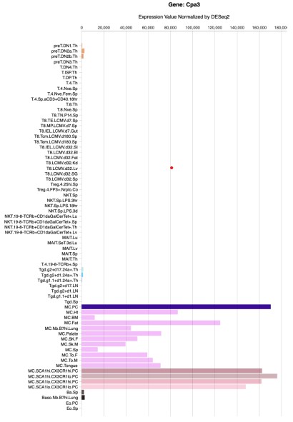
Regarding T cells, there is no evidence, and there are no reports, that Cpa3Cre/+ mice have defects in T cells (Feyerabend et al., Immunity 2011, Feyerabend et al., Cell Metabolism 2016). Cpa3 is weakly and transiently expressed early in the T cell lineage (Feyerabend et al., Immunity 2009; for expression levels in T cells versus mast cells, see Author response image 1). In summary, in contrast to the reviewer's claim, there are no known defects in T cell development or T cell functions in Cpa3Cre/+ mice. We think the reviewer needs to provide published evidence for his/her claim that Cpa3-deficient mice exhibit defects in T cells. We as authors are also obliged to support our claims scientifically, and rightfully so.
Author response image 1.
Generated from the Immgen database. Shown are RNAseq gene expression levels of diverse T-cell and mast cell populations.
Furthermore, it remains to be determined whether the altered gut microbiota observed in KitW/Wv mice is a consequence of impaired intestinal motility, whether a similar phenotype is observed in KitW-sh/W-sh mice, and whether comparable results occur in SCF-deficient models. Addressing these questions would provide greater clarity on the contribution of mast cells versus secondary factors in the observed phenotypes.
The purpose of our study was to verify or refute the key claim dating back to two 1996 Nature papers that mast cells play important roles against sepsis. We demonstrate here that this is not the case because mice without mast cells (Cpa3Cre/+ mice) were as resistant to sepsis as wild-type mice. Hence, mast cells are not involved in the immunity against sepsis, and 'secondary factors' are not involved in this simple experiment (both groups of mice, wild-type and Cpa3Cre/+ mice, were on the identical genetic background). Second, KitW/Wv mice are also as resistant to sepsis as wild-type mice when confronted with the identical intestinal slurry. Therefore, KitW/Wv mice have no immune deficit in response to sepsis. Hence, in our view, the underlying immunological question regarding the role of mast cells in sepsis has been conclusively addressed and answered by our data. We have changed the title to emphasize this central question.
The reviewer now asks us to delve even deeper into Kit biology and in particular intestinal pathophysiology in this and other Kit or steel mutants. While we share his/her interest in such questions, we fully disagree with the statement that 'addressing these questions would provide greater clarity on the contribution of mast cells versus secondary factors in the observed phenotypes.' We do not intend to enter the field of gut physiology or its link to microbiota, all the more because any results would not affect the central conclusion of our manuscript.
Given that KitW/Wv mice exhibit impaired peristalsis, is the observed increase in E. coli a consequence of this dysfunction?
See above
Previous studies with BMMC reconstitution experiments have indicated that mast cells are a source of TNF - how does this align with the current findings?
It does not align well. It is possible that cultured and transplanted mast cells (BMMC) produce TNF. Given that we did not find a reduction in TNF levels in the peritoneal lavage or serum in mice without mast cells undergoing sepsis, under physiological conditions mast cell-derived TNF does not seem to have a measurable impact on total TNF levels.
Reviewer #2 (Public review):
Summary:
This study presents a useful finding that the high susceptibility to CLP sepsis of Kitmutant mice is not due to mast cell deficiency, but to dysbiosis.
However, the present data are insufficient and incomplete to support the conclusion, and would benefit from more rigorous approaches. With the mechanism part strengthened, this paper would be of interest to researchers on mast cell biology and mucosal immunology.
We disagree with the view that our data are insufficient and incomplete. Our results demonstrate that mice lacking mast cells (Cpa3Cre/+ mice) are as resistant to sepsis as wild-type mice, demonstrating that mast cells do not play a detectable role in immunity against sepsis. Additionally, we show that KitW/Wv mice exhibit the same resistance to sepsis as wild-type mice when confronted with the identical intestinal slurry. This finding demonstrates that KitW/Wv mice have no immune deficit in response to sepsis. These central data are both sufficient and complete, given that our data fully address the potential role of mast cells in sepsis. Our study aimed to investigate the role of mast cells in sepsis, not to examine the mechanisms of dysbiosis or associated pathological phenotypes in Kit-mutant controls. We have changed the title to make this point.
Recommendations:
(1) The authors showed that E. coli increases in the cecum of Kit-mutant mice, which causes high CLP susceptibility. However, they did not provide any evidence E. coli is responsible for the high susceptibility.
We showed that E. coli CFUs were increased in the cecum of Kit-mutant mice, but we did not state that this causes CLP susceptibility. We wrote: 'Hence, KitW/Wv microbiota contains high levels of E. coli, which may underlie the observed pathogenicity'. We demonstrated that intestinal slurry from KitW/Wv mice is more pathogenic compared to intestinal slurry from wild-type mice. However, we did not search for or identify the bacterial species that causes this increased pathogenicity because we were addressing the role of mast cells in sepsis. We demonstrate an association of pathogenicity in sepsis experiments with cecal content of pathogenic bacteria (see also the new data on 16S rRNA sequencing). The same argument could be made for each bacterial species identified but this would be very complex experiments (both microbiologically and immunologically) given requirements for bacterial isolation, titration, and considerations of synergism. We therefore refrain from this undertaking.
In the Figure 3 experiments, the authors administered the same number of cecal bacteria and did not show the number of E. coli after the administration.
The samples were split and one aliquot was analysed by microbiology and the other aliquot was injected intraperitoneally. Fig. 3d shows the colony-forming units (for Lactobacilli and E coli) from aliquots of cecal slurry used in the intraperitoneal injection experiments shown in Fig. 3a-c. Hence, our data show the colony-forming units that were injected into the mice. It is unclear to us why this is not the key information rather than 'the number of E. coli after the administration'.
The authors should provide evidence showing that depletion of E. coli decreases susceptibility.
See response to point 1 above.
(2) The author should provide direct evidence of dysbiosis by, for example, shotgun sequencing of cecal and fecal contents.
We performed 16S rRNA sequencing of cecal contents and observed a dysbiotic shift towards an increase of Peptostreptococcaceae, Verrucomicrobiaceae, Coriobacteriaceae, Enterobacteriaceae, and Erysipelotrichaceae in KitW/Wv mice compared to Kit+/+ controls. None of these changes was observed when comparing the cecal microbiomes of Cpa3Cre/+ and Cpa3+/+ mice, indicating that the compositional shift in KitW/Wv mice is due to deficiency in Kit but not mast cells. Of note, as stated on page 14, the microbial changes we observed in KitW/Wv mice resemble dysbiotic patterns reported in chronic intestinal inflammation, experimental colitis, and impaired barrier function.
These new findings fully align and further support with our earlier conclusion that KitW/Wv mice harbour pro-pathogenic microbiota.
The new results are display in a new Figure 4, and described on pages 10-11 and discussed on pages 13-14.
(3) In case the authors find dysbiosis, they should analyze the mechanisms by which Kit mutation causes dysbiosis.
We have no intention to further explore Kit biology and in particular the intestinal pathophysiology caused by the Kit mutation because any results would not affect the central conclusion of our manuscript (see title). The review process and the revision shall center on making the core of a paper as conclusive as possible, and not widen a paper by requests 'tangential to the main conclusion' (Kaelin Jr. Nature 2017).
References:
Caruso, R., Ono, M., Bunker, M. E., Núñez, G. & Inohara, N. Dynamic and Asymmetric Changes of the Microbial Communities after Cohousing in Laboratory Mice. Cell Rep. 27, 3401-3412.e3 (2019).
Feyerabend, T. B. et al. Deletion of Notch1 Converts Pro-T Cells to Dendritic Cells and Promotes Thymic B Cells by Cell-Extrinsic and Cell-Intrinsic Mechanisms. Immunity 30, 67–79 (2009).
Feyerabend, T. B. et al. Cre-Mediated Cell Ablation Contests Mast Cell Contribution in Models of Antibody- and T Cell-Mediated Autoimmunity. Immunity 35, 832–844 (2011).
Feyerabend, T. B., Gutierrez, D. A. & Rodewald, H.-R. Of Mouse Models of Mast Cell Deficiency and Metabolic Syndrome. Cell Metab 24, 1–2 (2016).
Kaelin Jr, W. G. Publish houses of brick, not mansions of straw. Nature 545, 387– 387 (2017).
Moore, R. J. & Stanley, D. Experimental design considerations in microbiota/inflammation studies. Clin. Transl. Immunol. 5, e92 (2016).
Piliponsky, A. M. et al. Basophil-derived tumor necrosis factor can enhance survival in a sepsis model in mice. Nat. Immunol. 20, 129–140 (2019).
Ridaura, V. K. et al. Gut Microbiota from Twins Discordant for Obesity Modulate Metabolism in Mice. Science 341, 1241214 (2013).
Recommendations for the authors:
Reviewer #1 (Recommendations for the authors):
Suggestions for improved or additional experiments, data, or analyses:
(1) The study examines only a limited range of gut bacterial species, making it difficult to determine the specific contribution of E. coli expansion to the observed phenotype. A more comprehensive microbial profiling (e.g., 16S rRNA sequencing or metagenomics) would significantly strengthen the conclusions (e.g., Cpa3-mast cell deficient mice, Kit WWv, Kit W-sh/W-sh mice).
As mentioned above, we now performed 16S rRNA sequencing of cecal contents from KitW/Wv and Cpa3Cre/+ mice and their respective littermates. Addressing microbiota in KitW-sh/W-sh mice would not add information relevant for our paper.
(2) The role of impaired peristalsis in KitW/Wv mice as a contributor to microbial dysbiosis and increased E. coli burden should be further explored. Complementary studies using KitW-sh/W-sh or SCF-deficient mice could clarify whether the observed microbiota changes are unique to the W/Wv model.
We changed the title of the manuscript to emphasize our (unchanged) focus on the immunological role of mast cells in protecting against bacterial sepsis. The responses of Cpa3Cre mice clearly ruled out a role of mast cells to these infectious conditions. Our observation of altered microbiota in KitW/Wv mice is consistent with their increased CLP susceptibility. We cite the known peristalsis deficit of KitW/Wv mice as a possible explanation for the microbiota alterations. In the future, other investigators may find it interesting to elucidate the link between Kit mutations and dysbiosis. As stated further above, in our view, these additional questions and potential data have no bearings on the conclusions of our paper.
(3) In the cohousing experiments, no data are provided to confirm whether microbiota normalization was achieved between groups. Including microbial composition data pre- and post-cohousing would improve the reliability of the interpretation.
Cohousing made the susceptibility of KitW/Wv and Kit+/+ mice comparable. Detailed analysis of the extent of microbiota normalization would only make sense to ultimately determine specific taxa or combinations thereof that are responsible for the increased susceptibility of KitW/Wv mice, a question that was never the goal of this study.
(4) The use of Cpa3 Cre/+ mice introduces potential confounders, as these mice also have defects in basophils and T cells. Functional validation or additional models (e.g., Mas-TRECK or Mcpt5-Cre mice) could help isolate the mast cell-specific effects.
See our detailed explanation above (Reviewer #1 Public review). It is incorrect to claim that Cpa3Cre/+ mice have defects in T cells. The reviewer needs to provide published evidence for his/her claim that Cpa3-deficient mice exhibit defects in T cells. We as authors are also obliged to support our claims scientifically, and rightfully so.
(5) Clarification is needed regarding the role of mast cell-derived TNF. Given previous reports using BMMC reconstitution that implicate mast cells as a source of TNF, reconciling these findings with the current study's results would strengthen interpretation.
We also disagree here. Experiments in normal unmanipulated mice are inevitably superior to Kit mutants after BMMC reconstitution which is an artificial system. The transplanted cells do not mature normally and don’t settle in their natural niches. We mentioned in the discussion that there is conflicting literature derived from different models and mice.
But we do not share the expectation that results obtained with Kit-independent models, that differ from previous studies using Kit mutants, necessarily require reconciliation. The experiments are simply incomparable and the most physiologically relevant experiment will pave the way. Research on mast cell functions based on BMMC-reconstituted Kit mutants has meanwhile proven to be unreliable.
(6) A clearer delineation between mast cell-dependent and microbiota-mediated mechanisms in the discussion sections would enhance readability and impact.
We restructured the discussion and distinguished between Kit-dependent and mast cell-dependent phenotypes. We also discussed in detail the observed microbiota differences and their influences for the outcomes in the different sepsis experiments.



