
Peer review process
Revised: This Reviewed Preprint has been revised by the authors in response to the previous round of peer review; the eLife assessment and the public reviews have been updated where necessary by the editors and peer reviewers.
Read more about eLife’s peer review process.Editors
- Reviewing EditorMathieu WolffCNRS, University of Bordeaux, Bordeaux, France
- Senior EditorKate WassumUniversity of California, Los Angeles, Los Angeles, United States of America
Reviewer #1 (Public review):
Summary:
Badarnee and colleagues analyse fMRI data collected during an associative threat-learning task. They find evidence for parallel processes mediated by the mediodorsal, LGn and pulvinar nuclei of the thalamus. The evidence for these conclusions is promising, but limited by a lack of clarity regarding the preprocessing and statistical methods.
Strengths:
The approach is inventive and novel, providing information about thalamocortical interactions that are scant in the current literature.
Comments on revised version.
No further comments. The authors have adequately addressed my concerns.
Reviewer #3 (Public review):
Summary:
The present work was aimed at investigating the specific contributions of thalamic nuclei to associative threat learning and extinction. Using fMRI, it examined activation patterns across pulvinar divisions, the lateral geniculate nucleus (LGN), and the mediodorsal thalamus (MD) during threat acquisition, extinction, and recall. It goals was to uncover whether distinct thalamic systems support different modes of learning-automatic survival mechanisms versus more deliberate processes-and to propose a hierarchical pulvinar model of fear conditioning. The manuscript also tried to refine current neuroanatomical models of threat learning and memory, highlighting the role of thalamic nuclei in it.
Strengths:
(1) Valuable theoretical elaboration and modeling regarding the differential role of pulvinar subdivisions on feedforward (inferior, lateral) and higher-order integration (anterior), and their functional interplay with other relevant subcortical and cortical structures in associative threat and extinction learning.
(2) Large sample sizes and multipronged analytical approaches were used for hypothesis testing.
(3) Exhaustive literature review in the field of associative threat, as well as regarding the role of thalamic nuclei and other brain structures in it.
Comments on revised version:
My three main concerns remain insufficiently addressed or only partially addressed in the study limitations. First, using 3 mm isotropic voxels limits anatomical and functional specificity when investigating small thalamic nuclei. Higher-resolution fMRI protocols and probabilistic thalamic atlases that allow subject-specific segmentation have been available for several years and could have been used to improve anatomical precision. Second, although analyses in normalized space are widely accepted for group-level analyses, normalization can distort the size and boundaries of small thalamic nuclei. Analyses in native space, using subject-specific segmentation, would help address this limitation, particularly given the relatively low functional resolution. Finally, standard preprocessing procedures and the inclusion of motion parameters as covariates do not fully exclude residual motion-related effects or potential differences in motion across experimental conditions. The authors do not report additional motion quality-control procedures, such as excluding participants with excessive motion or substantial translational or rotational drift. Taken together, these concerns prevent me from upgrading my assessment.
Author response:
The following is the authors’ response to the previous reviews.
eLife Assessment:
This study provides valuable insights into the role of thalamic nuclei in associative threat and extinction learning, underpinned by a large dataset and rigorous, multipronged analyses. The evidence provided is solid, supporting the main conclusions. Minor analytical refinements notwithstanding, the manuscript will be of broad interest to researchers in learning and memory, fear, thalamic circuitry, and related mental health conditions.
We sincerely thank the editors and the reviewer for their careful evaluation of the revised manuscript and for their constructive comments. We greatly appreciate the time and effort invested in reviewing our work, and we have carefully considered each suggestion to further improve the clarity, presentation, and interpretation of the manuscript.
Author’s note:
We note that several comments refer to figure numbers and manuscript text from an earlier version of the manuscript (e.g., Figure 7 in the earlier version corresponds to Figure 8 in the current revised manuscript). For clarity, our responses below refer to the corresponding figures and sections in the current revised manuscript while addressing each comment individually.
Public Reviews:
Reviewer #1 (Public review):
Summary:
Badarnee and colleagues analyse fMRI data collected during an associative threat-learning task. They find evidence for parallel processes mediated by the mediodorsal, LGn and pulvinar nuclei of the thalamus. The evidence for these conclusions is promising, but limited by a lack of clarity regarding the preprocessing and statistical methods.
We thank the reviewer for these encouraging comments and for recognizing the novelty of our approach and its potential contribution to advancing our understanding of thalamocortical interactions during associative threat learning. We also appreciate the reviewer’s constructive suggestions regarding the methodological description, which have helped us improve the clarity of the manuscript.
Strengths:
The approach is inventive and novel, providing information about thalamocortical interactions that are scant in the current literature.
Weaknesses:
(1) There are not sufficient details present to allow for the direct interrogation of the methods used in the study.
Thank you for raising this point. As described in our previous revision, we substantially expanded the Experimental Procedure section by adding three dedicated subsections describing the fear learning procedure, shock calibration, and participant instructions. These additions included details regarding the stimuli, experimental context, stimulus and shock durations, average inter-trial interval, pseudorandomized stimulus presentation, and the procedure used to determine each participant’s shock intensity.
In the current revision, we have further clarified the Methods by specifying electrical stimulator that we used and specified the usage of the gel and placement of the electrodes. We believe these additions provide sufficient methodological detail to allow readers to understand and replicate the experimental procedure (page 40):
“Electrical stimulation was delivered using a BIOPAC stimulation system with BIOPAC electrodes and conductive gel. The electrodes were attached to the palm of the participant's right hand and positioned in close proximity without making contact with one another. The electric stimulation began…”
(2) The figures do not contain sufficiently granular details, making it challenging to determine whether the observed effects were robust to individual differences.
Thank you for this suggestion. To better illustrate the underlying data distribution and between-participant variability, we revised Fig. 1 and 4-6 by adding individual participant data points for the block-level analyses and kernel density estimate (KDE) distributions for the trial-level analyses. These additions allow readers to better assess the distribution of the data and the extent to which the reported effects are consistent across participants while maintaining figure readability. An example of the revised Fig. 1.
Comments on revisions:
I continue to recommend the plotting of individual data points. While there may be individual variance, it is important to quantify this in publication so that future studies can appreciate the uncertainty surrounding test statistics.
Thank you for this recommendation. As described in our response to Weakness 2 above, we revised the figures to better illustrate the underlying data distribution and between-participant variability. Specifically, we added individual participant data points for the block-level analyses and kernel density estimate (KDE) distributions for the trial-level analyses. We believe these revisions improve the transparency of the underlying data while maintaining figure readability. An example of a revised figure is included above.
Reviewer #2 (Public review):
Summary:
The authors quantify human fMRI BOLD responses in pulvinar and mediodorsal thalamic nuclei during a fear conditioning and extinction task across two days, in a large sample size (hundreds of participants). They show that the BOLD responses in these areas differentiate the conditioned (CS+) and safety (CS-) stimulus. Additionally this changes with repeated trials which could be a neural correlate of fear learning. They show that the anterior pulvinar is most correlated with the MD, and that this is not due to anatomical proximity. They perform graph analysis on the pulvinar sub nuclei which suggests that the medial pulvinar is a hub between the sensory (lateral/inferior) and associative (anterior) pulvinar. They show different patterns of thalamic activity across conditioning, extinction, recall, and renewal.
We thank the reviewer for this thoughtful summary of our work and for recognizing the scope of the study and the value of the findings. We appreciate the reviewer’s careful evaluation and constructive feedback, which helped improve the clarity and interpretation of the manuscript throughout the revision process. Below, we respond to each of the points raised under the Weaknesses and Comments on revisions sections and describe the corresponding revisions made to the manuscript.
Strengths:
The data has a large sample size (n=293 in some measures, n=412 in others). This is a validated human fear conditioning/extinction task that Dr Milad's group has been working with for several years. Few labs have investigated the thalamus activity during fear conditioning and extinction, particularly with a large sample size. There is an independent replication of the pulvinar network structure (Fig. 3), which suggests that the processing in the more sensory-related inferior and lateral pulvinar is relayed to the anterior pulvinar (and possibly thereby to more action-related prefrontal areas) via an intermediate step in the medial pulvinar - potentially a novel discovery but that needs more validation.
Weaknesses:
(1) The authors cannot make causal claims about their results based on correlational neuroimaging evidence. Causal claims should be pared back. E.g. Sentence 1 in results "The anterior pulvinar and MD contribute to early associative threat learning, as evidenced by increased functional activation in response to CS+ compared to CS- at the block level (Fig. 1b-c)." needs to be reworded to something like 'the anterior pulvinar and MD have increased functional activation... This suggests that these areas may contribute to early associate threat learning"
We agree with the reviewer that causal claims cannot be made based on correlational neuroimaging evidence. Accordingly, we carefully revised the manuscript throughout to avoid causal language and instead use wording that reflects statistical associations and interpretations. Representative examples of these revisions are provided below.
Representative examples of these revisions are shown below:
Page 7: “At the block level, both the anterior pulvinar and MD showed increased activation to CS+ vs. CS− (anterior pulvinar: t(292) = 4.41, p = 0.00001, d = 0.25; MD: t(292) = 6.41, p = 5.83x10⁻¹⁰, d = 0.37; Fig. 1b–c), suggesting a possible involvement of these regions in early associative threat learning.”
Page 13: “A data-driven approach suggests a hierarchical computational organization of pulvinar divisions during associative threat learning.”
Page 13: “we integrated findings from complementary analyses to propose a computational framework describing the functional relationships among pulvinar divisions during associative threat learning.”
Page 13-14: “The network plot identified a statistical association between the lateral and anterior pulvinar, together with pathways linking the lateral and inferior pulvinar to the anterior pulvinar through the medial pulvinar (Fig. 3b).”
Page 14: “The medial pulvinar statistically mediates the functional relationships within the pulvinar network and is associated with elevated centrality values. Together, these findings are consistent with a possible hub-like functional role of the medial pulvinar, consistent with higher-order integration of CS+ information, relative to the more sensory-related functional profiles in the inferior and lateral pulvinar. This possible hierarchical organization is consistent with previous evidence showing that the inferior and lateral pulvinar are associated with processing basic sensory information (Berman and Wurtz, 2010, 2008; Cortes et al., 2024), while the medial pulvinar is implicated in higher-order processing, including attention and working memory (Homman-Ludiye and Bourne, 2019).”
Page 15: “This data-driven analyses provide a possible new perspective on the functional specialization of pulvinar divisions during associative threat learning. Based on these observations, we hypothesized a computational model in which activation in the medial pulvinar statistically mediates the functional relationships between the inferior and lateral pulvinar and the anterior pulvinar (Fig. 3e). To evaluate this hypothesis, we performed a mediation analysis and assessed the robustness of the model using k-fold cross-validation.”
Page 25: “The vmPFC connectivity may reflect involvement of prefrontal processes related to evaluation and regulation of threat-related information.”
Page 27: “The medial pulvinar may statistically mediate the functional relationships between the inferior and lateral divisions and the anterior pulvinar, consistent with a hierarchical computational model.”
Page 29: “Together, these findings are consistent with the possibility that the anterior pulvinar and MD contribute in parallel to associative threat learning, extending the two-system.”
Page 32: “Using statistical mediation analysis, we found that activation in the medial pulvinar statistically mediated the relationship between activation in the inferior/lateral pulvinar and activation in the anterior pulvinar. This statistical mediation is consistent with, but does not establish, a hierarchical computational organization.”
(2) Fig .1 The fact that the difference in BOLD activity between CS+ and CS- goes away on the third trial is not addressed. This is a very large effect in the data.
Thank you for raising this important point. We have addressed this issue in both the Discussion and the Limitations sections, as follows:
Discussion (page 31):
“The attenuated CS+ vs. CS− differentiation on the third trial specifically in the pulvinar may reflect a decreased requirement for differential thalamic engagement once the initial association has been acquired. Notably, because the MD sustained the BOLD response to the CS+ in the third trial which may indicate involvement of this nucleus in continued processing or maintenance of the learned association. This aligns with the well-established MD-PFC circuit involved in cognitive processes (Wolff and Halassa, 2024). Additionally, in a previous study using a similar paradigm, we observed sustained CS+ vs. CS− differentiation on the third trial in the nucleus reuniens, as well (Tuna et al., 2025). These findings suggest that trial-dependent learning dynamics may vary across thalamic nuclei rather than reflecting a uniform thalamic learning signal. Together, while our paradigm does not inherently distinguish between different stages of learning, such as early acquisition and stabilization, our findings are consistent with stronger associative learning-related engagement during the first two trials, with a reduced differential response by the third trial that may reflect the involvement of different neural processes.”
Limitations (page 36):
“The reduced CS+ vs. CS− differentiation observed in the pulvinar subdivisions during the third trial may reflect a transition from the rapid associative learning processes engaged during the first two trials to a different stage of threat processing. However, the present experimental design does not allow us to determine the specific psychological or neural mechanisms underlying this change. Importantly, this attenuation was not observed uniformly across thalamic nuclei, suggesting that it does not reflect a general thalamic response but may instead indicate nucleus-specific functional dynamics. Future studies specifically designed to dissociate different stages of threat learning will be required to determine the functional significance of this trial-dependent effect.”
(3) Fig. 3 Could the observed network structure be due to anatomical proximity? Perhaps the authors should do an analogous analysis to what they did in Fig. 2 for this intra-pulvinar analysis. This analysis doesn't take into account the indirect connections through corticothalamic and thalamocortical connections with visual cortex and the pulvinar. There is an implicit assumption that there are interconnections between the pulvinar sub nuclei, but there are few strong excitatory projections between these sub nuclei to my knowledge. If visual areas are included in the graph, it would make things more complex, but would probably dramatically change the story. In this way, the message is somewhat constructed or arbitrary.
We thank the reviewer for this thoughtful suggestion. We performed an analysis analogous to that presented in Figure 2. Specifically, we computed partial correlations between pulvinar divisions while controlling for activation in the other thalamic nuclei included in the study (LGN and MD). The relationships between pulvinar divisions remained significant after accounting for shared variance with these nuclei (Supplementary Figure 4), suggesting that the observed associations are not solely explained by activation in neighboring thalamic regions. This analysis is particularly relevant for the LGN because of its close anatomical proximity to the pulvinar and its central role within the visual thalamic system.
Additionally, we statistically compared the strength of the partial correlations among all pairs of pulvinar divisions (Author response table 1). These analyses demonstrated substantial heterogeneity in the correlation strengths, with several pairwise differences remaining significant after FDR correction, indicating that the functional relationships among pulvinar divisions are heterogeneous rather than uniformly correlated, making a simple anatomical-proximity explanation less likely.
Author response table 1.
Pairwise comparisons demonstrate heterogeneous functional relationships among pulvinar subdivisions after controlling for MD and LGN activation.
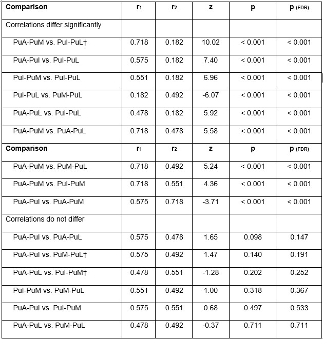
Note. All values are partial correlations controlling for MD and LGN activation (N = 293). Differences between correlations were tested using Steiger’s test for dependent correlations measured in the same participants. The sample size entered into the test was 293 - 2 = 291 as two variables (MD and LGN) were partialled out. The test itself then uses 291 - 3 = 288, the standard adjustment applied whenever correlations are converted to Fisher z scores. Steiger provides two versions of the same equation: one for comparisons in which the two correlations share a subdivision, and one for comparisons in which they share none (marked †). Analyses were conducted using the cocor package in R.
The p-values for the 15 comparisons were corrected using the Benjamini-Hochberg false discovery rate (FDR) procedure; 9 of 15 comparisons remained significant after correction. Correlations ranged from 0.182 to 0.718. PuA = anterior pulvinar; PuI = inferior pulvinar; PuM = medial pulvinar; PuL = lateral pulvinar.
The two correlations being compared share no subdivision.
Finally, we agree that pulvinar function is embedded within broader corticothalamic and thalamocortical circuits involving visual cortical regions. However, a comprehensive characterization of pulvinar-visual cortical interactions at the level of specific pulvinar divisions was beyond the scope of the current study. To acknowledge this limitation and avoid implying that the observed relationships represent isolated direct interactions among pulvinar divisions, we revised the conceptual model in Fig. 8 by adding bidirectional dashed gray arrows between the pulvinar divisions and visual cortex (see Fig 8). Additionally, we clarified this point in the Figure notes and the study limitations:
Limitations (page 36):
“The observed relationships among pulvinar divisions during conditioning are purely functional and do not distinguish direct inter-nuclear interactions from indirect coupling mediated by corticothalamic and thalamocortical pathways, including visual cortical regions. Thus, the proposed functional pulvinar model may reflect indirect corticothalamic and thalamocortical loops, currently undocumented internuclear interactions, or a combination of these mechanisms.”
(4) In the results section describing Fig. 4-7, there are no statistics supporting the claims made. There needs to be a set of graphs comparing the results across the study sessions and days, with statistical comparisons between the different experiments to confirm differences.
We thank the reviewer for this thoughtful comment. As stated in the scope of the study (page 6 in the manuscript), our objective was to characterize thalamic activation and connectivity within each learning phase separately. Accordingly, our statistical analyses were designed to test CS+ vs. CS− differentiation within each phase rather than to directly compare activation or connectivity across phases (e.g., conditioning, extinction, extinction recall, and threat renewal).
This phase-specific approach was selected because each learning phase probes a distinct psychological process with different experimental conditions. Therefore, direct comparisons of activation or connectivity between phases would address a different research question than the one investigated in the present study were not part of our predefined hypotheses or statistical design.
To address the reviewer’s concern, we carefully revised the Results and Discussion to make this analytical framework explicit and to avoid wording that could be interpreted as implying direct statistical comparisons between learning phases. Representative examples of revisions made throughout the manuscript to clarify that the statistical analyses and interpretations are phase-specific (CS+ vs. CS− within each phase) and do not involve direct statistical comparisons across learning phases.
Representative examples of these revisions are shown below:
Page 17: “Pulvinar divisions and MD show engagement during extinction learning and context-dependent threat memory processing.”
Page 21: “The threat renewal phase, in which participants were exposed to contextual cues similar to those used during threat conditioning, was associated with.”
Page 23: “At the block level, activation was greater for CS+ than CS− within each phase separately: conditioning…”
Page 23: “Collectively, these findings suggest that the LGN exhibited early trial-dependent responses within each learning phase separately, consistent with its proposed role in feedforward visual processing.”
Page 28: “Figure 8b title: Conceptual model of thalamic-dependent ‘toggle switching’ between safe and threat memory retrieval across contextual cues”
“Legend: (b) Conceptual model illustrating a possible thalamic-dependent “toggle switch” underlying context-dependent safety and threat memory expression. The observed MDdACC and anterior pulvinar-vmPFC connectivity patterns are consistent with possible preferential engagement of prefrontal circuits during extinction recall and threat renewal, respectively. This framework represents a theoretical integration of the present findings rather than evidence of causal neural mechanisms. It is based on the findings observed within each phase separately and should not be interpreted as a direct comparison between phases.”
Page 25: “Interestingly, within each phase separately, the findings suggest that thalamic connectivity patterns vary with contextual conditions during extinction recall and threat renewal. Specifically, the MD and anterior pulvinar showed distinct CS+E vs. CS− connectivity patterns during these phases, respectively.”
(5) FIg. 7 does not include the major corticothalamic and thalamocortical projections from early, mid-level, and higher visual cortex to the different pulvinar nuclei. I doubt that there are strong direct projections between the pulvinar nuclei, rather the functional connections are probably mediated through interconnections with cortical visual areas.
Thank you for pointing out this important point. As described in our response to Weakness 3 above, we agree that pulvinar function is embedded within broader corticothalamic and thalamocortical circuits involving visual cortical regions. To better acknowledge this, we revised the conceptual model by adding bidirectional dashed gray arrows between the pulvinar and visual cortex and retained a note in the figure legend clarifying that the schematic is not intended to represent a comprehensive anatomical connectivity map. We also emphasize in the manuscript that the proposed intrapulvinar relationships should be interpreted as a functional/computational model based on statistical dependencies rather than evidence of direct monosynaptic anatomical connections. An updated version of the figure is provided. See Fig 8.
(6) Stylistic: There are a lot of hypotheses and interpretations presented in this primary literature paper which may be better suited for a review or perspective piece.
Thank you for this comment. Our intention here is to integrate the empirical findings within an existing conceptual framework rather than present isolated observations. We believe that placing our results in the context of current models helps highlight their potential implications while remaining grounded in the experimental data. Nevertheless, we reviewed the Discussion again and ensured that speculative statements are clearly identified as hypotheses or possible interpretations and are appropriately qualified throughout the manuscript.
(7) In the discussion there is an assumption that the fMRI BOLD responses to CS+ and CS- need to be different to indicate that an area is processing these distinctly, but the BOLD signal can only detect large scale changes in overall activity. It's easy to imagine that an area could be involved in processing these two stimuli distinctly without showing an overall difference in the gross amount of activity.
Thank you for raising this important limitation. We agree that the absence of differential BOLD responses does not necessarily imply the absence of stimulus-specific neural processing. We explicitly acknowledged this limitation in the manuscript (page 35):
“Although our findings suggest distinct patterns of thalamic involvement during threat learning, fMRI cannot fully capture the complexity of thalamic organization. Pulvinar divisions, MD, and LGN each contain diverse neuronal subtypes and finer anatomical subdivisions that may serve distinct functions. Importantly, the absence of CS+ vs. CS− differences in BOLD activation should not be interpreted as a lack of stimulus-specific processing, as such distinctions may occur without detectable changes in overall BOLD activation…”
We reviewed this section again and believe it appropriately addresses the limitation raised by the reviewer.
(8) There is strong evidence that the BOLD responses to the threat-related and safety-related stimuli are different, modest evidence for their claims of learning/plasticity in these pathways, and circumstantial evidence supporting their hypothesized graph network models. Overall most of the claims made in the discussion are better considered possible interpretations rather than proven findings - this is not a criticism, as these experiments and subject matter are extremely complex.
We thank the reviewer for this thoughtful perspective. We agree that our findings provide varying levels of evidence across different aspects of the study and that the proposed network models should be interpreted as hypotheses informed by our empirical observations rather than definitive mechanistic conclusions. Accordingly, throughout the current revisions, we have carefully revised the Results and Discussion sections to distinguish empirical findings from interpretations by using more cautious language and explicitly acknowledging the limitations of our experimental approach. All revisions are in the manuscript, and representative examples of the changes can be found above.
(9) This study continues to validate the power and utility of this in human fear conditioning/extinction paradigm, and extends this paradigm to investigating fear learning beyond the traditional limbic system pathways. It's possible that their models for the pulvinar nuclei interconnections could guide future neuromodulation or DBS studies that could provide more causal evidence for their hypotheses.
We thank the reviewer for these encouraging comments and for recognizing the potential utility of this paradigm and the possible implications of our findings for future mechanistic and neuromodulation studies.
Comments on revisions:
The reviewers addressed my major concerns appropriately in the modified manuscript. As long as the MRI analysis concerns of Reviewer 3 are satisfied (MRI analysis is not my expertise), I am satisfied with the modified manuscript.
We sincerely thank the reviewer for the positive assessment of our revised manuscript and for recognizing that the major concerns were appropriately addressed. We appreciate the reviewer's constructive feedback throughout the review process, which has helped improve the quality and clarity of the manuscript.
Reviewer #3 (Public review):
Summary:
The present work was aimed at investigating the specific contributions of thalamic nuclei to associative threat learning and extinction. Using fMRI, it examined activation patterns across pulvinar divisions, the lateral geniculate nucleus (LGN), and the mediodorsal thalamus (MD) during threat acquisition, extinction, and recall. It goals was to uncover whether distinct thalamic systems support different modes of learning-automatic survival mechanisms versus more deliberate processes-and to propose a hierarchical pulvinar model of fear conditioning. The manuscript also tried to refine current neuroanatomical models of threat learning and memory, highlighting the role of thalamic nuclei in it.
We thank the reviewer for this accurate summary of the aims and scope of our study. We also appreciate the thoughtful and constructive feedback provided throughout the review process, which has helped us improve the methodological description, interpretation of the findings, and overall clarity of the manuscript. Below, we respond to each of the points raised under the Weaknesses and Recommendations sections.
Strengths:
(1) Valuable theoretical elaboration and modeling regarding the differential role of pulvinar subdivisions on feedforward (inferior, lateral) and higher-order integration (anterior), and their functional interplay with other relevant subcortical and cortical structures in associative threat and extinction learning.
(2) Large sample sizes and multipronged analytical approaches were used for hypothesis testing.
(3) Exhaustive literature review in the field of associative threat, as well as regarding the role of thalamic nuclei and other brain structures in it.
Weaknesses:
(1) The manuscript has improved methodologically and analytically after the review. Several weaknesses remain, in my opinion, but still findings are valuable and the evidence can be considered as convincing.
(a) fMRI data have low resolution (3 cubic mm), which certainly limits the examination of small nuclei such as the ones investigated here, and especially the examination of the LGN and inferior pulvinar.
Thank you! We have acknowledged this limitation in the manuscript (page 35):
“Pulvinar divisions, MD, and LGN each contain diverse neuronal subtypes and finer anatomical subdivisions that may serve distinct functions. Importantly, the absence of CS+ versus CS− differences in BOLD activation should not be interpreted as a lack of stimulus-specific processing, as such distinctions may occur without detectable changes in overall BOLD activation. Future advances in higher-resolution imaging and human brain atlases may further improve anatomical and functional precision.”
(b) fMRI was normalized to standard space. Analyzing the data in individual-subject space would have given you the options of avoiding altering every participant's brain and of using more precise atlases than the normalized AAL for ROI selection.
We thank the reviewer for this thoughtful suggestion. Analyses performed in native subject space and standardized MNI space each have methodological advantages. In the present study, we adopted a standardized MNI-space pipeline because it provides consistent anatomical localization and ROI definition across participants, facilitating group-level analyses and comparisons across individuals. We have added this consideration to the Limitations section (page 35).
“The present analyses were performed in standardized MNI space to enable consistent group-level analyses and ROI definition across participants. Although this is a widely accepted approach in human neuroimaging, future studies may examine the generalizability of the present findings using complementary approaches, including analyses performed in native subject space.”
(c) Motion during scanning was poorly controlled. Including the motion parameters as covariates of no interest in the GLM/analysis does not fully guarantee that motion is not influencing the results, and that motion is not differentially influencing some experimental conditions more than others.
We thank the reviewer for highlighting the importance of controlling for head motion in fMRI analyses. We agree and acknowledge that no preprocessing strategy can completely eliminate all potential residual motion-related effects in fMRI data.
For the activation analyses, the data were preprocessed using the standard fMRIPrep (v20.0.2) pipeline, which included motion correction through rigid-body realignment prior to statistical modeling. The first-level SPM analyses included the six estimated head-motion parameters (three translations and three rotations) as nuisance regressors. White matter and cerebrospinal fluid signals were also included as nuisance regressors to reduce non-neuronal sources of variance.
For the functional connectivity analyses, we applied CONN’s denoising pipeline, which included regression of motion parameters, regression of identified outlier scans (scrubbing), component-based physiological noise correction (CompCor), and temporal filtering prior to generalized psychophysiological interaction (gPPI) analyses. The combination of motion correction, nuisance regression, and additional denoising procedures, provides a multistep approach to reducing potential motion-related and non-neuronal confounds in the reported findings. We have further clarified these processes in the manuscript (pages 44-45 in the manuscript):
“Functional connectivity was computed using the CONN toolbox (version 22.a) for MATLAB (NietoCastanon and Whitfield-Gabrieli, 2022; Whitfield-Gabrieli and Nieto-Castanon, 2012). Anatomical images were segmented into gray matter, white matter, and CSF. Functional images were preprocessed following CONN’s default pipeline, including realignment, slice-timing correction, and coregistration. Functional images were then denoised using a component-based noise correction approach (CompCor), including regression of estimated motion parameters, regression of identified outlier scans using artifact detection procedures (scrubbing), and regression of white matter and CSF signals. The denoised BOLD time series were subsequently band-pass filtered between 0.008 and 0.09 Hz.
Following these denoising procedures – including regression of motion parameters, artifact-related outlier scans (scrubbing), physiological noise correction (CompCor), and temporal filtering – condition-dependent connectivity effects were inferred from subject-level generalized psychophysiological interaction (gPPI) contrast estimates rather than from raw time-series correlations…”
Recommendations for the authors:
Reviewer #3 (Recommendations for the authors):
(1) Shock methods: how long was the shock? Which electrical stimulator was used? Where were the shock electrodes placed? Was gel used?
Thank you for pointing this out. The duration of the electrical stimulation was already specified in the manuscript. In the current revision, we have further clarified the shock delivery procedure by adding details regarding the stimulation system, electrodes, use of conductive gel, and electrode placement. These details have now been added to the Experimental Procedures section (page 40):
“…Electrical stimulation was delivered using a BIOPAC stimulation system with BIOPAC electrodes and conductive gel. The electrodes were attached to the palm of the participant's right hand and positioned in close proximity without making contact with one another. The stimulation began…”
(2) Fig. 3G: needs details about the independent sample. Was this cohort held out for cross-validation? etc.
Thank you for raising this point. We added this information to Fig. 3 notes and to the results section.
Fig. 3 notes:
“(g) Replication of the mediation model in an independent sample. This independent cohort (N = 114) was collected as part of a separate research project that included the same Day 1 fear-conditioning paradigm used in the present study. The cohort was used to assess whether the mediation findings could be replicated independently.”
Results (page 15):
“...we applied the mediation analysis to an independent cohort (N = 114) collected as part of a separate research project using the same Day 1 fear-conditioning paradigm. The mediation findings were largely replicated in this independent cohort (Fig. 3f-3g).”


