Peer review process
Revised: This Reviewed Preprint has been revised by the authors in response to the previous round of peer review; the eLife assessment and the public reviews have been updated where necessary by the editors and peer reviewers.
Read more about eLife’s peer review process.Editors
- Reviewing EditorIlse DaehnIcahn School of Medicine at Mount Sinai, New York, United States of America
- Senior EditorDavid RonUniversity of Cambridge, Cambridge, United Kingdom
Reviewer #1 (Public review):
Summary:
The authors used an in vitro microfluidic system where HUVECs are exposed to high, low or physiologic (normal) shear stress to demonstrate that both high and low shear stress for 24 hours resulted in decreased KLF6 expression, decreased lipid peroxidation and increased cell death which was reversible upon treatment with Fer-1, the ferroptosis inhibitor. RNA sequencing (LSS vs normal SS) revealed decreased steroid synthesis and UPR signaling in low shear stress conditions, which they confirmed by showing reduced expression of proteins that mitigate ER stress under both LSS and HSS. Decreased KLF6 expression after exposure to HSS/LSS was associated with decreased expression of regulators of ER stress (PERK, BiP, MVD) which was restored with KLF6 overexpression. Overexpression of KLF6 also restored SLC7A11 expression, Coq10 and reduced c11 bodipy oxidation state- all markers of lipid peroxidation and ferroptosis. The authors then used vascular smooth muscle cells (atherosclerotic model) with HUVECs and monocytes to show that KLF6 overexpression reduces the adhesion of monocytes and lipid accumulation in conditions of low shear stress.
Strengths:
(1) The use of a microfluidic device used to simulate shear stress while keeping the pressure constant when varying shear stress applied is improved and more physiologic compared to traditional cone and shearing devices. Similarly, the utilization of both low and high shear stress in most experiments is a strength.
(2) This study provides a link between disturbed shear stress and ferroptosis, which is novel, and fits nicely with existing knowledge that endothelial cell ferroptosis promote atherosclerosis. This concept was also recently reported Sept 2025 when a publication also demonstrated that LSS trigger ferroptosis in vascular endothelial cells (PMID: 40939914), which partly validates these findings.
Weaknesses:
(1) While HUVECs are commonly used in endothelial in vitro studies, it would be preferable to confirm the findings using an arterial cell line such as human coronary artery cells when studying mechanisms of early atherosclerosis. Furthermore, physiologic arterial shear stress is higher than venous shear stress, and different vascular beds have varying responses to altered shear stress and as such, the up and downregulated pathways in HUVECs should be confirmed in an arterial system.
(2) The authors provide convincing evidence of disturbances in shear stress inducing endothelial ferroptosis with assays for impaired lipid peroxidation and increased cell death that was reversed with a ferroptosis inhibitor. However more detailed characterization of ferroptosis with iron accumulation assays, as well as evaluating GPX4 activity as a consequence of the impaired mevalonate pathway, and testing for concomitant apoptosis in addition to ferroptosis would add to the data.
(3) The authors state that KLF2 and KLF4 are not amongst the differentially expressed genes downregulated by reduced shear stress, which is contrary to previous data, where both KLF2 and KLF4 are well studied to be upregulated by physiologic laminar shear stress. While this might be due to the added pressure in their microfluidic system, it also might be due to changes in gene expression over time. In this case, a time course experiment would be needed. It is possible that KLF2, KLF4 and KLF6 are all reduced in low (and high) shear stress and cooperatively regulate the endothelial cell phenotype. Both KLF2 and KLF4 have been shown to be protective against atherosclerosis.
Comments on revisions:
The authors have failed to respond to all the preceding critiques with supporting experimental data. Recommend a reassessment of the initial critiques.
Author response:
The following is the authors’ response to the original reviews
Public Reviews:
Reviewer #2 (Public review):
Points to be addressed:
(1) As a statistical test, the authors report having used unpaired t-tests; however, often three groups are compared for which t-tests are inadequate. This is faulty as, amongst other things, it does not take multiple comparison testing into account.
We have adopted the reviewers' suggestions and conducted a variance analysis (ANOVA) to reanalyze the experimental results with three or more different condition groups. At the same time, we have retained the t-test results for experiments with only two condition groups.
(2) Both B-Actin and GAPDH seem to have been used for protein-level normalization. Why? The Figure 2HL first panel reports B-actin, whereas the other three report GAPDH. The same applies to Figures 3E-F, where both are shown, and it is not mentioned which of the two has been used. Moreso, uncropped blots seem to be unavailable as supplementary data for proper review. These should be provided as supplementary data.
In Figures 2G and 3E-F, β-actin and GAPDH both have been used for protein level normalization. The main issue is the mixed use of these two housekeeping proteins, without taking consistency into account in advance. In addition, the expression levels of these two proteins show no significant differences in response to different fluid shear stresses. The uncropped blot images have been organized and provided in the supplementary data.
(3) LSS and MSS were compared based on transcriptomic analysis. Conversely, RNA sequencing was not reported for the HSS. Why is this data missing? It would be valuable to assess transcriptomics following HSS, and also to allow transcriptomic comparison of LSS and HSS.
In the current study, we have only conducted the transcriptomic comparative analysis between LSS and MSS conditions, mainly considering that most of current researches focuses on the endothelial dysfunction and atherosclerosis under LSS. Since our HSS condition is overall about 24 dyn/cm2, which is also recognized within the normal physiological range in some reports. Moreover, the transcriptomic data are primarily used to identify the targets in our study. Interestingly, for these selected genes, they share the same trend involved in endothelial cell ferroptosis induced by LSS and HSS. At the same time, we strongly agree with the reviewer’s claim that the RNA sequencing results under HSS are also valuable. Therefore, in the future, we are planning to perform the transcriptomic sequencing analysis under the HSS or higher level of shear stress, aiming to discover new insights.
(4) Actual sample sizes should be reported rather than "three or more". Moreso, it would be beneficial to show individual datapoints in bar graphs rather than only mean with SD if sample sizes are below 10 (e.g., Figures 1B-H, Figure 2G, etc.).
After rechecking our original data, All analyzed results were from three biological replicates, so they are uniformly marked as 'n=3' in the article. According to the reviewer's suggestion, the position of each data point has been added in the chart of the statistical results along with the standard deviation bars.
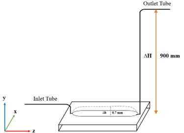
(5) The authors claim that by modifying the thickness of the middle layer, shear stress could be modified, whilst claiming to keep on-site pressure within physiological ranges (approx. 70 mmHg) as a hallmark of their microfluidic devices. Has it been experimentally verified that pressures indeed remain around 70 mmHg.
It is a very interesting question. In this article, the cross-sectional areas of different tunnel-like channel is related to the thickness of the middle layer, resulting in different level of shear stress. Since all flow rates under three conditions keep same at 1.6 ml/min, the average pressure is calculated to be around 70 mmHg based on our previously reported formula (PMID: 37662690). To address the reviewer's question about the actual pressure values, we used a water-filled tube connected to a chip and measured the height of the water surface in the elevated end relative to the chip position, as shown in the Author response image 1. As expected, when the height of the middle layer bulging to the same value (0.7 mm) as under the LSS condition, the water level reaches to 900 mm, which is corresponding to about 70 mmHg.
Author response image 1.
Schematic diagram of on-chip pressure detection
(6) A coculture model (VSMC, EC, monocytes) is mentioned in the last part of the results section without any further information. Information on this model should be provided in the methods section (seeding, cell numbers, etc.). Moreover, comparison of LSS vs LSS+KLF6 OE and HSS vs HSS+KLF6 OE is shown. It would benefit the interpretation of the outcomes if MSS were also shown. It would also be beneficial to demonstrate differences between LSS, MSS, and HSS in this coculture model (without KLF6 OE).
The specific methods for constructing the co-culture models (vascular smooth muscle cells, endothelial cells, monocytes) mentioned in the results section have been introduced in our previous paper. For the convenience for reading this article, we have added a brief description in the section of “Methods and materials” in this paper, including cell seeding and numbers. In this study, the results of LSS vs LSS+KLF6 OE and HSS vs HSS+KLF6 OE are presented to verify the role of KLF6 in LSS- or HSS-induced promotion of early atherosclerotic events. In our previously published paper (PMID: 37662690), we have showed the effects of three different shear stresses on the atherosclerotic events (shown in Fig. 4 in that paper). Those results have demonstrated that both LSS and HSS significantly promote early atherosclerotic events compared with the MSS.
(7) The experiments were solely performed with a venous endothelial cell line (HUVECs). Was the use of an arterial endothelial cell line considered? It may translate better towards atherosclerosis, which occurs within arteries. HUVECs are not accustomed to the claimed near-physiological pressures.
The human umbilical vein endothelial cell (HUVEC) is a commonly used cell line for many in vitro studies of vascular endothelium under fluid shear stress conditions. Although human arterial endothelial cells (HAECs) may be more suitable than HUVECs for responding to physiologically relevant pressure, HUVECs are more easy to obtain and maintain. However, we are going to order HAECs and will use them to validate the conclusion for the potential translatability.
Recommendations for the authors:
Reviewer #2 (Recommendations for the authors):
(1) Information on seeding of the microfluidic device is absent in the methods section (i.e., seeding, cell density, passage number, confluence, etc.). Moreso, treatment with Fer-1 is not reported in the methods section.
We have described the cell seeding information in‘Preparation of cell culture in the microfluidic chip’ and the Fer-1 treatment in ‘Cell death assay’ in the Method section.
(2) Figure 3F has "MSS", "HSS", and "LSS+KLF6" as groups on the x-axis; the latter should probably be "HSS+KLF6".
Thank you for pointing out this error in Figure 3F. We have made the correction.
(3) Data should be made available in online repositories rather than "making it available upon reasonable request". As it was not provided, the sequencing data could not be reviewed. In addition, it was stated that a preprint was available on BioRxiv, but I could not find it.
Thank you for the suggestion. We have uploaded the RNA-seq data to the NCBI GEO database, which was publicly available on December 9, 2025.


