
Peer review process
Revised: This Reviewed Preprint has been revised by the authors in response to the previous round of peer review; the eLife assessment and the public reviews have been updated where necessary by the editors and peer reviewers.
Read more about eLife’s peer review process.Editors
- Reviewing EditorAnne WestDuke University, Durham, United States of America
- Senior EditorLu ChenStanford University, Stanford, United States of America
Reviewer #1 (Public review):
Summary:
This is an important study that describes the consequences of the DNMT3A mutation in human neuronal development for the first time. The selective impact of DNMT3A function on GABAergic interneurons is interesting and an important feature of future therapeutics. The claims made in that manuscript are supported by strong evidence for the most part. And the data are of high quality in general and presented well.
Strengths:
The strengths of the work include 1. Characterization of multiple DNMT3A loss-of-function alleles, including two misense variants, R882H, P904L, and a deletion allele. The missense mutation lines both include an ideal control with the same genetic background. The CRISPRi-mediated DNMT3A knockdown has also been included. The study identifies the mTOR-PI3K pathway as a factor of overgrowth issues found in the mutant organoid. In bulk mRNA sequencing and whole-genome bisulfite sequencing, identify hypomethylated genomic regions associated with gene expression repression. Again, this is more pronounced in the ventral organoid compared to the dorsal organoid. In addition, the extensive electrophysiological characterizations with a high-density microelectrode array support the more mature status of mutant interneurons.
Weaknesses:
Although a strong study overall, some weaknesses are noted. These include:
(1) The lack of validation data for the generated iPSCs and hESCs, such as the chromosomal contents, ploidy, and pluripotency states
(2) Other weaknesses relate to data interpretation and insufficient discussion of related matters, as detailed in the recommendations to the authors.
(3) Also, some errors are noted and detailed in the recommendation section.
Comments on the latest version:
I have reviewed the revised manuscript and the authors' responses to the reviewers' comments. They addressed the comments adequately.
Reviewer #2 (Public review):
Summary:
Chapman, Determan et al. investigate how pathogenic mutations in DNMT3A which cause of Tatton-Brown-Rahman Syndrome (TBRS) disrupt human cortical developmental processes using a comprehensive panel of human pluripotent stem cell models spanning DNMT3A loss-of-function severity. The authors aim to identify the cellular and molecular mechanisms underlying TBRS-associated brain overgrowth and intellectual disability, and to test whether mechanistic convergence exists between TBRS and other overgrowth-intellectual disability disorders (OGIDs) caused by mutations in EZH2 (Weaver syndrome) or PIK3CA pathway components. Their central conclusion is that GABAergic interneuron development is selectively vulnerable to DNMT3A mutation where reduced DNA methylation causes premature de-repression of neuronal and synaptic genes, driving precocious neuronal maturation and hyperactivity sufficient to disrupt neuronal network synchrony. This report adds to a growing literature supporting the vulnerability of GABAergic interneurons in NDDs and further provides a mechanistic view of this vulnerability potentially convergent across OGIDs. The mechanistic claims around H3K27me3 compensation and mTOR-based therapeutic convergence, while promising, rest on more preliminary evidence and would benefit from the distinction between correlation and mechanism being made more explicit in the text. Overall, this is a compelling study with rigorous experimental design and novel findings with potential impact across better understanding OGID pathophysiology.
Strengths:
(1) A major strength of this work is the breadth and rigor of the disease modeling approach. Four independent TBRS model systems are used in tandem: a patient-derived iPSC line with isogenic CRISPR-corrected control (R882H), a knock-in hESC model (P904L) with its wild-type isogenic, patient deletion iPSC lines (Del1/2), and CRISPRi knockdown models (G1/G2), collectively spanning a range of DNMT3A loss-of-function that correlates with phenotypic severity. This allelic series design substantially strengthens causal inference beyond what any single isogenic pair could provide.
(2) The multi-omic integration across matched developmental stages provides a strong mechanistic foundation for the cellular phenotyping and provides significantly enhanced novelty. RNA-seq, whole-genome bisulfite sequencing, and H3K27me3 CUT&Tag are combined in the same cell types and timepoints show that DNMT3A loss reduces CG methylation at neuronal and synaptic gene loci, leading to premature transcriptional activation.
(3) The selective vulnerability of ventral (GABAergic) versus dorsal (glutamatergic) progenitors is one of the study's most important findings. This lineage specificity is consistently observed across all model systems and in both 2D and organoid formats, where ventral NPCs show increased proliferation, premature neuronal gene expression, and increased neurogenesis, while dorsal NPCs are largely unaffected at the transcriptomic and cellular level despite exhibiting comparable DNA methylation changes. This adds to a body of emerging work showing GABAergic interneuron vulnerability in NDDs where ubiquitously expressed genes such as chromatin modifiers are perturbed and provides additional molecular insights into potential mechanisms of "resilience" of dorsal populations.
(4) The functional characterization follows a logical progression from single-neuron electrophysiology (demonstrating GABAergic hyperactivity with increased action potential amplitude and firing rate) to network-level analysis using high-density multi-electrode arrays. The HD-MEA experimental design - pairing TBRS or control GABAergic neurons with a constant background of control iGlut neurons - cleanly isolates GABAergic dysfunction as the driver of network hypersynchrony.
Weaknesses:
(1) The concomitant induction of proliferation and differentiation in TBRS V-NPCs is conceptually striking, since these are generally considered antagonistic developmental programs. The authors clarify that neuronal and synaptic gene de-repression is the more prominent direct consequence of mCG loss, while PIK3/AKT/mTOR pathway upregulation is not itself directly linked to differentially methylated regions, suggesting an indirect relationship between DNMT3A LOF and increased proliferative signaling. This framing is reasonable, but the mechanism linking DNMT3A mutation to mTOR activation remains unresolved, and the manuscript would benefit from being explicit about this gap. Relatedly, the rapamycin rescue, while demonstrated across multiple models including 904 and Del1 (Supplementary Fig. S3e-f), remains limited to proliferation readouts. Whether mTOR inhibition also rescues the downstream neurogenesis, maturation, or network phenotypes is an important open question that the authors appropriately frame as motivation for future work.
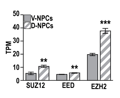
(2) The claim that H3K27me3 compensates for mCG loss is supported by prior work (Lii et al. 2022), which demonstrated increased PRC2 component expression and H3K27me3 gain at sites of DNA methylation loss in Dnmt3a knockout mouse neurons, and by data showing that PRC2 subunits (SUZ12, EED, EZH2) are significantly more highly expressed in D-NPCs than V-NPCs. Together, these findings provide a plausible molecular basis for why dorsal progenitors may be better equipped to maintain repression when DNA methylation is lost, and they make the EZH2 overexpression rescue in V-NPCs more interpretable. Yet, a formal distinction related to two competing, potentially underlying mechanisms, between active compensation, in which EZH2 is recruited to specific loci in response to methylation loss, and functional redundancy, in which higher baseline Polycomb occupancy in dorsal cells simply becomes the dominant repressive mark once mCG is reduced, has not been resolved.
Reviewer #3 (Public review):
Summary:
In this manuscript, the authors investigated TBRS etiology by using new human pluripotent stem cell models, modeling varying levels of TBRS-associated loss of DNMT3A function. They identified increased lineage-specific proliferation of precursors in TBRS ventral MGE-like progenitors, which they propose was related to increased signaling through the PIK3/AKT/mTOR pathway. Furthermore, they show that reduced DNA methylation during MGE-like progenitor differentiation into GABAergic interneurons can cause a premature expression of neuronal and synaptic genes, triggering precocious neuronal maturation. In conclusion, they propose that TBRS-derived GABAergic neurons exhibit hyperactivity that can alters the development and structure of neuronal networks.
Strengths:
Overall, the data presented is convincing, from an early developmental point of view, given that the iPSC-derived 2D cultures or organoids used do not get to reach a mature state. Nonetheless, the data clearly show the effects that deleterious mutations in TBRS can cause during the period of neurogenesis, which was missing in the field.
Comments on revised version.
The authors have responded to the reviewer's comments satisfactorily, and the manuscript has been much improved.
Author response:
The following is the authors’ response to the original reviews.
Public Reviews:
Reviewer #1 (Public review):
Summary:
This is an important study that describes the consequences of the DNMT3A mutation in human neuronal development for the first time. The selective impact of DNMT3A function on GABAergic interneurons is interesting and an important feature of future therapeutics. The claims made in that manuscript are supported by strong evidence for the most part. And the data are of high quality in general and presented well.
Strengths:
The strengths of the work include: Characterization of multiple DNMT3A loss-of-function alleles, including two misense variants, R882H, P904L, and a deletion allele. The missense mutation lines both include an ideal control with the same genetic background. The CRISPRi-mediated DNMT3A knockdown has also been included. The study identifies the mTOR-PI3K pathway as a factor of overgrowth issues found in the mutant organoid. In bulk mRNA sequencing and whole-genome bisulfite sequencing, identify hypomethylated genomic regions associated with gene expression repression. Again, this is more pronounced in the ventral organoid compared to the dorsal organoid. In addition, the extensive electrophysiological characterizations with a high-density microelectrode array support the more mature status of mutant interneurons.
Weaknesses:
Although a strong study overall, some weaknesses are noted. These include:
(1) The lack of validation data for the generated iPSCs and hESCs, such as the chromosomal contents, ploidy, and pluripotency states.
We thank the reviewer for their constructive feedback. We previously validated our 882 models with whole genome sequencing and teratoma formation upon mouse fat pad injection, while the parental human embryonic stem cell line (WA01 hESCs) used for P904L variant knock-in was validated by our Genome Engineering Stem Cell (GESC) core upon derivation of that variant knock-in model. We have now added both karyotyping and pluripotency staining (SOX2/OCT4) for all other hPSC lines as (new) Supplementary Figure S17 and included further description in our Methods section under “hPSC Model Generation and Culture” (pg. 19, lines 6-7).
(2) Other weaknesses relate to data interpretation and insufficient discussion of related matters, as detailed in the recommendations to the authors.
We thank the reviewer for their insightful suggestions and have detailed our responses in the “recommendations to the authors” section.
(3) Also, some errors are noted and detailed in the recommendation section.
We thank the reviewer for catching these errors and have since corrected them, with detailed responses below.
Reviewer #2 (Public review):
Summary:
Chapman, Determan et al. investigate how pathogenic mutations in DNMT3A, which cause Tatton-Brown-Rahman Syndrome (TBRS), disrupt human cortical developmental processes using a comprehensive panel of human pluripotent stem cell models spanning DNMT3A loss-of-function severity. The authors aim to identify the cellular and molecular mechanisms underlying TBRS-associated brain overgrowth and intellectual disability, and to test whether mechanistic convergence exists between TBRS and other overgrowth-intellectual disability disorders (OGIDs) caused by mutations in EZH2 (Weaver syndrome) or PIK3CA pathway components. Their central conclusion is that GABAergic interneuron development is selectively vulnerable to DNMT3A mutation, where reduced DNA methylation causes premature de-repression of neuronal and synaptic genes, driving precocious neuronal maturation and hyperactivity sufficient to disrupt neuronal network synchrony. This report adds to a growing literature supporting the vulnerability of GABAergic interneurons in NDDs and further provides a mechanistic view of this vulnerability, potentially convergent across OGIDs. The mechanistic claims around H3K27me3 compensation and mTOR-based therapeutic convergence, while promising, rest on more preliminary evidence and would benefit from the distinction between correlation and mechanism being made more explicit in the text. Overall, this is a compelling study with a rigorous experimental design and novel findings with a potential impact on a better understanding of the OGID pathophysiology.
Strengths:
(1) A major strength of this work is the breadth and rigor of the disease modeling approach. Four independent TBRS model systems are used in tandem: a patient-derived iPSC line with isogenic CRISPR-corrected control (R882H), a knock-in hESC model (P904L) with its wild-type isogenic, patient deletion iPSC lines (Del1/2), and CRISPRi knockdown models (G1/G2), collectively spanning a range of DNMT3A loss-of-function that correlates with phenotypic severity. This allelic series design substantially strengthens causal inference beyond what any single isogenic pair could provide.
(2) The multi-omic integration across matched developmental stages provides a strong mechanistic foundation for the cellular phenotyping and provides significantly enhanced novelty. RNA-seq, whole-genome bisulfite sequencing, and H3K27me3 CUT&Tag are combined in the same cell types, and timepoints show that DNMT3A loss reduces CG methylation at neuronal and synaptic gene loci, leading to premature transcriptional activation.
(3) The selective vulnerability of ventral (GABAergic) versus dorsal (glutamatergic) progenitors is one of the study's most important findings. This lineage specificity is consistently observed across all model systems and in both 2D and organoid formats, where ventral NPCs show increased proliferation, premature neuronal gene expression, and increased neurogenesis, while dorsal NPCs are largely unaffected at the transcriptomic and cellular level despite exhibiting comparable DNA methylation changes. This adds to a body of emerging work showing GABAergic interneuron vulnerability in NDDs where ubiquitously expressed genes such as chromatin modifiers are perturbed, and provides additional molecular insights into potential mechanisms of "resilience" of dorsal populations.
(4) The functional characterization follows a logical progression from single-neuron electrophysiology (demonstrating GABAergic hyperactivity with increased action potential amplitude and firing rate) to network-level analysis using high-density multi-electrode arrays. The HD-MEA experimental design - pairing TBRS or control GABAergic neurons with a constant background of control iGlut neurons - cleanly isolates GABAergic dysfunction as the driver of network hypersynchrony.
Weaknesses:
(1) The concomitant induction of proliferation and differentiation in TBRS V-NPCs is conceptually striking, since these are generally considered antagonistic developmental programs. The authors partially address this tension by noting that DNMT3A LOF alone is insufficient to initiate neuronal differentiation, i.e., V-NPCs upregulate neuronal and synaptic genes while retaining progenitor identity, implying that transcriptomic priming and commitment to differentiation are decoupled. However, the relationship between the proliferative phenotype and the epigenetic priming phenotype remains mechanistically unresolved. The manuscript documents mTOR pathway upregulation at the protein level and identifies shared DEGs that include proliferative regulators, but it does not establish whether mTOR-driven proliferation and mCG-loss-driven neuronal gene de-repression/enhanced differentiation are causally linked or represent two independent consequences of DNMT3A LOF.
We thank the reviewer for their comment and agree that this phenotype, whereby progenitors exhibited both increased proliferation and hallmarks of gene expression associated with neuronal differentiation is striking and interesting, given that these are typically antagonistic paradigms during normal development.
We documented that these phenotypes involve upregulated expression of both neuronal/synaptic and proliferative genes in V-NPCs (Figure 2d), with concomitant loss of repressive DNA methylation at regulatory elements associated with these genes (Figure 2f, Supplementary Data 5). In this work, DNMT3A mutation had a more prominent role in de-repressing neuronal and synaptic gene expression to promote hallmarks of neuron differentiation, while playing a relatively less central role in direct regulation of proliferation genes, as seen from the relative prominence of neuronal/synaptic- versus proliferation-related GO terms in our Supplementary Data 5 table (pg. 6, lines 16-19).
To examine the mechanisms underlying increased V-NPC proliferation in our TBRS models, we assessed a potential relationship with the PIK3/AKT/mTOR pathway, as this is implicated in increased proliferation resulting from DNMT3A-associated mutation in myeloid leukemia (Dai et al., 2017, PMID: 28461508). In our work, DNMT3A mutation increased the expression and/or phosphorylation of mTOR signaling pathway targets specifically in V-NPCs (Figure 1q-r, Supplementary Figure S3a-d). However, while TBRS mutation directly affected repressive DNA methylation at a suite of cell proliferation-related genes, these did not include the PIK3/AKT/mTOR pathway genes themselves, suggesting an indirect relationship between altered DNA methylation and increased mTOR signaling.
We have since incorporated discussion of how DNMT3A-mediated gene repression and levels of PIK3/AKT/mTOR pathway signaling may be interacting, providing a framework for future studies to identify how these related OGID gene mutations may converge mechanistically (pg. 5, lines 19-21; pg. 16, lines 8-10).
(2) Relatedly, the rapamycin rescue experiment is a valuable proof-of-concept for the PIK3/AKT/mTOR convergence but is limited to a single dose in a single model (882) with a single readout (Ki67+ proliferation). Given the prominence of mTOR pathway convergence in the manuscript as a potential shared therapeutic avenue across OGIDs, the data supporting this claim are somewhat preliminary. It remains unknown whether mTOR inhibition rescues downstream phenotypes (neurogenesis, gene expression, neuronal maturation) or whether less severe TBRS models respond similarly. This might also help tackle the first comment above. e.g., if mTOR inhibition rescued proliferation but not the transcriptomic priming, that would support two independent mechanisms.
We thank the reviewer for their comment. We explored both the overall levels and phosphorylation of proteins involved in PIK3/AKT/mTOR signaling in the 882, 904, Del1, Del2, and KO V-NPC models (Figure 1q-r, Supplementary Figure S3a-d), finding specific increases of all proteins. We showed that rapamycin addition reversed the increased proportion of KI67+ proliferating cell nuclei resulting from 882 mutation in V-NPCs in main Figure 1s, while demonstrating that rapamycin also reduced the proportion of KI67+ nuclei observed in both less severe 904 and Del1 V-NPC models (Supplementary Figure S3e-f).
We agree that understanding whether rapamycin treatment can rescue TBRS neuronal phenotypes would be very interesting, as previous work on Tuberous Sclerosis Complex has utilized rapamycin and other mTOR inhibitors to effectively reverse TSC-related alterations of neuronal morphology and neuronal hyperexcitability (Buttermore et al., 2025, PMID: 40792287). Future studies examining convergent mechanisms and therapeutics for OGIDs should examine how similarly targeting this and related pathways rescues altered neuronal morphology, maturation, and function, as we have demonstrated that TBRS mutation has subsequent consequences for V-IN differentiation, maturation, and function. This point has been detailed in the discussion section on pages 15-16.
(3) The claim that H3K27me3 compensates for mCG loss is an important mechanistic point, but the current data do not distinguish between active compensation, in which EZH2 is recruited in response to methylation loss, and functional redundancy, in which H3K27me3 is independently established and becomes the dominant repressive mark once DNA methylation is reduced. The EZH2 knockdown/inhibition experiments show that H3K27me3 is sufficient to maintain repression at hypo-DMR sites, but they do not establish that H3K27me3 gain is itself a response to methylation loss. Because H3K27me3 profiling was performed only in the severe 882 model, it is also unclear whether H3K27me3 gain scales with DNMT3A LOF severity, as a compensatory model would predict. Finally, the EZH2 overexpression rescue is performed in V-NPCs, whereas the compensation model is developed primarily in D-NPCs, making it difficult to assess whether the same mechanism operates in the lineage where it was originally inferred.
We thank the reviewer for the opportunity to clarify our findings and experimental reasoning. A previous study using a conditional Dnmt3a knockout mouse model (Li et al., 2022, PMID: 35604009) demonstrated increased expression of multiple PRC2 components following the loss of Dnmt3a. This study demonstrated that sites which lost DNA methylation gained H3K27me3 in postnatal neurons upon Dnmt3a loss. Therefore, we hypothesize that the gain of H3K27me3 likely occurs in response to loss of DNMT3A methylation.
While we did not perform CUT&Tag for H3K27me3 in our less severe models, we did validate gene expression changes following EZH2 knockdown and inhibition in both the R882H (Figure 4g-h) and P904L (Supplementary Figure S8b) models, finding that gene expression was unchanged in the model with the less severe DNMT3A mutation (P904L). Based upon these findings, we hypothesized that compensatory H3K27me3 may occur only upon severe DNMT3A loss, as seen in the dominant-negative R882H model. Furthermore, as H3K27me3 compensation was more prominent in D-NPCs, we hypothesized that this might be sufficient to prevent de-repression and aberrant neuronal gene repression upon loss of DNMT3A-mediated repression in D-NPCs. However, since TBRS mutation caused the most prominent de-repression of neuronal gene expression in V-NPCs, we also tested whether EZH2 overexpression could reverse this, finding that it partially suppressed this dysregulated neuronal gene expression. To better clarify this logic and the findings, we have made text edits to this results section and referenced Li et al., 2022 in both the results (pg. 9, lines 16-21; pg. 10, lines 4-7,10-12) and discussion (pg. 16, lines 12-15).
(4) The narrative framing of dorsal neuron development as unaffected by DNMT3A LOF is somewhat at odds with the data presented. The 882 D-NPCs show substantial DNA methylation changes, and TBRS D-INs exhibit what the authors describe as "substantive transcriptomic differences" involving persistent expression of pluripotency and progenitor genes, which seems to be a distinct but potentially significant phenotype. The impact of DNMT3A loss between ventral and dorsal lineages might be more accurately framed as divergent in nature rather than specific to a certain population.
We thank the reviewer for their comment. While TBRS mutations appear to have a significantly stronger effect on V-NPCs and subsequently V-INs, both transcriptomic and methylation alterations do also occur upon TBRS mutation in D-NPCs and D-INs, as noted in Supplemental Figure S4d, S11, and Supplemental Data 2. However, we observed substantially greater molecular alterations in V-NPCs/V-INs, a lack of overt cellular phenotypes in D-NPCs where assayed, and a lack of functional consequences in matured D-INs, suggesting a more significant requirement for DNMT3A in regulating the differentiation and subsequent maturation of cortical inhibitory interneurons during embryonic and early pre-natal development, the developmental periods that we can readily model in hPSC-derived neurons.
It should also be noted that these hPSC differentiation models do not recapitulate post-natal deposition of non-CpG (mCA) DNA methylation, a mechanism disrupted postnatally by TBRS-associated mutations in our prior work in murine models (Harrison Gabel; e.g. Beard et al., 2023, PMID: 37952155), which we have now added in the results section (pg. 7, lines 8-11). Therefore, we hypothesize that if we could sufficiently mature D-INs to a state that modeled postnatal development and recapitulated this non-CpG methylation, we might be able to detect cellular and functional phenotypes in later stage D-INs. To avoid misinterpretation, we have altered the language in the results section to confirm that there are both transcriptomic and methylation changes in our D-NPCs/D-INs, but that these are not accompanied by cellular phenotypes or neuronal dysfunction (pg. 6, lines 1-2; pg. 7 line 3; pg. 7, lines 20-23; pg. 8, lines 1-2; pg. 8, lines 18-23; pg. 9, lines 1-4).
(5) SST stainings are not entirely convincing. They appear mostly nuclear, and some instances localized to rosettes in organoids, whereas the protein is largely confined to processes and is expected to be found outside progenitor-rich zones like rosettes.
We agree that the perinuclear SST staining detected in these young ventral telencephalic-patterned organoids at day 30 differs somewhat from the more process-localized and cytosolic signal seen in later stage organoids in other studies. This may be related to the use of different commercial SST antibodies across studies but also likely reflects SST immunoreactivity in newborn neurons near the onset of SST expression. For example, immature SST-immunoreactive neurons in the early postnatal rat cortex exhibit predominant SST staining in perinuclear cytoplasm and short processes (e.g. Fig. 3 in Lee et al, PMID: 9664223) while acquiring more cytosolic and process-localized staining as postnatal neuron maturation occurs. Evaluation of immunopositivity for other markers of neurogenesis (ASCL1) and immature neurons (TUJ1) is also congruent with these findings for SST, with TBRS-associated mutations increasing in the fraction of cells in V-NPCs/V-ORGs that express these three markers.
Reviewer #3 (Public review):
Summary:
In this manuscript, the authors investigated TBRS etiology by using new human pluripotent stem cell models, modeling varying levels of TBRS-associated loss of DNMT3A function. They identified increased lineage-specific proliferation of precursors in TBRS ventral MGE-like progenitors, which they propose was related to increased signaling through the PIK3/AKT/mTOR pathway. Furthermore, they show that reduced DNA methylation during MGE-like progenitor differentiation into GABAergic interneurons can cause a premature expression of neuronal and synaptic genes, triggering precocious neuronal maturation. In conclusion, they propose that TBRS-derived GABAergic neurons exhibit hyperactivity that can alters the development and structure of neuronal networks.
Strengths:
Overall, the data presented is convincing, from an early developmental point of view, given that the iPSC-derived 2D cultures or organoids used do not get to reach a mature state. Nonetheless, the data clearly show the effects that deleterious mutations in TBRS can cause during the period of neurogenesis, which was missing in the field.
Weaknesses:
(1) Li et al., 2022 (referred to in the manuscript) seems to already show the interplay between H3K27me3 and Dnmt3a discussed in this study i.e., that in the absence of DNA methylation, there is an expansion of polycomb-like repression. These data should be better acknowledged in the paragraph 'Repressive H3K27me3 compensates for severe loss of DNA methylation' (page 9), given it supports the data presented in this manuscript and suggests this as a common mechanism in the interplay between these two repressive marks, as it is well established in the literature.
We thank the reviewer for this suggestion. We have now added Li et al., 2022 to both the results section (pg. 9, lines 16-20) and our discussion section (pg. 16, lines 12-13).
(2) The authors should acknowledge that the omics data come from a mixed population of cells.
We thank the reviewer for their comment. We have validated that the established 2-D differentiation methods we used in this study generate cell populations with >85-90% enrichment for the desired progenitor and neuronal cell type, based upon marker expression, but acknowledge that these are bulk -omics data obtained from cells that may represent a mixed population and have now detailed this in the methods section under “Sequencing” (pg. 21, lines 16-18).
(3) The authors are encouraged to further discuss whether the overgrowth observed in ventral GABAergic cultures or organoids compares to the overgrowth observed in diseased patients. One expects MRIs to have been performed in patients and that these could be harnessed to discern if overgrowth occurs in the cortex or ventral regions of the brain.
We thank the reviewer for their suggestion and do note that at least one published study documents increased cortical thickness in the MRIs of TBRS patients (Jiménez de la Peña et al., 2024, PMID: 37795572); however, to our knowledge studies have not examined regional or cell type-selective overgrowth of cortical tissue in TBRS patients. Future clinical studies examining the nature of the neuronal progenitor overgrowth and resulting consequences for patient brain imaging would be of interest to better understand TBRS-associated etiology of brain overgrowth and its manifestations.
Recommendations for the authors:
Reviewer #1 (Recommendations for the authors):
(1) A potential explanation for the ventral organoid's sensitivity compared to the dorsal can be provided. I understand that the PRC2-mediated methylation loss was more pronounced in the ventral than in the dorsal. However, this raises the question: why is this so? Are the PRC2 components more highly expressed in ventral organoids or in GABAergic neurons than in glutamatergic neurons?
We thank the reviewer for this question. Upon examining RNA-sequencing data for control V-NPCs and D-NPCs, we found that expression of the PRC2 subunits SUZ12, EED, and EZH2 is significantly elevated in D-NPCs relative to V-NPCs (see Author response image 1). This could contribute to restoring epigenetic repression in D-NPCs in the context of DNMT3A mutation, to yield the more limited transcriptomic changes and greater gain of H3K27me3 we observed in TBRS D-NPCs, despite their comparable levels of global mCG loss). This could also account for our finding that overexpression of EZH2 in V-NPCs could normalize some gene expression changes observed in the TBRS models.
Author response image 1.
(2) DNMT3A is a major enzyme that mediates non-CpG methylation during synaptogenesis. There is no discussion of this phenomenon or any related analysis. I suppose the brain organoids may be too immature to detect non-CpG methylation. Even then, this can be described in the results sections.
We thank the reviewer for this comment. While prior work has examined an important role for DNMT3A in postnatal deposition of non-CpG methylation (Christian et al. 2020; Beard et al. 2023), we found that our hPSC models, as the reviewer suggests, were too immature to detect appreciable levels of non-CpG methylation. Accordingly, we have focused our work here on neurodevelopmental requirements for DNMT3A; this work demonstrated that many of the functional alterations of neuronal network activity originate from altered neurogenesis and neuronal maturation of cortical GABAergic interneurons. We have now included a description of the important role of non CpG methylation by DNMT3A during postnatal development and have indicated that the immaturity of hPSC-derived neurons precludes our ability to study non-CpG methylation using these models in the results (pg. 7, lines 8-11) with present language in the discussion (pg. 15, lines 8-11).
(3) Given the Figure 4 results showing H3K27 methylation and EZH2 knockdown rescue the DNMT3A mutation, the authors argue that EZH2 and DNMT3A regulate a similar set of genes and that the two diseases are related. To claim this, there should be data showing that gene misregulation upon loss of EZH2 and DNMT3A is similar.
Given our data, it is premature to draw direct relationships with the dysregulated genes we detected in TBRS versus the molecular basis of Weaver Syndrome; therefore, we have attempted to better reflect the potential but currently unproven relationship between these OGIDs and altered the respective language in the results section (pg. 10, lines 10-12) to reflect this. Understanding convergent mechanisms of OGIDs involving both DNMT3A (TBRS) and EZH2 (Weaver Syndrome) gene mutations will be a compelling topic for future studies, as our work here suggests that they may regulate similar gene suites.
(4) Stronger phenotypes were observed in the R882H mutant compared to the P904L mutant. However, this cannot be translated to the functionality of the mutant protein or disease severity because one is on iPSCs and another is made in hESCs. The limitation should be noted to avoid misleading.
We agree that the severity of each pathogenic mutation can be influenced by its presence on a different iPSC vs hESC background; accordingly, our study design indicated the hPSC background for each model and employed paired isogenic controls to clearly define the consequences of each TBRS mutation relative to a model with an identical genetic background but lacking the mutation. Our finding that the R882H mutation resulted in more severe epigenomic and transcriptomic consequences than the P904L mutation is congruent with findings made in prior work (Beard et al., 2023 PMID: 37952155; Russler-Germain et al., 2014 PMID: 24656771).
(5) It is unclear what measurements were done for DNMT3A level quantification shown in Figure 1e- f.
Protein quantification for DNMT3A models was performed by western blot, shown in Supplemental Fig. S16. We have now included reference to whole blots in the methods section under “Cellular Phenotyping” (page 20 line 13).
(6) The results section of the paper for Figures 6 and 7 cites the wrong figure numbers and panels.
We thank the reviewer for catching these errors and have corrected them in the revised manuscript draft.
(7) Some typos for PI3K (meaning PIK3) in several places, including the Abstract.
We thank the reviewer for catching these errors and have corrected them in the revised manuscript draft.
Reviewer #2 (Recommendations for the authors):
(1) There is a figure numbering discrepancy in the manuscript - the text references six main figures, but the figure pages include seven, with the MEA network data apparently mislabeled.
We thank the reviewer for catching these errors and have corrected them in the new manuscript draft.
(2) The nomenclature "D-IN" for dorsal immature neurons is potentially misleading, as "IN" conventionally denotes interneurons, which these glutamatergic cells are not.
While we agree that IN could be interpreted as interneurons, this nomenclature is defined at an early point in the manuscript and was used to allow readers to easily identify compare findings made in D-NPCs and D-INs (and, as a counterpart, findings made in V-NPCs versus V-INs).



