
Peer review process
Not revised: This Reviewed Preprint includes the authors’ original preprint (without revision), an eLife assessment, public reviews, and a provisional response from the authors.
Read more about eLife’s peer review process.Editors
- Reviewing EditorP Robin HiesingerInstitute for Biology Free University Berlin, Berlin, Germany
- Senior EditorMa-Li WongState University of New York Upstate Medical University, Syracuse, United States of America
Reviewer #1 (Public review):
In the manuscript by Fabian-Fine et al., the authors employ neuroanatomy to investigate aquaporin-4 expression in cells they consider tanycytes and their supposed involvement in tau tangles and amyloid-beta plaques in the hippocampus. This study includes samples from three mice and two Alzheimer's disease (AD) patients.
My key concern and question is whether the cells presented in the manuscript are tanycytes. Tanycytes are specialized ependymoglial cells located in the circumventricular organs and are known to express specific markers. Importantly, they are not myelinated cells, which is a crucial distinction that the authors do not address.
Additionally, the methodologies described in the manuscript lack clarity and controls. For instance, the use of Cdh5-GCaMP882 mice is not adequately justified. It is unclear what these mice contribute to the study's objectives, particularly concerning the aim of investigating waste removal processes in the brain. Moreover, the rationale behind the purported "fluorophore uptake experiments" is unclear and appears to involve the uptake of fluorophore-labeled goat anti-rabbit secondary antibody, which seems implausible to me.
The hypotheses and claims presented in this manuscript are not sufficiently substantiated and are conceptually unclear. The notion that amyloid beta and tau proteins play structural roles in a hypothesized "tanycytes"-derived canal network is not sufficiently supported by the evidence. Furthermore, the study lacks rigorous data to convincingly establish the proposed interactions between these proteins and the processes of waste internalization.
In conclusion, due to conceptual and methodological issues, I consider the current evidence as inadequate to support the primary claims.
Reviewer #2 (Public review):
Summary:
In this study, the authors propose the existence of an AQP4-positive tanycyte-associated canal system in the hippocampus and suggest that this system participates in waste clearance and contributes to Alzheimer's disease pathology. Using histological, ultrastructural, immunohistochemical, and RNA-based approaches, the manuscript attempts to reinterpret amyloid-β plaques and tau-associated structures as components of a tanycyte-derived waste-internalization system. The work is conceptually ambitious and raises observations that may stimulate discussion regarding glial organization and waste clearance in the diseased brain.
Strengths:
A strength of the manuscript is the combination of imaging modalities and anatomical observations across mouse and human tissue. Some of the reported morphological features are intriguing and may warrant additional investigation. The study also attempts to integrate structural observations with broader hypotheses regarding neurodegeneration and Alzheimer's disease.
Weaknesses:
The central interpretation depends almost entirely on identifying the observed hippocampal structures as tanycytes, and the evidence supporting this conclusion remains insufficient. Tanycytes are classically associated with ventricular regions in circumventricular organs, particularly in the third ventricle and median eminence region, yet the manuscript does not provide sufficiently specific anatomical or molecular evidence to convincingly distinguish the described structures from astrocytic, ependymal, radial glial-like, oligodendroglial, myelin-associated, vascular-associated, or degenerative elements. The marker profile used throughout the study, particularly the reliance on AQP4 labeling and Luxol-positive structures, is not sufficiently selective to establish tanycyte identity, especially in pathological tissue where reactive glial changes may occur.
This becomes particularly important because the manuscript repeatedly interprets Luxol-positive and myelin-associated structures as tanycytic processes or "myelin-derived tanycyte protrusions," despite tanycytes not being known to produce myelin. Alternative explanations are not sufficiently explored. Some of the canal-like structures shown in Figure 4 also resemble vascular profiles, and additional vessel markers would be necessary to exclude this possibility.
Several of the proposed structures and mechanisms are also difficult to reconcile with established cell biology and neuroanatomy. The introduction of new terminology such as "tanysomes," "waste receptacles," and "toroids" further extends the interpretation beyond what is currently demonstrated experimentally.
The discussion and integration of the existing literature on tanycytes are also insufficient. Tanycytes themselves are not clearly introduced; the manuscript does not adequately discuss what is currently established regarding tanycyte anatomy, ventricular localization, morphology, and function. Foundational literature defining tanycyte biology, including work from the Prévot group or others, is largely absent despite its central importance to the field. Because the manuscript proposes a substantial departure from established neurobiological concepts, it is particularly important that previous literature be discussed comprehensively and critically. The current version does not sufficiently contextualize the proposed model within the existing literature on tanycyte, AQP4, glymphatic, and Alzheimer's disease, making it difficult to evaluate what is genuinely novel versus what is merely being reinterpreted. It is also not entirely clear what is genuinely new here compared with the authors' previous work, particularly reference 11, which appears to present a highly similar conceptual framework.
More broadly, several of the manuscript's mechanistic conclusions extend well beyond the available evidence. The proposal that amyloid-β plaques and tau pathology represent hypertrophic tanycyte-derived waste structures is provocative and potentially interesting, but currently remains largely correlative and speculative. At several points, it becomes difficult to distinguish direct observations from broader mechanistic interpretation. The manuscript itself acknowledges that the proposed glial-canal hypothesis contradicts the current understanding of nervous system organization and states that ultrastructural serial-section analysis would be required to unambiguously determine the origin of the myelinated profiles described. This point is critical because the study's central conclusions depend on the assumption that these structures are tanycyte-derived. At present, this interpretation remains insufficiently demonstrated, which substantially limits the strength of the broader pathological and mechanistic conclusions proposed throughout the manuscript.
Although access to human material is understandably limited, the study appears to include only one male and one female AD patient, making it difficult to assess the reproducibility or frequent these structures are across individuals and pathological conditions. The manuscript would benefit from clearer characterization of prevalence, reproducibility, and variability across samples.
Overall, the manuscript presents an unconventional and thought-provoking model that may stimulate discussion. However, the evidence currently provided does not convincingly establish tanycyte identity for the described hippocampal structures, and several of the broader disease-related interpretations would require substantially stronger anatomical and molecular evidence before the proposed model can be convincingly supported.
Author response:
Reviewer #1 (Public review):
My key concern and question is whether the cells presented in the manuscript are tanycytes. Tanycytes are specialized ependymoglial cells located in the circumventricular organs and are known to express specific markers. Importantly, they are not myelinated cells, which is a crucial distinction that the authors do not address.
We agree with the reviewer that tanycytes that have been described in the third ventricle have not been reported to be myelinated. However, our study was conducted on the hippocampal formation that borders the ventral horn of the lateral ventricles. We will include images of the myelin-forming ependymal cells that we refer to as tanycytes
Additionally, the methodologies described in the manuscript lack clarity and controls.
We will expand on our methods section and include controls.
For instance, the use of Cdh5-GCaMP882 mice is not adequately justified. It is unclear what these mice contribute to the study's objectives, particularly concerning the aim of investigating waste removal processes in the brain. Moreover, the rationale behind the purported "fluorophore uptake experiments" is unclear and appears to involve the uptake of fluorophore-labeled goat anti-rabbit secondary antibody, which seems implausible to me.
Most experiments described in this manuscript were carried out on human brain. However, functional studies will have to be carried out on rodent brain. We will thus process rodent tissue as well to test whether our observed findings are consistent between human and rodent. The animals used for these experiments were raised for bladder research and are wild type regarding neuronal and glial cells, particularly using the Cy3 channel. Utilizing these brains for our experiments has allowed us to test rodent tissue at both light- and electron-microscopic levels without having to sacrifice additional animals. The consistency of our findings between human and rodent brain further supports that the calcium indicator in the vascular system of these mice did not affect neurons or glial cells.
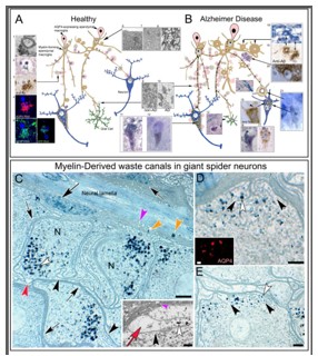
Regarding the uptake experiment: When we initially discovered that myelin-forming macroglia form waste-internalizing glial canals within neuronal in spider brain it was unclear where the AQP4-immunoreactive cells were located. The somata of the myelin-forming cells lacked AQP4 immunoreactivity. Suspecting a synergistic interaction between the myelin-forming and AQP4 expressing cells whereby the myelinforming cells create the canal structure that sequesters waste from the neuron and the AQP4-expressing cells create a convective flow toward the waste-internalizing structures. However, unable to locate the somata of these cells, we submerged a freshly dissected spider brain with the attached surrounding tissue intact in physiological spider saline and slowly added blue vital dye solution to test which cells would internalize the dye. We then identified the (blue) cells in the lining of the dorsally located tubular system that we routinely detached from our brain preparations explaining why we were unable to locate these cells. Immunolabeling of this tubular system revealed the cells that reside in the lining of this tubular system (see Author response images 1 and 2) the original (Figure 8) shows their long slender processes. Interestingly, this system is continuous with the stomatogastric system.
Author response image 1.
Shows the proposed canal system in spiders
Author response image 2.
AQP4-immunoreactive cells in the spider primitive ventricular system that we localized due to similar uptake experiments we have conducted in mouse brain.
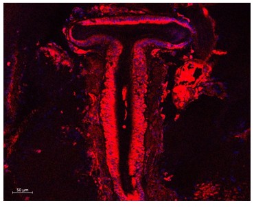
We have utilized this method in mouse brain to test the validity of our postulation, that ependymal tanycytes internalize the presented fluorochrome from extracellular spaces and test which areas and structures may be involved in this uptake. As demonstrated in this experiment, the alveus, and a fine network of cell processes within the brain parenchyma show fluorescence, indicative that they internalize the fluorochrome from extracellular spaces. We used goat-coupled fluorochrome to further test with a FITCcoupled secondary antibody that the observed fluorescence is indeed due to uptake of the goat-coupled secondary antibody and not due to intrinsic autofluorescence. Control preparations lacked this fluorescence. To further test our postulation that the uptake is indeed AQP4-mediated we have applied an AQP4-blocker, which showed a significantly reduced fluorochrome uptake compared to the controls without this blocker.
As we state in the text, we are aware of the limitations of this experimental design, however, like in our spider experiments we consider these findings helpful as they likely show an overview of the cellular network in the hippocampus that governs waste-uptake and may help identify suitable target areas for similar studies on organotypic tissue cultures utilizing two-photon microscopy.
The hypotheses and claims presented in this manuscript are not sufficiently substantiated and are conceptually unclear. The notion that amyloid beta and tau proteins play structural roles in a hypothesized "tanycytes"-derived canal network is not sufficiently supported by the evidence. Furthermore, the study lacks rigorous data to convincingly establish the proposed interactions between these proteins and the processes of waste internalization. In conclusion, due to conceptual and methodological issues, I consider the current evidence as inadequate to support the primary claims.
We respectfully disagree with this comment and hope that the inclusion of additional evidence together with the clear visibility of this canal system in the spider brain will encourage the reviewer to investigate this possibility themselves. We cannot ignore large amounts of amyloid beta-immunolabeled receptacles emanating from tanysomes in swell-bodies and declare them fixation artifacts, particularly when we demonstrate the expression of Presenilin 1 and APP in swell bodies. We furthermore encourage the reviewer to revisit myelinated cells in the brain in both depictions in the available literature and actual brain preparations. We have not been able to locate actual electronmicrographs of longitudinal sections through neurons that show myelination past the axon hillock at the EM-level consistent with our current understanding of myelination. The only depiction of this form of myelination we found were schematic drawings. We will include several new images that show such longitudinal sections through neurons that are easily obtained and we have numerous additional images that we are happy to share. In all our preparations (mouse, rat and human) the myelination pattern is consistent with the images we will depict in new figures 1 and 2. Not to bring attention to this inconsistency would be dishonest scientific conduct.
Reviewer #2 (Public review):
Summary:
In this study, the authors propose the existence of an AQP4-positive tanycyte-associated canal system in the hippocampus and suggest that this system participates in waste clearance and contributes to Alzheimer's disease pathology. Using histological, ultrastructural, immunohistochemical, and RNA-based approaches, the manuscript attempts to reinterpret amyloid-β plaques and tau-associated structures as components of a tanycyte-derived waste-internalization system. The work is conceptually ambitious and raises observations that may stimulate discussion regarding glial organization and waste clearance in the diseased brain.
Strengths:
A strength of the manuscript is the combination of imaging modalities and anatomical observations across mouse and human tissue. Some of the reported morphological features are intriguing and may warrant additional investigation. The study also attempts to integrate structural observations with broader hypotheses regarding neurodegeneration and Alzheimer's disease.
Weaknesses:
The central interpretation depends almost entirely on identifying the observed hippocampal structures as tanycytes, and the evidence supporting this conclusion remains insufficient. Tanycytes are classically associated with ventricular regions in circumventricular organs, particularly in the third ventricle and median eminence region, yet the manuscript does not provide sufficiently specific anatomical or molecular evidence to convincingly distinguish the described structures from astrocytic, ependymal, radial glial-like, oligodendroglial, myelin-associated, vascular-associated, or degenerative elements. The marker profile used throughout the study, particularly the reliance on AQP4 labeling and Luxol-positive structures, is not sufficiently selective to establish tanycyte identity, especially in pathological tissue where reactive glial changes may occur.
As mentioned in our response to reviewer 1 we have now included additional experimental evidence that demonstrates the myelinated ependymal cells and additional gene expression experiments.
This becomes particularly important because the manuscript repeatedly interprets Luxolpositive and myelin-associated structures as tanycytic processes or "myelin-derived tanycyte protrusions," despite tanycytes not being known to produce myelin. Alternative explanations are not sufficiently explored. Some of the canal-like structures shown in Figure 4 also resemble vascular profiles, and additional vessel markers would be necessary to exclude this possibility.
Several of the proposed structures and mechanisms are also difficult to reconcile with established cell biology and neuroanatomy. The introduction of new terminology such as "tanysomes," "waste receptacles," and "toroids" further extends the interpretation beyond what is currently demonstrated experimentally.
The discussion and integration of the existing literature on tanycytes are also insufficient. Tanycytes themselves are not clearly introduced; the manuscript does not adequately discuss what is currently established regarding tanycyte anatomy, ventricular localization, morphology, and function. Foundational literature defining tanycyte biology, including work from the Prévot group or others, is largely absent despite its central importance to the field. Because the manuscript proposes a substantial departure from established neurobiological concepts, it is particularly important that previous literature be discussed comprehensively and critically. The current version does not sufficiently contextualize the proposed model within the existing literature on tanycyte, AQP4, glymphatic, and Alzheimer's disease, making it difficult to evaluate what is genuinely novel versus what is merely being reinterpreted. It is also not entirely clear what is genuinely new here compared with the authors' previous work, particularly reference 11, which appears to present a highly similar conceptual framework.
More broadly, several of the manuscript's mechanistic conclusions extend well beyond the available evidence. The proposal that amyloid-β plaques and tau pathology represent hypertrophic tanycyte-derived waste structures is provocative and potentially interesting, but currently remains largely correlative and speculative. At several points, it becomes difficult to distinguish direct observations from broader mechanistic interpretation. The manuscript itself acknowledges that the proposed glial-canal hypothesis contradicts the current understanding of nervous system organization and states that ultrastructural serialsection analysis would be required to unambiguously determine the origin of the myelinated profiles described. This point is critical because the study's central conclusions depend on the assumption that these structures are tanycyte-derived. At present, this interpretation remains insufficiently demonstrated, which substantially limits the strength of the broader pathological and mechanistic conclusions proposed throughout the manuscript.
Although access to human material is understandably limited, the study appears to include only one male and one female AD patient, making it difficult to assess the reproducibility or frequent these structures are across individuals and pathological conditions. The manuscript would benefit from clearer characterization of prevalence, reproducibility, and variability across samples.
Overall, the manuscript presents an unconventional and thought-provoking model that may stimulate discussion. However, the evidence currently provided does not convincingly establish tanycyte identity for the described hippocampal structures, and several of the broader disease-related interpretations would require substantially stronger anatomical and molecular evidence before the proposed model can be convincingly supported.
We agree with the reviewer that it is important to correctly investigate and describe cellular structure. The first author of this manuscript is a 30-year veteran of published cellular ultrastructure and the three-dimensional reconstruction of cells and entire cell networks. We have spent the last five years to try and confirm our current understanding of cellular structure, and we are unable to reproduce our current identification of cell structure. One example is mentioned in response to reviewer 1. We are unable to find electonmicrographs in publications that show myelination of neurons consistent with the countless schematic depictions available online and in the literature. This includes publications about myelination. Our findings are all consistent with the new images we will include in our revised manuscript. Longitudinal sections through neurons are easily obtained and neurons can be followed well beyond the axon hillock.
A second example that is inconsistent with our current understanding of cells and biochemical processes in cells are ‘astrocytes’ and ‘reactive astrocytes’. We will show in the revised manuscript swell-bodies have no defined cytoplasm that every cell requires to fulfil basic cellular functions required for survival. It is very apparent to a structural expert that swell-bodies lack cytoplasm. The ‘consistency’ of this lacking cytoplasm is ‘inconsistent’ with fixation artifacts. In Author response image 3 we demonstrate that immunolabeling for AQP4 shows two types of structures. (1) Immunolabeled structures that are void of immunolabeling in their lumina and show receptacle-like immunoreactivity along the outside (panels I,J,L,M image below) consistent with swellbodies as indicated by the provided amyloid beta-stained and Luxol H&E-stained swellbodies that are associated with immunolabeled receptacle-shaped structures on the outside (panels K,N). A structurally trained eye quickly recognizes that these are not immunolabeled cells but are consistent with swell-bodies and referred to as ‘reactive astrocytes’ in the literature. We show in panel O what an immunolabeled cell looks like, with slender processes and immunolabeled cytoplasm. In this image in panels G1-4 we demonstrate that swell-bodies contain AQP4 mRNA explaining why they are immunoreactive for this protein. It is important that we bring attention to these details and do not randomly describe structure just based on a signal. This is exactly the point we make and I hope that the reviewer recognizes our expertise in cellular neuroscience and in particular in recognizing cellular structure.
Again, I urge the scientific community not to dismiss our findings but to actually study the validity of our findings.
Author response image 3.
In the revised manuscript we will include additional experiments that we have carried out, that have also increased the sample number of tested human brain. We have in total so far investigated 13 different human brain samples, six of which are AD-affected.
We will revise our discussion to explain our observations in context with the literature better and include so much evidence in support of our hypothesis that it would be unreasonable to dismiss all this compelling and logical evidence.
This research was not planned; it resulted from our accidental discovery in the spider system when our animals struggled with early onset neurodegeneration that we needed to address. This is when we recognized the waste-internalizing role of myelin in giant spider neurons. As we will discuss in the revised manuscript, such systems are highly conserved throughout evolution, and this is what made us realize that the only images of myelinated neurons we could find were either schematic drawings, single cross sections through myelinated cell profiles or very high magnification insets that also did not show the actual neuron that is myelinated.
One can argue that there are both myelinated and unmyelinated axons. However, the varicose projections clearly originate in the ependymal lining and double-label for AQP4. This is consistent with our postulation and inconsistent with our current understanding of myelination.
I sincerely urge the neuroscience community to re-visit myelination in the brain, we have done this for the past five years with extensive experience in this field and the only hypothesis that is supported by our findings is presented in this manuscript. Please do not dismiss these findings, the spiders have uncovered a waste canal system in the brain and putting this system in place will help us to gain a better understanding regarding neurodegenerative diseases. Lastly, and maybe most importantly, understanding how this system works in spiders and how the myelin is structurally anchored to microtubule that were missing in our degenerating spiders has allowed us to identify the cause for this sudden neurodegeneration and rescue our tropical, cold-blooded spider colony by installing a new heating system and raising the room temperature so that the coldsensitive microtubules no not dissociate anymore.
We would like to thank the reviewers to strengthen the content of this manuscript with their critical comments, we hope that our revision will help clarify some doubts.
(1) Pasquettaz R, Kolotuev I, Rohrbach A, Gouelle C, Pellerin L, Langlet F. Peculiar protrusions along tanycyte processes face diverse neural and nonneural cell types in the hypothalamic parenchyma. Journal of Comparative Neurology. 2021;529(3):553. doi: 10.1002/cne.24965. PubMed PMID: edsgcl.646848213.



