
Peer review process
Not revised: This Reviewed Preprint includes the authors’ original preprint (without revision), an eLife assessment, public reviews, and a provisional response from the authors.
Read more about eLife’s peer review process.Editors
- Reviewing EditorMatthew RowanEmory University, Atlanta, United States of America
- Senior EditorMichael TaffeUniversity of California, San Diego, San Diego, United States of America
Reviewer #1 (Public review):
Summary:
The manuscript by Montenegro and colleagues reports a uniquely significant set of compelling results from a carefully designed study. The findings are fundamental and should substantially advance our understanding of whether prenatal exposure to high levels of alcohol produces neural changes that are precursors to the development of Alzheimer-like pathology in an animal model of Fetal Alcohol Spectrum Disorder (FASD). The quest was to test whether prenatal exposure to high-dose alcohol in the mouse would result in selective damage that would result in Alzheimer disease-like cellular disorder and mnemonic impairment. Both outcomes emerged and were exacerbated in relevant transgenic mice. The untoward effect on memory endured and even worsened with age.
Strengths:
The authors noted the importance of using a validated animal model to test their hypotheses related to AD-like outcomes because human postmortem data are unavailable and even in vivo data are limited to younger FASD cohorts. The authors further noted limitations, which also anticipate experiments that could chart the temporal course of the effect and windows of prenatal alcohol exposure that result in damage or resilience. In short, as the authors state on lines 435-7, "These data provide the first experimental evidence that developmental alcohol exposure impacts core proteolytic pathways central to AD/ADRD pathogenesis."
Weaknesses:
Addressing the following would clarify several points in an already well-written paper:
(1) It would be useful to have a timeline of the study, much like the one provided for the water maze protocol. On that timeline, please include the sample sizes examined, ages at exposure, and other pertinent procedures, indicating which animals remained alive for testing, etc.
(2) What were the attrition rates for each study group?
(3) What are the human age equivalents of the maternal mice?
(4) It would be helpful to see a graph of the BECs of each animal relative to the doses given. That would clarify how the alcohol exposure amount and timing are the same and where they are different for all exposed mice, given that the alcohol levels were somewhat different by group, as noted in the Methods. Were these BEC differences at all related to group differences in outcome measures or memory performance?
Reviewer #2 (Public review):
Summary:
In this study, the impact of prenatal alcohol (PAE) on amyloid precursor protein (APP) C-terminal fragments and notch intracellular domain (NICD) levels in adulthood is measured in 3xTg-AD mice.
Prenatal alcohol alters gamma secretase activity with development and aging. This could have implications for Alzheimer's disease risk in populations without inherited Alzheimer's risk genetics.
Strengths:
Strengths include the model, the use of orthogonal approaches, and the rigorous, high-quality data.
Weaknesses:
Some figures lack prenatal alcohol treatment in the 3xTg-AD mice.
Some overstatements should be tempered. For instance, one cannot conclude that the changes in CTFs are driving the changes in learning and memory (as suggested in the last line of the abstract) without a direct intervention testing this. For instance, though PAE caused a more robust learning deficit at 6 mo in WT, the impact on CTFs was less than it was at 3 mo. PAE did not significantly change CTFs or learning/memory in 3xTg-AD mice at 4 months, suggesting the genotype effect takes over at this point. The text should be adjusted to reflect this.
Conclusion:
In summary, this is a rigorous assessment of the long-term impacts of PAE on CTFs and learning/memory in adult WT and 3xTg-AD mice.
Reviewer #3 (Public review):
Summary:
The goal of this study was to test the hypothesis that prenatal alcohol exposure (PAE) can affect Alzheimer's disease (AD) pathogenesis using biochemical proxies, histology, and a behavioral paradigm sensitive to AD-related memory decline. Major strengths include the breadth of techniques used, consideration of different AD-related molecular markers, and the use of different ages as well as appropriate controls. The authors largely achieved their aims to show that PAE does affect amyloid precursor fragments (CTFs) and notch signaling very early on as well as long-term effects in adulthood, which may uncover a previously underappreciated mechanism that may contribute to AD-related neuropathology and behavioral outcomes during lifespan.
Strengths:
(1) Several techniques are used to address molecular, behavioral, and histological PAE-related changes.
(2) There is use of appropriate controls and an AD-relevant mouse model.
(3) Different ages are used to address age-related and long-term effects in AD and control mice.
(4) The novelty of results shows early changes in amyloid-related processes, affected by PAE.
Weaknesses:
(1) It is unclear as to whether there are sex differences, particularly in the adult cohort.
(2) More clarity is needed on sample size per cohort and whether mice that were used for anatomy and biochemical analyses were previously used for behavior. Including a table and noting any overlap would be useful.
(3) In many instances, two-way ANOVAs with treatment (PAE vs vehicle) and genotype as factors will be useful to report (e.g., Figure 1).
(4) In Figure 5 and line 253, it is stated that older mice have more severe deficits, but there are no direct statistical comparisons with younger AD mice.
(5 Lines 270-271 refer to mice as "presymptomatic", but these mice do have behavioral symptoms. Do the authors mean no neuropathology yet? Any data showing lack of robust neuropathology would be useful.
Author response:
Public Reviews:
Reviewer #1 (Public review):
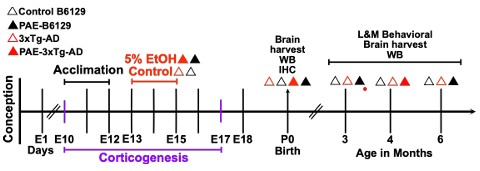
(1) It would be useful to have a timeline of the study, much like the one provided for the water maze protocol. On that timeline, please include the sample sizes examined, ages at exposure, and other pertinent procedures, indicating which animals remained alive for testing, etc.
We agree with the reviewer that a timeline would be helpful for clarifying the PAE exposure paradigm and the subsequent sample collection and processing. Sample sizes vary across the different analyses; therefore, we have indicated the sample size for each experiment in the corresponding figure. To further improve clarity, we will add Author response image 1, which will include a schematic of the overall experimental timeline, including the PAE regimen, collection time points, and sample processing, as shown below.
Author response image 1.
(2) What were the attrition rates for each study group?
We thank the reviewer for raising this important point. There was no attrition of animals within the experimental groups; the number of animals included at the beginning and end of the study remained the same. However, we did observe a reduction in litter size following prenatal alcohol exposure (PAE). In our 3xTg-AD colony, litters typically consisted of approximately 8 pups under control conditions, whereas PAE litters occasionally contained 4–6 pups. Thus, the reduction in animal numbers reflects decreased litter size associated with PAE rather than attrition during the study. We would also like to clarify that the primary scope of this study was not to provide a terminal/end-point analysis of disease progression, but rather to investigate the emergence of Alzheimer’s disease (AD)-related phenotypes during early adulthood following PAE. Accordingly, our longitudinal experimental design focused on identifying the earliest molecular, synaptic, behavioral, and neuropathological alterations that emerge during this period. This approach allowed us to examine whether PAE accelerates or precipitates the onset of AD-related symptomatology in the 3xTg-AD model, rather than following the animals through advanced disease stages. We will clarify this rationale in the revised manuscript.
(3) What are the human age equivalents of the maternal mice?
We appreciate the reviewer’s question regarding the age of the maternal mice. The dams used in our study were young adult females (2 to 3 months of age) at the time of breeding. Because chronological age does not translate linearly between mice and humans, particularly during development and reproductive maturation, we have avoided assigning a precise human-age equivalent. Based on established comparative developmental frameworks and calculations, these animals represent a 20 years old young-adult in the reproductive stage, rather than an advanced maternal-age condition [1]. We will clarify this point in the revised manuscript.
(4) It would be helpful to see a graph of the BECs of each animal relative to the doses given. That would clarify how the alcohol exposure amount and timing are the same and where they are different for all exposed mice, given that the alcohol levels were somewhat different by group, as noted in the Methods. Were these BEC differences at all related to group differences in outcome measures or memory performance?
We appreciate the reviewer’s suggestion to provide a more detailed representation of the BEC data. We agree that displaying the BECs for individual animals would provide additional clarity regarding the consistency of alcohol exposure across groups. We have therefore included the individual BEC values in the new Figure 1. We observed some variability in BECs between the 3xTg-AD and B6129 groups, as noted in the Methods. Importantly, however, the average alcohol consumption was comparable between the two genotypes, indicating that the difference in BECs was not due to differences in the amount of alcohol consumed. All dams in the PAE groups reached BECs above 0.08 g/dL, the commonly used legal blood alcohol concentration limit in the United States, supporting the use of our paradigm as a binge-like alcohol exposure model. We further examined whether the variability in BECs was associated with the differences observed in outcome measures, including memory performance. We did not find evidence that the differences in BECs accounted for the group differences in behavioral or molecular outcomes. Thus, although some intergroup variability in BECs was present, the overall alcohol exposure was comparable, and the observed phenotypic differences were not attributable to differences in alcohol consumption.
Reviewer #2 (Public review):
(1) Some figures lack prenatal alcohol treatment in the 3xTg-AD mice.
We appreciate the reviewer’s observation and agree that the rationale for the different experimental groups across the figures should be clarified. The primary focus of this study is to characterize the effects of prenatal alcohol exposure (PAE) in wild-type B6129 mice, with the 3xTg-AD mice serving primarily as a disease-model reference to determine whether the effects observed following PAE in wild-type animals overlap with or resemble features of AD pathology. Accordingly, the initial figures focus on the effects of PAE in B6129 mice and include the non-exposed 3xTg-AD group as a reference for the AD phenotype. The last two figures specifically address the effects of PAE in the 3xTgAD model, with the 3xTg-AD mice becoming the experimental subject of interest rather than serving solely as a disease reference. For this reason, the PAE-3xTg-AD group is not included in the earlier figures, whereas it is included in the final two figures where the effect of PAE on the AD model is directly evaluated. We will clarify this experimental rationale in the revised manuscript and figure legends.
(2) Some overstatements should be tempered. For instance, one cannot conclude that the changes in CTFs are driving the changes in learning and memory (as suggested in the last line of the abstract) without a direct intervention testing this. For instance, though PAE caused a more robust learning deficit at 6 mo in WT, the impact on CTFs was less than it was at 3 mo. PAE did not significantly change CTFs or learning/memory in 3xTg-AD mice at 4 months, suggesting the genotype effect takes over at this point. The text should be adjusted to reflect this.
We appreciate the reviewer’s careful consideration of this point. We agree that the relationship between APP CTF accumulation and learning and memory deficits should not be interpreted as causal in the absence of a direct intervention experiment. We were careful in choosing the wording throughout the manuscript to describe these findings as associated changes rather than evidence of a causal interaction. Our data demonstrate the presence of APP CTF accumulation and learning and memory deficits following PAE, but they do not establish that CTF accumulation directly drives the behavioral phenotype. We therefore will temper the language in the Abstract and throughout the manuscript to avoid overstatement. We also acknowledge that the relationship between these phenotypes is not necessarily linear across age: although PAE produced a more pronounced learning deficit at 6 months in B6129 mice, the magnitude of APP CTF accumulation was greater at the earlier time point. Importantly, we consider the possibility that the greater APP CTF accumulation observed at earlier ages may represent an early molecular insult whose functional consequences become evident later in life. In this context, the temporal dissociation between the molecular and behavioral phenotypes could be consistent with a “two-hit” model, in which an early-life insult induced by PAE creates or primes a pathological vulnerability that subsequently manifests as cognitive dysfunction with ageing [2]. We recognize, however, that this interpretation remains a hypothesis and would require longitudinal mechanistic studies to establish. This temporal relationship may also contribute to the broader concept of early-life origins of AD/ADRD, suggesting that prenatal environmental exposures may initiate molecular alterations during neurodevelopment that remain detectable or predispose the brain to later-life dysfunction. Similarly, the absence of significant changes in APP CTFs or learning and memory in 4-month-old 3xTg-AD mice suggests that the effects of the AD genotype may become dominant at this stage. Consistent with this interpretation, we state in the Discussion that future studies are needed to identify and experimentally test the direct molecular pathways affected by PAE that ultimately contribute to learning and memory impairment. We will revise the text accordingly to make this distinction clear while highlighting the potential significance of an early molecular insult preceding the later emergence of behavioral phenotypes.
Reviewer #3 (Public review):
(1) It is unclear as to whether there are sex differences, particularly in the adult cohort.
We appreciate the reviewer’s comment regarding potential sex differences. We agree that considering sex as a biological variable is important, particularly for the adult cohorts. To address this point, we will include identifying marks for male and female animals separately in our plots where sample size permits. This will allow the reader to better evaluate potential sex-dependent effects of PAE and to determine whether the observed phenotypes are consistent across sexes. We will also clarify this approach in the revised manuscript.
(2) More clarity is needed on sample size per cohort and whether mice that were used for anatomy and biochemical analyses were previously used for behavior. Including a table and noting any overlap would be useful.
We appreciate the reviewer’s suggestion and agree that greater clarity regarding the sample sizes and use of animals across analyses is important. The sample size for each cohort and experimental group is indicated in the corresponding figures, and we will make this information more explicit in each figure legend to facilitate interpretation. Animals that underwent behavioral testing were subsequently used for biochemical analyses, allowing us to examine molecular changes in the same animals in which behavioral phenotypes were characterized. In contrast, for the neonatal cohort, we performed both anatomical and biochemical analyses, to assess the distribution and extent of APP CTF accumulation across the brain during this early developmental period. This approach was selected to provide a broader assessment of the spatial distribution of APP CTF accumulation at birth. We will clarify these experimental details in the revised Methods and figure legends.
(3) In many instances, two-way ANOVAs with treatment (PAE vs vehicle) and genotype as factors will be useful to report (e.g., Figure 1).
We appreciate the reviewer’s suggestion regarding the use of two-way ANOVA with treatment and genotype as factors. However, we respectfully disagree that this approach is appropriate for all of the analyses presented in this manuscript. Our experimental design and the specific biological questions addressed in each experiment were not uniform across cohorts. In particular, the primary objective of the study was to characterize the effects of PAE in B6129 mice, with the 3xTg-AD mice serving primarily as a disease-model reference, while the effects of PAE in the 3xTg-AD model were specifically examined in the later experiments. Therefore, combining genotype and treatment as factors across all datasets would not always reflect the experimental questions or the structure of the cohorts. In addition, some experiments did not include all four groups, making a two-way ANOVA inappropriate for those analyses. Nevertheless, we agree that a two-way ANOVA may be informative for experiments in which both genotype and treatment are fully represented and the experimental design supports this analysis. We will therefore consider and apply two-way ANOVA, where appropriate, to those datasets, including evaluation of the main effects of genotype and treatment and their interaction. We will clarify the statistical approach and its rationale in the revised Methods and figure legends.
(4) In Figure 5 and line 253, it is stated that older mice have more severe deficits, but there are no direct statistical comparisons with younger AD mice.
We appreciate the reviewer’s observation. We agree that, in the absence of a direct statistical comparison between age groups, the statement that older mice have “more severe deficits” may be too strong. Our intention was to describe the apparent progression of the phenotype across age rather than to imply that we had statistically demonstrated an age-dependent increase in severity. We have therefore revised the text in Figure 5 and at line 253 to use more cautious language, describing the greater magnitude of the observed deficits in older mice without implying a direct statistical comparison between age groups. We agree that a formal conclusion regarding age-dependent progression would require a statistical analysis, which we will include in the revised manuscript.
(5) Lines 270-271 refer to mice as "presymptomatic", but these mice do have behavioral symptoms. Do the authors mean no neuropathology yet? Any data showing lack of robust neuropathology would be useful.
We appreciate the reviewer’s careful observation. We agree that the term “presymptomatic” was not sufficiently precise, particularly because the mice already exhibit measurable behavioral alterations at this age. Our intention was not to suggest that these animals were free of phenotypic abnormalities, but rather that they were at an early stage of disease progression, before the emergence of robust neuropathological features. We have therefore revised the terminology to avoid referring to these mice as “presymptomatic.” Instead, we describe them as being in an early stage of disease development, characterized by emerging behavioral and molecular alterations but without the extensive cardinal neuropathology typically associated with later stages of the 3xTg-AD phenotype. We agree that the distinction between behavioral symptoms and neuropathological progression is important. In this study, our focus was on the emergence of early AD-related phenotypes during young adulthood, rather than on establishing the absence of neuropathology. We have therefore avoided making a definitive claim regarding the lack of neuropathology and have revised the text to more accurately reflect the scope of our data.
Additional References
(1) Dutta, S. & Sengupta, P. Men and mice: Relating their ages. Life Sci. 152, 244–248 (2016).
(2) Gunn, J. S. et al. Exploring the ‘Multiple-Hit Hypothesis’ of Neurodegenerative Disease: Bacterial Infection Comes Up to Bat’. Frontiers in Cellular and Infection Microbiology | www.frontiersin.org 1, 138 (2019).



