
Peer review process
Not revised: This Reviewed Preprint includes the authors’ original preprint (without revision), an eLife assessment, public reviews, and a provisional response from the authors.
Read more about eLife’s peer review process.Editors
- Reviewing EditorFrank KirchhoffUlm University Medical Center, Ulm, Germany
- Senior EditorJoshua SchifferFred Hutch Cancer Center, Seattle, United States of America
Reviewer #1 (Public review):
Howell et al investigate the functional capacities of CD16dim CD56dim NK cells, including their activity against HIV-1-infected T cells. The authors provide an extensive characterization of the functional activity of different NK cell subsets derived from peripheral blood. CD16 is an important receptor expressed on NK cells, and previous studies have demonstrated that CD16 expression changes depending on the activation status of NK cells - one important regulator of CD16 expression is proteolytic shedding/cleavage of CD16 on activated NK cells by the metalloprotease ADAM17. In vitro activation of NK cells, for example in response to K562 cells or other target cells, results in a rapid downregulation of the expression of CD16 on the surface of NK cells, unless an ADAM17 inhibitor is added. This is an important point to consider in the interpretation of the presented results. Overall, the manuscript includes many data in nine figures plus supplemental figures, and would benefit from some focusing of the results.
(1) Figure 1
The observation that CD16dim NK cells responded more strongly by degranulation to K562 cells and HIV-1-infected cells could be due to the shedding of CD16 following activation. In other words, more strongly activated NK cells express higher levels of CD107 but also shed CD16, resulting in higher CD107 expression in CD16low NK cells. The authors should investigate this, for example by performing the degranulation assays shown in Figure 1 in the presence and absence of an ADAM17 inhibitor.
(2) Figure 2
The authors sorted CD16dim and bright NK cells for these experiments and observed higher lysis of HIV-1-infected CD4+ T cells. Important controls should be included in these experiments - how strong was the lysis of HIV-1-uninfected CD4+ T cells by these different NK cell subsets? It also appears that the results shown were derived using NK cells from one donor, and "representative of two independent sort experiments performed with separate donors, each yielding similar results". Why are the authors now showing the respective data? One or two experiments appear too few to come to these conclusions. To support the broad conclusions drawn by the reviewers, the experiments should be performed in a larger number of individuals.
(3) Figure 3
It appears that experiments were performed again using bulk NK cell populations, and superior degranulation and killing frequencies by CD16dim NK cells might reflect different levels of activation again, as described above for Figure 1. The same applies to Figure 4 - lower degranulation events in CD16bright NK cells are consistent with lower activation of these cells, resulting in less CD16 downregulation. Also, it is not clear to the reviewer why CD107a expression and killing frequencies decrease with higher effector-to-target ratios (Figure 3).
(4) Pages 19-25
It would be helpful if the authors could provide some conclusions regarding their findings - it is very difficult for the reader to follow the many reported frequencies and p-values. What does this actually mean? Overall, the results appear to follow prior observations that licensed (KIR3DL+) NK cells respond more strongly than unlicensed (KIR3DL1neg) NK cells. The consistent observation within these different subanalyses that CD16dim NK cells degranulate more than CD16bright NK cells is probably the result of activation-induced CD16 downregulation in these assays, as mentioned above. Providing two-way ANOVA analysis results for these very many observations would furthermore require, in the opinion of the reviewer, adjustments for multiple comparisons.
(5) Figures 5 and 6
These figures demonstrate that NK cell-mediated activation by HIV-1-infected cells depends on NKG2D ligands and can be inhibited by blocking this interaction - this is consistent with data presented by the Barker group and others previously, and does not provide new information.
(6) Figure 7
The authors extended their functional analyses of NK cells to ADCC function. It is very well established that CD16 is downregulated in the context of ADCC following activation of NK cells. Consistent with this, higher degranulation is observed by CD16dim NK cells.
(7) The data using ADAM17 inhibition in the final figures of the manuscript
These data are of interest, but should be presented in a more structured way. First of all, does the addition of ADAM17 inhibitors change the overall proportion of CD16bright and dim NK cells following activation, independent of whether these cells degranulate or not? Overall, the proportion of CD16dim NK cells that degranulate appears to be reduced in the presence of the ADAM inhibitor, which is consistent with reduced CD16 shedding and maintenance of CD16 expression on activated NK cells - and this is supported by the increase in CD107a-positive NK cells that express CD16 (Figure 8a). Overall, the differences between CD16bright and dim NK cells in their level of activation appear to disappear in the presence of an ADAM17 inhibitor, based on the data shown in Figure 8b, suggesting that CD16 downregulation is occurring in response to activation of NK cells as a consequence of CD16 shedding, and can be inhibited by an ADAM17 inhibitor.
Taken together, many of the data presented in the manuscript are consistent with the very well-established downregulation of CD16 expression on activated NK cells, suggesting that the observed association between reduced CD16 expression on CD56dim NK cells and enhanced effector functions is a consequence of higher activation of these NK cells.
Reviewer #2 (Public review):
Summary:
This study investigates the cytotoxic activity of human NK-cell subsets against autologous HIV-1-infected CD4 T cells and identifies CD56dimCD16dim NK cells as the dominant effector population. The authors propose that this subset possesses superior cytotoxic activity compared with CD56dimCD16bright NK cells and could therefore represent an attractive target for HIV cure strategies. While the study addresses an important and clinically relevant question, several of its major conclusions rely on assumptions that are not adequately supported by the experimental design. In particular, CD16 is treated as a stable phenotypic marker throughout most of the study despite its well-established and rapid downregulation following NK cell activation.
Strengths:
(1) The study addresses an important and clinically relevant question regarding which NK cell subset is responsible for the elimination of autologous HIV-1-infected cells. To the best of my knowledge, this is the first study directly comparing the anti-HIV functional activities of CD56dimCD16dim vs CD56dimCD16bright NK cells.
(2) The experiments performed with purified NK cell subset (Figure 2) provide some evidence that CD56dimCD16dim NK cells possess enhanced cytotoxic activity relative to CD56dimCD16bright NK cells. This experimental approach is considerably more convincing than the analyses performed on mixed NK cell populations and should be expanded throughout the study.
Weaknesses:
(1) The central conclusion is weakened by the use of CD16 as a stable phenotypic marker. CD16 is well established to be rapidly downregulated following NK-cell activation and target cell (K562 or infected cells) engagement through ADAM17-mediated shedding. NK cell shedding regulates NK cell effector functions by promoting target cell detachment, boosting serial killing capacity, and preventing overstimulation. Therefore, NK cells displaying a CD56dimCD16dim phenotype after co-culture cannot be assumed to represent a pre-existing subset with intrinsically superior cytotoxic activity, but may instead correspond to activated CD56dimCD16bright NK cells that have downregulated CD16 during the assay. Because the vast majority of the functional experiments classified NK cell subsets based on post-assay CD16 expression, it is difficult to distinguish intrinsic functional differences between NK cell subsets from activation-induced phenotypic conversion. This limitation affects the interpretation of most of the study's principal findings.
(2) The "killing frequency" analysis presented in Figure 3 is based on a mathematical estimate rather than a direct experimental measurement. Since total target cell killing is measured in mixed NK cell populations, it cannot be attributed to individual NK cell subsets. This experiment must be repeated using purified NK cell subsets.
(3) The serial degranulation assay presented in Figure 4 does not directly measure serial target cell killing and therefore does not support the conclusion that CD56dimCD16dim NK cells possess superior serial killing capacity. Furthermore, the increased serial degranulation observed in the CD16dim population could simply reflect activation-induced CD16 downregulation rather than an intrinsic property of this subset. This experiment should therefore be repeated using purified NK cell subsets.
(4) The finding that CD56dimCD16dim NK cells exhibit greater ADCC activity is somewhat counterintuitive given the central role of CD16 in mediating ADCC. Moreover, these experiments are likely confounded by activation-induced CD16 downregulation, which is expected to be even more pronounced during ADCC. Thus, the apparent superiority of the CD56dimCD16dim subset may simply reflect the conversion of activated CD56dimCD16bright NK cells into the CD16dim gate rather than intrinsically greater ADCC activity. To directly compare the intrinsic ADCC capacity of each subset, these experiments should be repeated using purified NK cell populations prior to target-cell stimulation.
Author response:
We thank the editors and all three reviewers for their careful and constructive evaluation of our manuscript. We recognize that a single concern, the possibility that cells classified as CD56dimCD16dim after co-culture represent activated CD56dimCD16bright cells that have shed CD16 rather than a pre-existing subset, underlies the majority of the comments. We therefore address this concern first, in a central response, and then respond to each reviewer and editor comment in turn. Where a comment relates to this shared concern, we point to the central response rather than repeating the argument.
The analytical and presentational revisions described below are complete: the statistical analyses have been re-run with corrections for multiple comparisons, the Discussion has been rewritten, the figures and supplemental tables have been renumbered and corrected, and existing data on pre-stimulation receptor expression and NKp30 have been incorporated. These will appear in the revised manuscript. The new experiments described will be completed within approximately six to eight weeks and provided with the revised manuscript.
Central are CD56dimCD16dim cells a pre-existing subset, or activated CD56dimCD16bright cells that have shed CD16?
We agree with the premise that CD16 is rapidly shed by ADAM17 upon activation, and that classifying subsets by post-assay CD16 expression alone cannot, on its own, distinguish a pre-existing subset from activation-induced conversion. For this reason, our conclusion does not rest on post-assay classification. The evidence below, from experiments already in the manuscript, argues against activation-induced conversion, and we will strengthen it with the expanded sorted-subset experiments described at the end.
Cells sorted before target exposure establish the advantage independently of any during-assay shedding (Fig. 2, unchanged in the revised manuscript).
The most direct evidence comes from subsets purified before the assay. In Fig. 2, NK cells were sorted into CD56dimCD16dim and CD56dimCD16bright populations before any exposure to target cells, and their cytolytic function was measured as specific lysis of autologous HIV-infected T cells. Purified CD16dim cells lysed infected targets approximately twice as efficiently as purified CD16bright cells across the effector-to-target range, reaching 77.58% versus 39.18% at 1:1. The difference was significant at 1:4 (p = 0.0008), 1:2 and 1:1 (both p < 0.0001); at the lowest ratio tested, 1:8, specific lysis was low in both subsets and the difference did not reach significance (p = 0.2121; Supplemental Table 4). The additional donors described below will allow this comparison to be made across a larger data set, including at the lowest ratios where specific lysis is low in both subsets.
Because the subsets are defined by sorting before target contact, and because the readout is direct target lysis rather than post-assay CD16 gating, this advantage cannot arise from activation-induced CD16 shedding during the assay. Public reviewer 2 and the peer reviewer both identified this experiment as the strongest evidence in the manuscript. Its interpretation is secure; what it requires is additional donors for statistical robustness, which we provide in the planned expansion below.
The two subsets respond to ADAM17 inhibition in opposite directions (Figs. 7 and 9, now Figures 6 and 8).
If CD56dimCD16dim cells were simply CD56dimCD16bright cells that had shed CD16, the two would be one population sampled at different points along a shedding continuum, and inhibiting ADAM17 would move them in the same direction. Instead, ADAM17 inhibition moves them in opposite directions. In the antibody-dependent degranulation assay (Fig. 7B, now Figure 6B), ADAM17 inhibition increased CD56dimCD16bright degranulation but decreased CD56dimCD16dim degranulation across VRC01 concentrations. The same opposition is seen when serial degranulation is resolved by the number of degranulation events per cell (Fig. 9, now Figure 8): ADAM17 inhibition increased multiple degranulation events in CD56dimCD16bright cells, with cells undergoing three events rising from 0.79% to 5.12%, while in CD56dimCD16dim cells it reduced them, with three events falling from 15.16% to 2.24% and the non-degranulating fraction rising from 61.56% to 92.13%.
A single population would not be expected to respond to the same perturbation in opposite directions, and these observations are difficult to reconcile with the CD16dim cells being activated CD16bright cells; rather, they point to two functionally distinct subsets with opposite dependence on ADAM17 activity. This is consistent with our model, in which CD16dim cells use ADAM17-mediated shedding to detach and serially re-engage, whereas CD16bright cells are hindered by the loss of CD16.
The CD16dim degranulation advantage is driven by NKG2D through a mechanism separable from ADAM17 (Fig. 8, now Figure 7).
The change in subset frequency on exposure to VRC01-treated infected cells (Fig. 8B, now Figure 7B) is abolished by anti-NKG2D even though VRC01 and ADAM17 remain present, indicating that this frequency shift is driven by NKG2D-dependent activation rather than by antibody-CD16 engagement alone.
The degranulation data in Fig. 8A (now Figure 7A) show that the two perturbations act differently in the two subsets. In CD56dimCD16dim cells, both reduce the response, and the combination reduces it further than either alone: from 12.60% under vehicle to 5.24% with anti-NKG2D (p < 0.0001), 5.05% with ADAM17 inhibition (p < 0.0001), and 2.03% with both (p < 0.0001 versus vehicle; p < 0.0001 versus anti-NKG2D alone; p = 0.0001 versus ADAM17 inhibition alone). If anti-NKG2D acted only by removing the activation trigger for ADAM17, that is, if NKG2D and ADAM17 lay on a single linear pathway, blocking the pathway at two points would not be expected to add to the effect of either alone. The further reduction therefore indicates that NKG2D and ADAM17 contribute through separable mechanisms.
In CD56dimCD16bright cells the two perturbations act in opposite directions. Anti-NKG2D reduced degranulation from 2.58% to 0.92% (p = 0.0263), whereas ADAM17 inhibition increased it to 3.99% (p = 0.0679). NKG2D therefore supports the response of CD56dimCD16bright cells while ADAM17 activity constrains it, the reverse of the pattern in CD56dimCD16dim cells, where ADAM17 activity is required. Two populations differing only in the extent to which they have shed CD16 would not be expected to respond to the same two perturbations in opposite ways.
Supporting evidence: pre-sorted subsets are stable and differ before stimulation.
Two further observations support a pre-existing subset. First, we have directly tracked the fate of each subset sorted before target exposure. NK cells were sorted into CD16bright and CD16dim subsets, exposed to HIV-infected cells for one hour, and reanalyzed for CD16 expression. One hour is the point at which we observe the highest frequency of degranulating cells in both subsets (Figure 3—Figure Supplement 1A in the revised manuscript), and therefore the point at which activation-induced shedding would be most likely to be detected. Sorted CD16bright cells remained predominantly CD16bright (approximately 62%); of those that lost CD16, most became CD16negative (approximately 35%) rather than CD16dim (approximately 4%). Sorted CD16dim cells likewise shifted predominantly to a CD16negative phenotype (approximately 75%). Activation-induced CD16 shedding therefore directs cells of both subsets toward the CD16negative gate rather than generating the CD16dim population from CD16bright cells. These data are presented in Author response image 1.
Author response image 1.
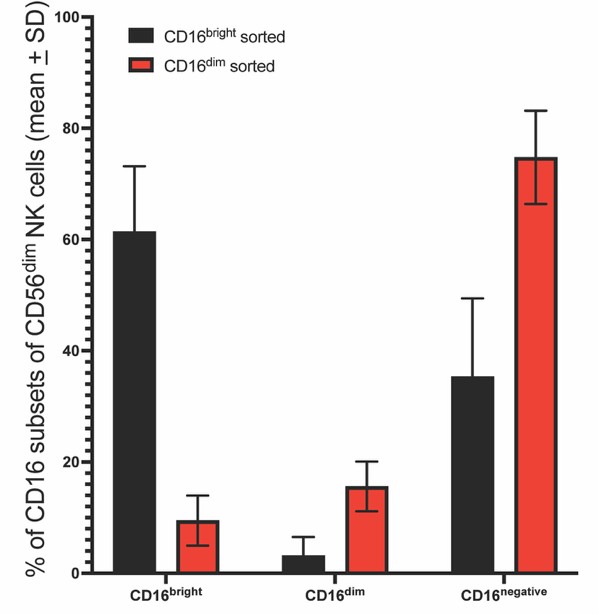
Phenotype of sorted CD56dimCD16bright and CD56dimCD16dim NK cells after exposure to HIV-infected T-cells. NK cells were sorted into CD56dimCD16bright and CD56dimCD16dim subsets, exposed to purified autologous productively HIV-1SHM-1-infected T cells for 1 hour at a 1:1 effector-to-target cell ratio, and reanalyzed for CD16 expression. Bars show the percentage of each sorted NK cell population (CD56dimCD16bright and CD56dimCD16dim) falling into the CD16bright, CD16dim, and CD16negative gates after exposure, as the mean ± standard deviation (SD) of three replicates. One hour is when the highest frequency of degranulating cells is observed in both subsets.
Second, the subsets differ before any stimulation. CD56dimCD16dim cells express higher NKG2D than CD56dimCD16bright cells before any target-cell contact. In the no-target condition, NKG2D was 1182 gMFI higher on CD56dimCD16dim cells (p < 0.0001), a difference of approximately 1.6-fold; across all conditions tested the subset means were 2521.5 versus 1772.2 gMFI, or 1.42-fold (two-way ANOVA: subset F(1, 20) = 1897, p < 0.0001, 70.97% of the total variation; Fig. 6, now Figure 5; Supplemental Table 18). This difference is specific to NKG2D: NKp46, measured on the same cells in the same wells, did not differ between the subsets in the no-target condition (mean difference 45.67 gMFI, p = 0.0668), and was higher on CD56dimCD16bright cells when targets were present. The subsets therefore differ in NKG2D density before activation, and not in activating receptor density generally.
Planned strengthening.
To place this beyond doubt, we will expand the sorted-subset experiments, performing the specific-lysis assay (Fig. 2) and the antibody-dependent degranulation assay on subsets purified before target exposure across additional donors, together with uninfected-target controls. If cell yields from the sort permit, we will also perform the serial degranulation assay on sorted subsets; because the CD56dimCD16dim subset constitutes fewer than 5% of CD56dim NK cells and the serial degranulation assay requires four sequential labelling and washing steps, we cannot commit to this in advance of the sort. These experiments require sorting and primary-cell work and will be completed within approximately six to eight weeks and provided with the revised manuscript.
We note that performing every functional assay in this study on sorted subsets is not feasible within the scope of this revision. Sorting the CD56dimCD16dim subset, which constitutes fewer than 5% of CD56dim NK cells, from a sufficient number of donors to repeat the full panel of assays would require resources beyond those currently available to us. We have therefore prioritized the specific-lysis and antibody-dependent degranulation assays, which bear most directly on the concern raised by the reviewers and the editor, and will extend the approach to the remaining assays as resources allow.
Public Reviews:
Reviewer #1 (Public review):
Overall organization
Overall, the manuscript includes many data in nine figures plus supplemental figures, and would benefit from some focusing of the results.
We agree, and we have reduced the main figures from nine to eight. The NKG2D ligand histograms, previously Figure 5A, have been removed for the reason given in our response to the peer reviewer, Recommendation 4. The degranulation against wild-type and ΔVpr-infected targets, previously Figure 5B, and the degranulation and killing frequency measurements in mixed populations, previously Figure 3, have been moved to the supplementary material. The inhibitory receptor analyses have been reduced from approximately 1,800 words and more than 80 reported p-values to approximately 700 words and 24, with the detail retained in the supplemental tables. Each Results section now opens with a statement of the principal finding before the supporting data, and the Discussion synthesizes what the findings mean rather than restating them.
Figure 1 and Figure 1—figure supplement 2
The observation that CD16dim NK cells responded more strongly by degranulation to K562 cells and HIV-1-infected cells could be due to the shedding of CD16 following activation. In other words, more strongly activated NK cells express higher levels of CD107 but also shed CD16, resulting in higher CD107 expression in CD16low NK cells. The authors should investigate this, for example by performing the degranulation assays shown in Figure 1 in the presence and absence of an ADAM17 inhibitor.
We thank the reviewer for raising this important point, which we recognize is shared by all three reviewers and the editors, and which we address in full in the central response above. We note for clarity that the K562 data are not in Figure 1; they are presented in Figure 1—figure supplement 2, both in the reviewed preprint and in the revised manuscript. They were included to reproduce a previously established finding (Amand et al., Front Immunol 2017;8:699), and were not intended as a central experimental claim; the mechanistic focus of this study is the response to HIV-infected cells. Notably, that same study addressed the question by sorting the CD56dim subsets before stimulation rather than gating after, and our study applies the same approach and extends it to the HIV-infected setting. Four lines of evidence argue against the interpretation that CD16dim degranulation reflects activation-induced CD16 shedding of CD16bright cells:
(1) In cells sorted before target exposure, purified CD16dim cells lyse HIV-infected targets approximately twice as efficiently as purified CD16bright cells across the effector-to-target range, significantly so at 1:4 and above (Fig. 2; Supplemental Table 4); because the subsets are defined before any activation and the readout is direct target lysis rather than post-assay CD16 gating, this advantage cannot arise from shedding during the assay.
(2) The two subsets respond to ADAM17 inhibition in opposite directions, both in the magnitude of degranulation (Fig. 7B, now Figure 6B) and in the number of serial degranulation events per cell (Fig. 9, now Figure 8): ADAM17 inhibition increased degranulation in CD16bright cells but decreased it in CD16dim cells.
(3) Blocking NKG2D together with ADAM17 reduced CD16dim degranulation below either treatment alone (Fig. 8A, now Figure 7A), indicating that the CD16dim advantage is driven by NKG2D through a mechanism separable from ADAM17-mediated shedding.
(4) The subsets also differ before any stimulation, with NKG2D 1182 gMFI higher on CD16dim cells in the no-target condition (p < 0.0001) and no corresponding difference in NKp46 under the same condition (Fig. 6, now Figure 5).
We will further strengthen these findings by expanding the sorted-subset experiments across additional donors, as described in the central response.
Figure 2
The authors sorted CD16dim and bright NK cells for these experiments and observed higher lysis of HIV-1-infected CD4+ T cells. Important controls should be included in these experiments - how strong was the lysis of HIV-1-uninfected CD4+ T cells by these different NK cell subsets? It also appears that the results shown were derived using NK cells from one donor, and "representative of two independent sort experiments performed with separate donors, each yielding similar results". Why are the authors now showing the respective data? One or two experiments appear too few to come to these conclusions. To support the broad conclusions drawn by the reviewers, the experiments should be performed in a larger number of individuals.
We thank the reviewer for these constructive points.
(1) Uninfected-target control. We agree this is an important control and will include lysis of uninfected autologous CD4 T cells by the sorted CD16dim and CD16bright subsets in Figure 2, confirming that the observed lysis is specific to HIV-infected targets. We note that the corresponding CD107a degranulation controls against uninfected targets are presented in Figure 1—Figure Supplement 4C of the revised manuscript.
(2) Number of donors and presentation of data. We agree that the conclusions require more than the representative donor shown. As described in the central response, we will expand these sorted-subset experiments to a larger number of individuals and will present the data from all donors rather than a single representative experiment. Because they require cell sorting and primary-cell work, these experiments will be completed within approximately six to eight weeks and provided with the revised manuscript.
Figures 3 and 4
It appears that experiments were performed again using bulk NK cell populations, and superior degranulation and killing frequencies by CD16dim NK cells might reflect different levels of activation again, as described above for Figure 1. The same applies to Figure 4 - lower degranulation events in CD16bright NK cells are consistent with lower activation of these cells, resulting in less CD16 downregulation. Also, it is not clear to the reviewer why CD107a expression and killing frequencies decrease with higher effector-to-target ratios (Figure 3).
(1) Activation-induced shedding in bulk experiments (Figs. 3 and 4, now Figure 2—figure supplement 1 and Figure 3). We agree that these figures use bulk NK cell populations gated by CD16, and we address the underlying shedding concern in full in the central response. The concern that the lower serial degranulation of CD16bright cells in the direct-killing assay simply reflects lower activation and therefore less shedding is addressed directly by our ADAM17-inhibition data. At 0 µg/mL VRC01, that is, in the absence of antibody, ADAM17 inhibition already affects the two subsets differently rather than in the same direction (Figs. 7B and 7C, now Figures 6B and 6C), as would be expected if they were one population differing only in activation level. This differential response is also seen across the antibody-dependent conditions in Figs. 7B and 9 (now Figures 6B and 8). As described in the central response, we will additionally repeat the specific-lysis and antibody-dependent degranulation measurements on subsets purified before target exposure across additional donors, and the serial degranulation assay as well if cell yields from the sort permit.
(2) Decrease in CD107a and killing frequency at higher effector-to-target ratios (Fig. 3, now Figure 2—figure supplement 1). This reflects the nature of the readout. CD107a mobilization is measured per effector cell, as the percentage of NK cells that degranulate, and is therefore maximized when targets are in excess. At low effector-to-target ratios, nearly every NK cell can encounter and engage a target, yielding a high percentage of CD107a-positive cells; at high ratios, targets become limiting, so a large fraction of NK cells never contact a target and remain unstimulated, and the rapid destruction of the limited target pool further reduces the stimulus available to the remaining cells. This lowers the measured per-effector degranulation frequency even as the absolute number of targets killed is maintained, and it is distinct from a lysis assay, which measures the fate of the target population and accordingly rises with increasing effector-to-target ratio. The same per-effector readout behavior applies to the degranulation data shown in Figs. 5C and 6C (now Figures 4A and 4B, and Figure 5C). This explanation has been added to the revised Discussion.
Pages 19-25
It would be helpful if the authors could provide some conclusions regarding their findings - it is very difficult for the reader to follow the many reported frequencies and p-values. What does this actually mean? Overall, the results appear to follow prior observations that licensed (KIR3DL+) NK cells respond more strongly than unlicensed (KIR3DL1neg) NK cells. The consistent observation within these different subanalyses that CD16dim NK cells degranulate more than CD16bright NK cells is probably the result of activation-induced CD16 downregulation in these assays, as mentioned above. Providing two-way ANOVA analysis results for these very many observations would furthermore require, in the opinion of the reviewer, adjustments for multiple comparisons.
We thank the reviewer, and we have addressed this in three ways.
(1) Readability. We agree that these sections were difficult to follow as presented. We have rewritten them, opening each with a statement of the principal finding before the supporting statistics, and reducing the inhibitory receptor section from approximately 1,800 words and more than 80 reported p-values to approximately 700 words and 24, with the detail retained in the supplemental tables. The Discussion now synthesizes what the findings mean.
(2) CD16dim degranulation in these subanalyses. The consistent observation that CD16dim cells degranulate more than CD16bright cells across these subanalyses is addressed in full in the central response, where several lines of evidence, including subsets sorted before target exposure (Fig. 2) and the opposite responses of the two subsets to ADAM17 inhibition (Figs. 7B and 9, now Figures 6B and 8), argue against activation-induced CD16 downregulation as the explanation.
(3) Multiple comparisons. We agree, and we have re-analyzed these comparisons, applying the post-hoc test matched to each comparison structure: Dunnett's where every subset is compared against a single designated subset, Tukey's where all pairwise comparisons are of interest, and Šidák’s where a prespecified subset of comparisons is of interest. Adjusted p-values are reported throughout, and the design and post-hoc test used for each figure and panel are given in a new supplemental table. The streamlining described above has also reduced the number of comparisons reported in the main text.
Figures 5 and 6
These figures demonstrate that NK cell-mediated activation by HIV-1-infected cells depends on NKG2D ligands and can be inhibited by blocking this interaction - this is consistent with data presented by the Barker group and others previously, and does not provide new information.
We agree that the dependence of NK-cell recognition of HIV-infected cells on NKG2D and its ligands is established, including in our own earlier work (Ward et al., PLoS Pathog 2009;5(10):e1000613) and by others, and we do not present that dependence as a novel finding.
On review, the histograms in Figure 5A were reproduced from that earlier study and should not have been included without attribution. We have removed that panel and cited the original finding in its place. Figures 5B and 5C are both new results from this study, and both are retained. Figure 5B, which shows that NK cells degranulate in response to wild-type HIV-infected targets but not to ΔVpr-infected or uninfected targets, establishing that the degranulation response in this system depends on Vpr, becomes Figure 4—figure supplement 1 in the revised manuscript. Figure 5C, the NKG2D blockade experiment, becomes Figure 4A and 4B.
We would also distinguish Figure 6 (now Figure 5), which we consider a substantive finding rather than a restatement of the established NKG2D-ligand dependence. That figure shows that NKG2D expression differs at the level of the individual subsets, and that the difference is present before stimulation and is specific to NKG2D. In the no-target condition, NKG2D was 1182 gMFI higher on CD56dimCD16dim cells (p < 0.0001), approximately 1.6-fold, while NKp46 measured on the same cells in the same wells did not differ (mean difference 45.67 gMFI, p = 0.0668). This provides a candidate mechanism for the superior effector function of the CD56dimCD16dim subset, in addition to their serial-degranulation capacity, and it bears directly on the central question of whether the two subsets differ intrinsically rather than as a consequence of activation. The revised text presents the established NKG2D-ligand dependence as context while making the subset-level NKG2D difference, and its mechanistic significance, more prominent.
Figure 7
The authors extended their functional analyses of NK cells to ADCC function. It is very well established that CD16 is downregulated in the context of ADCC following activation of NK cells. Consistent with this, higher degranulation is observed by CD16dim NK cells.
We agree that CD16 is downregulated during antibody-dependent responses, and this is precisely why we included the ADAM17-inhibition experiments within Figure 7 (now Figure 6), to determine whether the higher degranulation of CD56dimCD16dim cells is a consequence of that shedding or a property of a distinct subset. As detailed in the central response, these experiments argue against the shedding interpretation. In Fig. 7B (now Figure 6B), inhibiting ADAM17 affects the two subsets in opposite directions: it increases the degranulation of CD56dimCD16bright cells while decreasing that of CD56dimCD16dim cells. If the CD16dim cells were simply CD16bright cells that had shed CD16, blocking shedding would be expected to move the two in the same direction; the opposite responses instead indicate two distinct populations with opposite functional dependence on ADAM17 activity. The same opposition is seen when serial degranulation is resolved by the number of events per cell (Fig. 9, now Figure 8). Thus, while CD16 downregulation during antibody-dependent responses is well established, these data indicate that the superior response of the CD56dimCD16dim subset is not explained by it. This interpretation is now explicit in the revised Discussion.
ADAM17-inhibition data (final figures)
These data are of interest, but should be presented in a more structured way. First of all, does the addition of ADAM17 inhibitors change the overall proportion of CD16bright and dim NK cells following activation, independent of whether these cells degranulate or not? Overall, the proportion of CD16dim NK cells that degranulate appears to be reduced in the presence of the ADAM inhibitor, which is consistent with reduced CD16 shedding and maintenance of CD16 expression on activated NK cells - and this is supported by the increase in CD107a-positive NK cells that express CD16 (Figure 8a). Overall, the differences between CD16bright and dim NK cells in their level of activation appear to disappear in the presence of an ADAM17 inhibitor, based on the data shown in Figure 8b, suggesting that CD16 downregulation is occurring in response to activation of NK cells as a consequence of CD16 shedding, and can be inhibited by an ADAM17 inhibitor.
We thank the reviewer for these suggestions, which we have used to present the ADAM17-inhibition data more clearly.
(1) Effect on subset proportions, independent of degranulation. This is shown in Fig. 8B (now Figure 7B). Because the two subsets differ greatly in baseline frequency, with CD56dimCD16bright cells constituting the large majority of CD56dim NK cells before stimulation, a change in raw bulk proportion is small and difficult to interpret, for example a shift from roughly 95% to 92.5% of the bright population. To place the two subsets on comparable footing, the panel reports, for each subset, the frequency following target exposure minus its frequency in the matched unstimulated condition. Presented this way, ADAM17 inhibition clearly reduces the activation-associated change in subset proportions, consistent with reduced CD16 shedding. This normalization is stated in the legend and is now described in the Results text so that the analysis is not overlooked.
(2) Interpretation of the ADAM17-inhibition data. We agree that CD16 downregulation occurs as a consequence of activation-induced shedding and is prevented by ADAM17 inhibition; this is not in dispute. We would, however, offer an additional observation that bears on whether the between-subset functional difference is itself a product of that shedding. In Fig. 8A (now Figure 7A), combining ADAM17 inhibition with NKG2D blockade reduces CD56dimCD16dim degranulation to 2.03%, below both anti-NKG2D alone at 5.24% and ADAM17 inhibition alone at 5.05% (p < 0.0001 and p = 0.0001 respectively). If the CD16dim advantage were solely a consequence of CD16 shedding, and if NKG2D blockade acted only by reducing that shedding, the combination could not reduce degranulation further than ADAM17 inhibition alone.
The same figure also shows that the two perturbations act in opposite directions within the CD56dimCD16bright subset: anti-NKG2D reduced their degranulation from 2.58% to 0.92% (p = 0.0263), whereas ADAM17 inhibition increased it to 3.99% (p = 0.0679). NKG2D therefore supports the response of CD56dimCD16bright cells while ADAM17 activity constrains it, the reverse of the pattern in CD56dimCD16dim cells. Together with the opposite responses of the two subsets to ADAM17 inhibition described in the central response, this indicates that NKG2D and ADAM17 contribute through separable mechanisms and that the two subsets are not one population at different stages of shedding. These data are now presented in a more structured form and the interpretation is explicit in the revised text.
Summary statement
Taken together, many of the data presented in the manuscript are consistent with the very well-established downregulation of CD16 expression on activated NK cells, suggesting that the observed association between reduced CD16 expression on CD56dim NK cells and enhanced effector functions is a consequence of higher activation of these NK cells.
We appreciate the reviewer articulating the central concern so clearly. We agree that CD16 downregulation on activated NK cells is well established and occurs in our assays; where we reach a different conclusion is on whether the enhanced function of the CD56dimCD16dim subset is a consequence of that downregulation. As set out in the central response, three observations argue that it is not: the advantage is present in cells sorted into subsets before any target contact, where post-assay CD16 changes cannot apply (Fig. 2); the two subsets respond to ADAM17 inhibition in opposite directions, both in magnitude (Fig. 7B, now Figure 6B) and in the number of serial degranulation events per cell (Fig. 9, now Figure 8), which is difficult to reconcile with their being one population at different activation levels; and blocking NKG2D together with ADAM17 reduces CD16dim degranulation below either alone (Fig. 8, now Figure 7), indicating that the advantage is driven by NKG2D through a mechanism separable from shedding. We therefore interpret the association between low CD16 and enhanced function not as activation-induced downregulation of a single population, but as a property of a distinct, pre-existing subset. This interpretation is stated and defended explicitly in the revised Discussion, and will be strengthened with the expanded pre-sorted experiments.
Reviewer #2 (Public review):
(1) The central conclusion is weakened by the use of CD16 as a stable phenotypic marker. CD16 is well established to be rapidly downregulated following NK-cell activation and target cell (K562 or infected cells) engagement through ADAM17-mediated shedding. NK cell shedding regulates NK cell effector functions by promoting target cell detachment, boosting serial killing capacity, and preventing overstimulation. Therefore, NK cells displaying a CD56dimCD16dim phenotype after co-culture cannot be assumed to represent a pre-existing subset with intrinsically superior cytotoxic activity, but may instead correspond to activated CD56dimCD16bright NK cells that have downregulated CD16 during the assay. Because the vast majority of the functional experiments classified NK cell subsets based on post-assay CD16 expression, it is difficult to distinguish intrinsic functional differences between NK cell subsets from activation-induced phenotypic conversion. This limitation affects the interpretation of most of the study's principal findings.
We thank the reviewer for this careful and well-articulated concern, which we recognize as the central issue of the review, and which we address in full in the central response above. We agree with the reviewer's premises: CD16 is rapidly shed by ADAM17 upon activation, and this shedding is itself functionally important, promoting target detachment, supporting serial engagement, and limiting overstimulation. Indeed, ADAM17-mediated shedding is integral to the serial degranulation mechanism we propose. We also agree that classifying subsets by post-assay CD16 expression alone cannot, on its own, distinguish a pre-existing subset from activation-induced conversion.
For this reason, our conclusion does not rest on post-assay classification. As detailed in the central response, the CD56dimCD16dim advantage is demonstrated in cells sorted into subsets before any target contact, where the readout is direct lysis rather than post-assay gating and where activation-induced shedding therefore cannot account for the difference (Fig. 2), an experiment both this reviewer and the peer reviewer identify as the strongest in the manuscript. This is reinforced by evidence that the two subsets are functionally distinct rather than one population caught at different stages of shedding: they respond to ADAM17 inhibition in opposite directions, both in the magnitude of degranulation (Fig. 7B, now Figure 6B) and in the number of serial degranulation events per cell (Fig. 9, now Figure 8); blocking NKG2D together with ADAM17 reduces CD16dim degranulation below either alone, indicating a mechanism separable from shedding (Fig. 8, now Figure 7); and the subsets differ before stimulation, with NKG2D 1182 gMFI higher on CD16dim cells and no corresponding difference in NKp46 (Fig. 6, now Figure 5). We will strengthen this further by expanding the sorted-subset experiments across additional donors, and the revised text rests the manuscript's conclusions explicitly on the pre-sorted data.
(2) The "killing frequency" analysis presented in Figure 3 is based on a mathematical estimate rather than a direct experimental measurement. Since total target cell killing is measured in mixed NK cell populations, it cannot be attributed to individual NK cell subsets. This experiment must be repeated using purified NK cell subsets.
We agree that the killing frequency in Fig. 3 (now Figure 2—figure supplement 1C) is a mathematical estimate rather than a direct measurement. We would add that this is intrinsic to the metric: killing frequency is a derived quantity whether calculated from mixed or purified populations, so repeating it on purified subsets would not convert it into a direct measurement. This analysis has been moved to the supplementary material, and its limitations are stated in the Discussion, namely that killing frequency is an estimate and should be interpreted as such. Direct, subset-resolved killing is instead provided by Figure 2, in which NK cells sorted before target exposure show that purified CD56dimCD16dim cells lyse HIV-infected targets more efficiently than purified CD56dimCD16bright cells; this is the measurement on which our conclusion regarding direct killing rests, and it is the experiment we will expand across additional donors.
(3) The serial degranulation assay presented in Figure 4 does not directly measure serial target cell killing and therefore does not support the conclusion that CD56dimCD16dim NK cells possess superior serial killing capacity. Furthermore, the increased serial degranulation observed in the CD16dim population could simply reflect activation-induced CD16 downregulation rather than an intrinsic property of this subset. This experiment should therefore be repeated using purified NK cell subsets.
We agree that Fig. 4 (now Figure 3) measures serial degranulation, not serial killing directly. The text has been revised throughout to describe this as serial degranulation rather than serial killing, so that our conclusions match what was measured. Regarding the concern that increased serial degranulation in CD16dim cells reflects activation-induced CD16 downregulation, we address this in the central response; the opposite responses of the two subsets to ADAM17 inhibition (Figs. 7B and 9, now Figures 6B and 8) argue against that interpretation. As the reviewer suggests, we will repeat the serial degranulation assay on subsets purified before target exposure if cell yields from the sort permit. We note that this assay requires four sequential labelling and washing steps and that the CD56dimCD16dim subset constitutes fewer than 5% of CD56dim NK cells, so the number of sorted cells recovered may be limiting; we will report the outcome either way.
(4) The finding that CD56dimCD16dim NK cells exhibit greater ADCC activity is somewhat counterintuitive given the central role of CD16 in mediating ADCC. Moreover, these experiments are likely confounded by activation-induced CD16 downregulation, which is expected to be even more pronounced during ADCC. Thus, the apparent superiority of the CD56dimCD16dim subset may simply reflect the conversion of activated CD56dimCD16bright NK cells into the CD16dim gate rather than intrinsically greater ADCC activity. To directly compare the intrinsic ADCC capacity of each subset, these experiments should be repeated using purified NK cell populations prior to target-cell stimulation.
We agree that the greater antibody-dependent response of CD56dimCD16dim cells is counterintuitive given the central role of CD16, and we regard it as an informative finding rather than an artifact. As set out in our revised Discussion, the surface density of gp120 on HIV-infected primary T-cells is approximately 6.4 × 10² molecules per cell (Vasiliver-Shamis et al., 2008), two orders of magnitude below high-density antigens such as CD20 on Raji cells at approximately 5 × 10⁴ molecules per cell (Lallemand et al., 2017). Antibody-dependent responses against HIV-infected cells therefore proceed under conditions of limiting antigen, and both CD56dim subsets face the same constraint. What differs between them is not the constraint but the NKG2D available to meet it: CD56dimCD16dim cells carry higher NKG2D before target contact and respond more strongly, despite their lower CD16. Consistent with a requirement for a second signal under these conditions, ADAM17 inhibition reduced CD56dimCD16dim degranulation even at 0 µg/mL VRC01 (Figs. 7B and 7C, now Figures 6B and 6C), where no antibody is present to engage CD16.
Regarding the concern that this superiority reflects conversion of CD16bright cells into the CD16dim gate, we address this in full in the central response; the opposite responses of the two subsets to ADAM17 inhibition, in both degranulation magnitude (Fig. 7B, now Figure 6B) and serial degranulation (Fig. 9, now Figure 8), argue against it. As the reviewer recommends, we will directly compare the intrinsic antibody-dependent capacity of each subset using cells purified before target-cell stimulation, extending the pre-sorted approach of Figure 2 to the antibody-dependent setting across additional donors.
Recommendations for the authors:
Reviewer #2 (Recommendations for the authors):
(1) As discussed in the public review, the majority of the functional assays should be repeated using purified NK cell subsets. This approach would eliminate the confounding effect of activation-induced CD16 downregulation and allow the intrinsic functional properties of each subset to be directly compared.
We agree, and this is the central experimental commitment of our revision. As described in the central response, we will repeat the specific-lysis and antibody-dependent degranulation assays using NK cell subsets purified before target-cell exposure, so that the intrinsic functional properties of each subset are compared directly and are not subject to activation-induced changes in CD16 expression. We will also perform the serial degranulation assay on sorted subsets if cell yields permit; that assay requires four sequential labelling and washing steps, and the CD56dimCD16dim subset constitutes fewer than 5% of CD56 dim NK cells, so we cannot commit to it in advance of the sort. This extends the pre-sorted approach already used in Figure 2, which the reviewer identifies as the strongest evidence in the manuscript, across additional donors and across the functional readouts. These experiments require cell sorting and primary-cell work and will be completed within approximately six to eight weeks and provided with the revised manuscript.
(2) The experiments performed with purified NK cell subsets in Figure 2 provide the strongest evidence supporting the authors' conclusion that CD56dimCD16dim NK cells exhibit greater direct cytotoxicity against HIV-infected target cells. These data are the most convincing in the manuscript because they are not confounded by post-assay changes in CD16 expression. However, unlike the other functional assays, no representative gating strategy or raw flow cytometry plots are provided, and the results appear to be based on a single representative experiment. Given the importance of these data to the manuscript's central conclusion, this experiment should be expanded to include biological replicates from additional donors, representative flow cytometry plots, and validation using additional HIV-1 infectious molecular clones.
We appreciate the reviewer identifying the sorted-subset experiments in Figure 2 as the strongest evidence for our conclusion, and we agree these data warrant expansion. In the revised manuscript we will:
(1) Expand the experiment to include biological replicates from additional donors, with all donors shown rather than a single representative experiment.
(2) Provide the representative gating strategy and flow cytometry plots for the sorted subsets. The reviewer is correct that these should be included, and we will add them, including for the expanded experiments.
(3) Validate the finding using additional HIV-1 strains. We note, for clarity, that the virus used throughout this study is a primary patient isolate (HIV-1SHM-1), as stated in the Materials and Methods, rather than an infectious molecular clone. We have now compared NK cell degranulation against autologous CD4positive T-cells productively infected with HIV-1SHM-1, with the X4-tropic infectious molecular clone HIV-1NL4-3, and with the R5-tropic laboratory-adapted strain HIV-1BaL, at three effector cell to target cell ratios. CD56 dim CD16 dim cells degranulated more than CD56 dimCD16bright cells against every virus at every ratio, in all nine comparisons at p < 0.0001. Both subsets responded less to HIV-1NL4-3 and HIV-1BaL than to HIV-1SHM-1, and did so in proportion: the ratio of CD56 dimCD16 dim to CD56 dim CD16bright degranulation ranged from 2.7 to 3.8 across all nine conditions. The magnitude of the response therefore varies with the virus, whereas the relationship between the two subsets does not. These data are included in the revised manuscript as Figure 1—figure supplement 3, with the statistical analysis in a supplemental table.
The experiments described in points 1 and 2 require cell sorting and primary-cell work and will be completed within approximately six to eight weeks and provided with the revised manuscript.
(3) The mechanism underlying the enhanced effector function of CD56dimCD16dim NK cells remains unclear. Although the phenotypic characterization presented in Figure 5 (and related supplement figures) is informative, NK cell receptor expression was assessed after target cell stimulation, when it may already have been altered by activation and CD16 downregulation. Receptor expression should therefore be evaluated prior to stimulation. In addition to NKG2D, the authors should also consider assessing additional activating receptors, notably NKp30, which has recently been implicated in the elimination of autologous HIV-1-infected cells (PMID: 41079618).
We agree that receptor expression should be assessed before stimulation, and in fact it is. In Fig. 6 (now Figure 5), the receptor gMFI data include the no-target condition, showing that CD56dimCD16dim cells express 1182 gMFI more NKG2D than CD56 dimCD16bright cells before any target-cell contact (p < 0.0001), approximately 1.6-fold, and therefore before any activation-induced change in receptor expression. NKp46, measured on the same cells in the same wells, did not differ between the subsets under the same condition (mean difference 45.67 gMFI, p = 0.0668), indicating that the difference is specific to NKG2D rather than a general difference in activating receptor density. The baseline condition is now labelled explicitly as 1:0 in the revised figure and described as such in the Results text.
Regarding NKp30, we have assessed this receptor. Degranulation did not differ between NKp30 positive and NKp30 negative cells within either CD56dim subset, whereas both CD56dimCD16dim groups exceeded both CD56dimCD16bright groups regardless of NKp30 status, indicating that the enhanced degranulation of the subset is not attributable to NKp30, paralleling our finding for NKp46. These NKp30 data are included in the revised manuscript as Figure 5—figure supplement 1, with the corresponding statistical analysis in a supplemental table. We note that this analysis addresses whether NKp30 accounts for the difference between the subsets; it does not exclude a role for NKp30 in NK-cell recognition of HIV-infected cells more generally, consistent with the study the reviewer cites, which we now discuss.
(4) In Figure 5, the histograms corresponding to the uninfected and ΔVpr conditions appear to be identical. If this is indeed the case, this represents a serious concern, as these are two distinct experimental conditions and should not be represented by the same flow cytometry plot. This raises the possibility of an inadvertent panel duplication. The authors should carefully verify the figure and replace the duplicated panel if necessary.
We thank the reviewer for this careful observation. On review, the histograms in Figure 5A were reproduced from our earlier study (Ward et al., PLoS Pathog 2009;5(10):e1000613) and should not have been included without attribution. We have removed that panel and cite the original finding in its place.
Figures 5B and 5C are both new results from this study, and both are retained. Figure 5B, which shows that NK cells degranulate in response to wild-type HIV-infected targets but not to ΔVpr-infected or uninfected targets, becomes Figure 4—figure supplement 1 in the revised manuscript. Figure 5C, the NKG2D blockade experiment, becomes Figure 4A and 4B.
(5) The ADCC experiments and calculation require additional methodological clarification, particularly the analyses presented in Figure 7C. Although NK cell degranulation is commonly used as a surrogate marker of ADCC, the data presented in Figure 7 do not appear to isolate the antibody-dependent component of the response. To specifically quantify ADCC-mediated degranulation, the degranulation induced by HIV-infected target cells alone (i.e., in the absence of VRC01) should be subtracted from that measured in the presence of VRC01. Notably, in Figure 7C (DMSO), the CD56dimCD16dim population appears to exhibit similar levels of degranulation in the absence and presence of VRC01, suggesting that antibody-dependent degranulation may be limited in this subset, which does not support the author's conclusions.
We thank the reviewer for raising this, and we agree that the antibody-dependent and antibody-independent components of the response should be distinguished. We would, however, respectfully argue against the subtraction as a means of doing so, and we believe the experiments already in the manuscript address the underlying question more directly.
The condition without VRC01 is not a background to be removed. It is the NKG2D-driven response of the same cells to the same infected targets, measured through the same degranulation machinery, and it is one of the principal findings of the study. Subtracting it treats the two components as though they were independent and additive, when both converge on a single immunological synapse and a single degranulation event per cell. The difference between the two conditions is therefore not the antibody-dependent response; it is the increment in total degranulation produced by adding antibody, which is a different quantity and one that carries no clean interpretation at the level of the individual cell.
The question the reviewer raises, whether the antibody-dependent component differs between the subsets, is answered directly by the two-way ANOVA of these data. Across the VRC01 titration, the effect of NK cell subset accounts for 85.91% of the total variation (F(1, 16) = 424.0, p < 0.0001) and the effect of VRC01 concentration for 10.18% (F(3, 16) = 16.74, p < 0.0001), while the subset × VRC01 interaction is not significant (F(3, 16) = 1.112, p = 0.3732) and accounts for 0.68% (Supplemental Table 20 in the revised manuscript). The absence of an interaction means that adding antibody raises the response of both subsets by a comparable amount, and that the difference between the subsets is the same at every VRC01 concentration tested. This is a statistical statement about the antibody-dependent component, obtained without subtracting one condition from another.
We agree with the implication the reviewer draws from this, and we state it plainly in the revised Discussion: the antibody-dependent increment is modest in both subsets. We attribute this to the very low surface density of gp120 on HIV-infected primary T-cells, approximately 6.4 × 10² molecules per cell (Vasiliver-Shamis et al., 2008), which is two orders of magnitude below high-density antigens such as CD20 on Raji cells, approximately 5 × 10⁴ molecules per cell (Lallemand et al., 2017). Under these conditions the antibody-dependent signal available to any NK cell is limited, and this applies equally to both subsets.
Where we differ from the reviewer is on the conclusion this supports. That the antibody-dependent increment is modest in both subsets does not weaken our central claim, which is comparative: at every VRC01 concentration tested, including in the presence of antibody, CD56dimCD16dim cells degranulate more than CD56dimCD16bright cells against antibody-coated HIV-infected targets. That comparison is what the manuscript reports, and it is unaffected by how the response is partitioned between its antibody-dependent and antibody-independent components.
We also note that the experiments in Figs. 7B and 7C (now Figures 6B and 6C) do isolate a component of the response experimentally rather than arithmetically. Inhibiting ADAM17 removes the contribution that depends on CD16 turnover, and it does so in opposite directions in the two subsets, reducing CD56dimCD16dim degranulation and increasing that of CD56dimCD16bright cells at every VRC01 concentration. These are direct experimental manipulations of the antibody-dependent pathway, and they are more informative than the arithmetic difference between two conditions.
Finally, we take the reviewer's point that the analyses in Fig. 7C require clearer explanation. In the revised manuscript we state explicitly what is plotted, namely the percentage of each CD16 subset among CD107a positive CD56dim NK cells, we describe the background subtraction that applies to all CD107a data in this study, and we report the statistical analysis of each panel in full.
(6) It is also unclear how the authors interpret the effects of ADAM17 inhibition. While ADAM17 inhibition increases the ADCC activity of the CD56dimCD16bright population, it simultaneously decreases that of the CD56dimCD16dim population. An alternative explanation is that inhibition of CD16 shedding prevents activated CD56dimCD16bright NK cells from transitioning into CD56dimCD16dim during the assay. This possibility should be discussed and experimentally addressed, as it provides a plausible alternative interpretation of the observed phenotype.
We thank the reviewer for articulating this alternative, which we address in full in the central response. We agree that the opposite effects of ADAM17 inhibition on the two subsets are central to interpreting these experiments, and we interpret them as evidence that the two are distinct populations rather than one transitioning into the other.
The reviewer's alternative, that ADAM17 inhibition prevents CD56dimCD16bright cells from transitioning into the CD56dimCD16dim gate, predicts that blocking shedding should reduce the CD56dimCD16dim population by cutting off its supply from CD16bright cells. Three observations argue against this being the explanation for the functional difference. First, the effect is not merely a change in population size but a change in per-cell function in opposite directions: ADAM17 inhibition increases the number of serial degranulation events in CD56dimCD16bright cells while decreasing them in CD56dimCD16dim cells (Fig. 9, now Figure 8), which is difficult to explain if the dim cells were simply bright cells prevented from converting. Second, blocking NKG2D together with ADAM17 reduces CD56dimCD16dim degranulation below either treatment alone (Fig. 8A, now Figure 7A); if NKG2D blockade acted only by reducing the shedding that drives the putative transition, the combination could not exceed the effect of ADAM17 inhibition alone. Third, we have tracked the fate of each subset sorted before target exposure: at one hour, when degranulation is maximal, only approximately 4% of sorted CD16bright cells were found in the CD16dim gate, while approximately 35% had moved to the CD16negative gate (Author response image 1 accompanying this response). Shedding therefore directs CD16bright cells past the CD16dim gate rather than into it.
We discuss this alternative explicitly in the revised Discussion and will address it further experimentally by repeating these assays on subsets purified before target exposure, where no transition can occur during the assay.
Reviewing Editor Comments:
The conclusion that CD56dimCD16dim NK cells are intrinsically superior effectors against HIV-infected target cells requires additional evidence because CD16 is rapidly downregulated following NK-cell activation. Throughout most of the study, NK-cell subsets are classified after target-cell encounter, making it difficult to distinguish pre-existing CD56dimCD16dim cells from activated CD56dimCD16bright cells that have undergone ADAM17-mediated CD16 shedding. The authors should repeat functional experiments using NK-cell subsets purified before target-cell exposure and determine the extent to which ADAM17 inhibition alters subset frequencies and functional readouts. These experiments are essential to establish whether the observed functional differences reflect intrinsic biology rather than activation-induced phenotypic conversion.
We thank the editor for this clear synthesis of the central concern, which we address in full in the central response above. In brief, our conclusion does not rest on post-encounter classification: the CD56dimCD16dim advantage is established in cells sorted into subsets before any target contact, using direct lysis as the readout (Fig. 2), and is reinforced by the opposite responses of the two subsets to ADAM17 inhibition in both degranulation magnitude (Fig. 7B, now Figure 6B) and serial degranulation (Fig. 9, now Figure 8), by the separable contributions of NKG2D and ADAM17 (Fig. 8, now Figure 7), and by pre-stimulation differences between the subsets, with NKG2D 1182 gMFI higher on CD16dim cells and no corresponding difference in NKp46 (Fig. 6, now Figure 5). We agree these questions are central and will repeat the functional experiments on subsets purified before target exposure, and the effect of ADAM17 inhibition on subset frequencies is presented explicitly in Figs. 7C and 8B (now Figures 6C and 7B).
Major conclusions should be supported by more rigorous experimental validation. In particular, the sorted NK-cell experiments should be expanded using multiple independent donors, include killing of uninfected target cells as controls and provide representative gating strategies and flow cytometry plots. Likewise, the current analyses of killing frequency, serial killing, and ADCC should be strengthened by direct measurements using purified NK-cell subsets rather than mathematical estimates or analyses performed in mixed NK-cell populations.
We agree and will strengthen the validation as follows. The sorted-subset experiments will be expanded across multiple independent donors, with all donors shown. Uninfected-target controls will be included for the sorted-cell lysis experiments (Fig. 2); the corresponding CD107a controls against uninfected targets are already presented in Figure 1—Figure Supplement 4C of the revised manuscript. Representative gating strategies and flow cytometry plots will be provided for the sorted-cell experiments, as for our other assays. Regarding direct measurement: the killing-frequency metric (Fig. 3, now Figure 2—figure supplement 1C) is a mathematical estimate whether derived from mixed or purified populations, and it has been moved to the supplementary material with this limitation noted in the Discussion, while direct, subset-resolved killing is provided by the pre-sorted lysis experiment (Fig. 2), which we will expand; the serial degranulation assay (Fig. 4, now Figure 3) is now described as serial degranulation rather than serial killing, and will be repeated on purified subsets if cell yields from the sort permit; and the antibody-dependent comparison will be performed on subsets purified before stimulation.
Some aspects of the data analysis and presentation require clarification. The authors should evaluate receptor expression before target-cell stimulation, clarify the ADCC analyses and interpretation of ADAM17 inhibition, verify the apparent duplicated flow-cytometry panel, apply appropriate statistical corrections for multiple comparisons where necessary, and streamline the presentation by emphasizing the principal conclusions rather than extensive descriptive analyses.
We have addressed each of these. Receptor expression before stimulation is shown in the gMFI data of Fig. 6 (now Figure 5) at the no-target condition, where CD56dimCD16dim cells carry 1182 gMFI more NKG2D than CD56dimCD16bright cells (p < 0.0001) with no corresponding difference in NKp46 (p = 0.0668); this condition is now labelled explicitly as 1:0 in the figure and described as such in the Results text. The antibody-dependent analyses and the interpretation of ADAM17 inhibition are clarified in the revised text, as detailed in our responses to the three reviewers and the central response.
On the duplicated panel: the histograms in Figure 5A were reproduced from Ward et al. (2009) and should not have been included without attribution. That panel has been removed and the original finding is cited in its place. Figures 5B and 5C are both new results and are retained, becoming Figure 4—figure supplement 1 and Figures 4A and 4B respectively.
We have applied appropriate multiple-comparison corrections, using the post-hoc test matched to each comparison structure and reporting adjusted p-values throughout; the design and post-hoc test used for each figure and panel are given in a new supplemental table. Finally, we have streamlined the presentation by opening each Results section with a statement of the principal finding before the supporting data, and by reducing the inhibitory receptor section from approximately 1,800 words and more than 80 reported p-values to approximately 700 words and 24, with the detailed data retained in the supplemental tables and their significance synthesized in the Discussion.
References
Amand M, Iserentant G, Poli A, Sleiman M, Fievez V, Sanchez IP, Sauvageot N, Michel T, Aouali N, Janji B, Trujillo-Vargas CM, Seguin-Devaux C, Zimmer J. 2017. Human CD56dimCD16dim cells as an individualized natural killer cell subset. Frontiers in Immunology 8:699. doi:10.3389/fimmu.2017.00699.
Lallemand C, Liang F, Staub F, Simansour M, Vallette B, Huang L, Ferrando-Miguel R, Tovey MG. 2017. A novel system for the quantification of the ADCC activity of therapeutic antibodies. Journal of Immunology Research 2017:3908289. doi:10.1155/2017/3908289.
Vasiliver-Shamis G, Tuen M, Wu TW, Starr T, Cameron TO, Thomson R, Kaur G, Liu J, Visciano ML, Li H, Kumar R, Ansari R, Han DP, Cho MW, Dustin ML, Hioe CE. 2008. Human immunodeficiency virus type 1 envelope gp120 induces a stop signal and virological synapse formation in noninfected CD4+ T cells. Journal of Virology 82:9445-9457. doi:10.1128/JVI.00835-08.
Ward J, Davis Z, DeHart J, Zimmerman E, Bosque A, Brunetta E, Mavilio D, Planelles V, Barker E. 2009. HIV-1 Vpr triggers natural killer cell-mediated lysis of infected cells through activation of the ATR-mediated DNA damage response. PLoS Pathogens 5(10):e1000613. doi:10.1371/journal.ppat.1000613.



