Peer review process
Not revised: This Reviewed Preprint includes the authors’ original preprint (without revision), an eLife assessment, public reviews, and a provisional response from the authors.
Read more about eLife’s peer review process.Editors
- Reviewing EditorMartin DenzelAltos Labs, Cambridge, United Kingdom
- Senior EditorDavid RonUniversity of Cambridge, Cambridge, United Kingdom
Reviewer #1 (Public Review):
In this manuscript, Yong and colleagues link perturbations in lysosomal lipid metabolism with the generation of protein aggregates resulting from proteosome inhibition. The main tool used is the ProteoStat stain to assess protein aggregate burden in native cells (i.e. cells under no exogenous or endogenous stress). They initially use CRISPR-based genome-wide screens to identify several genes that affect this aggregate burden. Interestingly, knockdown of genes involved in lysosomal acidification was a major signature which led to identification of other culprit lysosome-associated genes that included ones involved in lipid metabolism. Subsequent CRISPR screen focused on lipidomic analysis led to identification of sphingolipid and cholesterol esters as lipid classes with effects on proteostasis. Despite using various tools of lysosomal function, acidity, permeability, etc, the authors couldn't identify the link between lysosomal lipid metabolism and protein aggregate formation. Nevertheless, the interrelationship of these two processes was the overall conclusion of this manuscript.
Although this work is interesting and thought-provoking, their approach to identify novel pathways involved in proteostasis is limited and this weakens the contribution of the paper in its current form.
Reviewer #2 (Public Review):
It is certainly an interesting observation that lipid homeostasis influences proteostasis, although this need not be considered so surprising given that many fundamental cellular processes are interconnected. The paper is deserves to be read, but the level of general interest would be greatly enhanced if the authors were able to take the story further mechanistically. This might be too much of an ask, but they should go further in excluding one very attractive alternative model: effects on proteasome activity. This explanation should be addressed definitively because the transcription factor that regulates proteasome subunit gene expression (Nrf1/NFE2L1) is processed in the ER and is therefore well placed to be influenced by membrane conditions, and because it is shown here that proteasome inhibition increase ProteoStat puncta. Indeed, some years ago it was published that Nrf1/NFE2L1 is inhibited within the ER membrane by cholesterol, and a more recent paper showed that in C. elegans it is activated by oleic acid through effects on ER membrane homeostasis and lipid droplet formation. The authors address proteasome activity only by using a dye that is not referenced. Here a much more solid answer is needed. In general, most conclusions in the paper rely essentially solely on ProteoStat assays. The entire study would be greatly strengthened if the authors incorporated biochemical or other modalities to substantiate their results.
The presentation would be improved greatly if the authors provided diagrams illustrating the pathways implicated in their results, as well as their models. As it is the paper falls flat at the end of the results in the absence of a mechanism to explain their findings. Diagrams would be helpful for focusing the reader on what IS learned from the work, which is important.
Author Response:
Points from reviewer 1 (Public Review):
In this manuscript, Yong and colleagues link perturbations in lysosomal lipid metabolism with the generation of protein aggregates resulting from proteosome inhibition.
We apologize for any confusion in the explanation of the results. We found that both proteasome inhibition and, independently, perturbations to lysosomal lipid metabolism lead to accumulation of protein aggregates in the lysosome. There was no evidence of proteasome inhibition in the context of lysosomal lipid perturbations (Figure 4J).
Despite using various tools of lysosomal function, acidity, permeability, etc, the authors couldn't identify the link between lysosomal lipid metabolism and protein aggregate formation.
Indeed, despite testing numerous mechanistic hypotheses, we have yet to explain how perturbation of lysosomal lipid metabolism causes protein aggregates. However, we have demonstrated that lipids are both necessary (via epistasis and serum delipidation) and sufficient (media supplementation) to drive these phenotypes.
Although this work is interesting and thought-provoking, their approach to identify novel pathways involved in proteostasis is limited and this weakens the contribution of the paper in its current form.
We are glad the reviewer found the work to be thought-provoking. As a fundamental cellular process critical for longevity, we agree that the connections made here between lipids, lysosomes and protein aggregates are interesting and broaden the impact of cellular health on proteostasis. Though we have falsified multiple hypotheses for how perturbation of lysosomal lipid metabolism could influence protein aggregation, we agree that a major weakness of the current work is our limited mechanistic understanding of this process. We hope that by engaging the thoughtful and creative eLife readership, novel mechanistic hypotheses will emerge.
Points from reviewer 2 (Public Review):
This might be too much of an ask, but they should go further in excluding one very attractive alternative model: effects on proteasome activity. This explanation should be addressed definitively because the transcription factor that regulates proteasome subunit gene expression (Nrf1/NFE2L1) is processed in the ER and is therefore well placed to be influenced by membrane conditions, and because it is shown here that proteasome inhibition increase ProteoStat puncta.
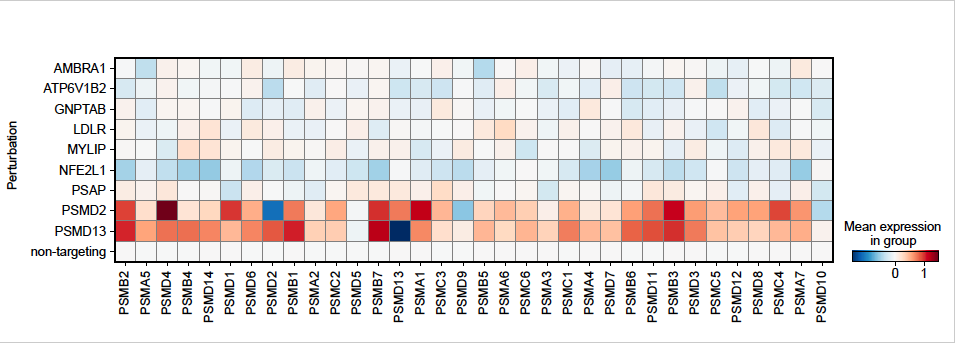
We appreciate the constructive suggestion to examine loss of proteasome expression as a relevant mechanism linking cellular dyslipidemia with proteostasis impairment. We analyzed the genome-wide perturb-seq data from Replogle et al. [1], which was performed in K562 cells cultured under similar conditions to our screen. As expected, perturbation of Nrf1/NFE2L1 reduced expression of proteasome subunits, whereas perturbation of proteasome subunits that increased proteostat staining (e.g. PSMD2, PSMD13) homeostatically increased expression of multiple proteasome subunits. In contrast, other top hits, including those related to lipid-related perturbations (e.g. MYLIP, PSAP) did not reduce the expression of genes encoding the proteasome (Author response image 1).
Author response image 1.
The relative expression of genes encoding proteasomal subunits for representative genes was re-plotted from genome-wide perturb-seq data in K562 cells [1]. Shown are hit genes that increase Proteostat staining along with non-targeting controls and the positive control gene NFE2L1. Proteasome expression was induced by proteasome impairment (PSMD2 and PSMD13) and repressed by NFE2L1 knockdown. Other hit genes related to lipid metabolism and lysosome function did not consistently impact the expression of proteasome subunits.
The authors address proteasome activity only by using a dye that is not referenced. Here a much more solid answer is needed.
We thank Reviewer #2 for bringing to our attention the missing reference for the proteasome activity probe we used (Me4BodipyFL-Ahx3Leu3VS). Both this probe [2] and its close derivative [3], BodipyFL-Ahx3Leu3VS, were fully characterized previously. We’ll include these references in the revision. In our hands, this probe behaved as expected under MG132 and Bortezomib treatment when quantified by flow cytometry (Fig. 4I), and by in-blot fluorescence scan (data will be included as supplementary in the revision). We further observed that HMGCR KD increased proteasome activity, consistent with what’s suggested by current literature. This validated our use of this probe and strongly suggested that proteasome activity was not perturbed by impaired lipid homeostasis.
In general, most conclusions in the paper rely essentially solely on ProteoStat assays. The entire study would be greatly strengthened if the authors incorporated biochemical or other modalities to substantiate their results.
We agree that orthogonal characterization of proteostasis impairment would be valuable. We chose the ProteoStat stain as a reporter of proteostasis because it is capable of integrating the aggregation states of multiple endogenously expressed proteins, and in the absence of exogenous stressors such as the overexpression of aggregation-prone proteins. With aging, a context where ProteoStat staining increases, hundreds of proteins exhibit reduced solubility [4], thus motivating the focus on endogenously expressed proteins. Despite the biochemical limitations, we think our work is differentiated from published screens focused on specific metastable proteins by our focus on regulators of endogenous proteostasis.
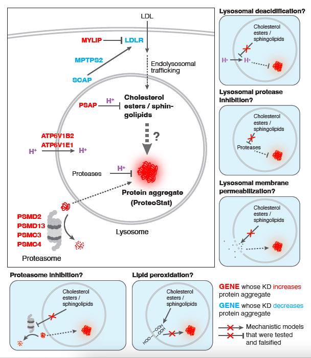
The presentation would be improved greatly if the authors provided diagrams illustrating the pathways implicated in their results, as well as their models.
We thank Reviewer #2 for the helpful suggestion. We have provided the suggested diagrams below (Author response image 2).
Author response image 2.
Mechanistic models linking screen hits to accrual of lysosomal protein aggregates, related to Figure 4. Perturbations that increased cholesterol and sphingolipid levels were evaluated for effects on lysosomal pH, lysosomal proteolytic capacity, lysosomal membrane permeability, lipid peroxidation and proteasome activity. None of these mechanisms appear to play a causal role in protein aggregation in response to elevated lipids.
Author Response References
-
Replogle, J. M. et al. Mapping information-rich genotype-phenotype landscapes with genome-scale Perturb-seq. Cell 185, 2559-2575.e28 (2022).
-
Berkers, C. R. et al. Probing the Specificity and Activity Profiles of the Proteasome Inhibitors Bortezomib and Delanzomib. Mol Pharmaceut 9, 1126–1135 (2012).
-
Berkers, C. R. et al. Profiling Proteasome Activity in Tissue with Fluorescent Probes. Mol. Pharmaceutics 4, 739–748 (2007).
-
David, D. C. et al. Widespread Protein Aggregation as an Inherent Part of Aging in C. elegans. Plos Biol 8, e1000450 (2010).


