Peer review process
Revised: This Reviewed Preprint has been revised by the authors in response to the previous round of peer review; the eLife assessment and the public reviews have been updated where necessary by the editors and peer reviewers.
Read more about eLife’s peer review process.Editors
- Reviewing EditorBrice BathellierCentre National de la Recherche Scientifique, Paris, France
- Senior EditorBarbara Shinn-CunninghamCarnegie Mellon University, Pittsburgh, United States of America
Reviewer #1 (Public Review):
The inferior colliculus (IC) is the central auditory system's major hub. It integrates ascending brainstem signals to provide acoustic information to the auditory thalamus. The superficial layers of the IC ("shell" IC regions as defined in the current manuscript) also receive a massive descending projection from the auditory cortex. This auditory cortico-collicular pathway has long fascinated the hearing field, as it may provide a route to funnel "high-level" cortical signals and impart behavioral salience upon an otherwise behaviorally agnostic midbrain circuit.
Accordingly, IC neurons can respond differently to the same sound depending on whether animals engage in a behavioral task (Ryan and Miller 1977; Ryan et al., 1984; Slee & David, 2015; Saderi et al., 2021; De Franceschi & Barkat, 2021). Many studies also report a rich variety of non-auditory responses in the IC, far beyond the simple acoustic responses one expects to find in a "low-level" region (Sakurai, 1990; Metzger et al., 2006; Porter et al., 2007). A tacit assumption is that the behaviorally relevant activity of IC neurons is inherited from the auditory cortico-collicular pathway. However, this assumption has never been tested, owing to two main limitations of past studies:
(1) Prior studies could not confirm if data were obtained from IC neurons that receive monosynaptic input from the auditory cortex.
(2) Many studies have tested how auditory cortical inactivation impacts IC neuron activity; the consequence of cortical silencing is sometimes quite modest. However, all prior inactivation studies were conducted in anesthetized or passively listening animals. These conditions may not fully engage the auditory cortico-collicular pathway. Moreover, the extent of cortical inactivation in prior studies was sometimes ambiguous, which complicates interpreting modest or negative results.
Here, the authors' goal is to directly test if the auditory cortex is necessary for behaviorally relevant activity in IC neurons. They conclude that surprisingly, task relevant activity in cortico-recipient IC neuron persists in absence of auditory cortico-collicular transmission. To this end, a major strength of the paper is that the authors combine a sound-detection behavior with clever approaches that unambiguously overcome the limitations of past studies.
First the authors inject a transsynaptic virus into the auditory cortex, thereby expressing a genetically encoded calcium indicator in the auditory cortex's postsynaptic targets in the IC. This powerful approach enables 2-photon Ca2+ imaging from IC neurons that unambiguously receive monosynaptic input from auditory cortex. Thus, any effect of cortical silencing should be maximally observable in this neuronal population. Second, they abrogate auditory cortico-collicular transmission using lesions of auditory cortex. This "sledgehammer" approach is arguably the most direct test of whether cortico-recipient IC neurons will continue to encode task-relevant information in absence of descending feedback. Indeed, their method circumvents the known limitations of more modern optogenetic or chemogenetic silencing, e.g. variable efficacy.
The authors have revised their manuscript and adequately addressed the major concerns. Although more in depth analyses of these rich datasets are definitely possible, the current results nevertheless stand on their own. Indeed, the work serves as a beacon to move away from the idea that cortico-collicular projections function primarily to impart behavioral relevance upon auditory midbrain neurons. This knowledge inspires a search for alternative explanations as to the role of auditory cortico-collicular synapses in behavior.
Reviewer #2 (Public Review):
Summary:
This study takes a new approach to studying the role of corticofugal projections from auditory cortex to inferior colliculus. The authors performed two-photon imaging of cortico-recipient IC neurons during a click detection task in mice with and without lesions of auditory cortex. In both groups of animals, they observed similar task performance and relatively small differences in the encoding of task-response variables in the IC population. They conclude that non-cortical inputs to the IC provide can substantial task-related modulation, at least when AC is absent.
Strengths:
This study provides valuable new insight into big and challenging questions around top-down modulation of activity in the IC. The approach here is novel and appears to have been executed thoughtfully. Thus, it should be of interest to the community.
Weaknesses:
There are however, substantial concerns about the interpretation of the findings and limitations to the current analysis. In particular, Analysis of single unit activity is absent, making interpretation of population clusters and decoding less interpretable. These concerns should be addressed to make sure that the results can be interpreted clearly in an active field that already contains a number of confusing and possibly contradictory findings.
Reviewer #3 (Public Review):
Summary:
This study aims to demonstrate that cortical feedback is not necessary to signal behavioral outcome to shell neurons of the inferior colliculus during a sound detection task. The demonstration is achieved in a very clear manner by the observation of the activity of cortico-recepient neurons in animals which have received lesions of the auditory cortex. The experiment shows that neither behavior performance nor neuronal responses are significantly impacted by cortical lesions except for the case of partial lesions which seem to have a disruptive effect on behavioral outcome signaling.
Strengths:
The demonstration of the main conclusions is based on state-of-the-art, carefully controlled methods and is highly convincing. There is an in depth discussion of the different effects of auditory cortical lesions on sound detection behavior.
Weaknesses:
The description of feedback signals could be more detailed although it is difficult to achieve good temporal resolution with the calcium imaging technique necessary for targeting cortico-recipient neurons.
Author response:
The following is the authors’ response to the original reviews.
Public Reviews:
We thank the reviewers for the detailed assessment of our work as well as their praise and constructive feedback which helped us to significantly improve our manuscript.
Reviewer #1 (Public Review):
The inferior colliculus (IC) is the central auditory system's major hub. It integrates ascending brainstem signals to provide acoustic information to the auditory thalamus. The superficial layers of the IC ("shell" IC regions as defined in the current manuscript) also receive a massive descending projection from the auditory cortex. This auditory cortico-collicular pathway has long fascinated the hearing field, as it may provide a route to funnel "high-level" cortical signals and impart behavioral salience upon an otherwise behaviorally agnostic midbrain circuit.
Accordingly, IC neurons can respond differently to the same sound depending on whether animals engage in a behavioral task (Ryan and Miller 1977; Ryan et al., 1984; Slee & David, 2015; Saderi et al., 2021; De Franceschi & Barkat, 2021). Many studies also report a rich variety of non-auditory responses in the IC, far beyond the simple acoustic responses one expects to find in a "low-level" region (Sakurai, 1990; Metzger et al., 2006; Porter et al., 2007). A tacit assumption is that the behaviorally relevant activity of IC neurons is inherited from the auditory cortico-collicular pathway. However, this assumption has never been tested, owing to two main limitations of past studies:
(1) Prior studies could not confirm if data were obtained from IC neurons that receive monosynaptic input from the auditory cortex.
(2) Many studies have tested how auditory cortical inactivation impacts IC neuron activity; the consequence of cortical silencing is sometimes quite modest. However, all prior inactivation studies were conducted in anesthetized or passively listening animals. These conditions may not fully engage the auditory cortico-collicular pathway. Moreover, the extent of cortical inactivation in prior studies was sometimes ambiguous, which complicates interpreting modest or negative results.
Here, the authors' goal is to directly test if auditory cortex is necessary for behaviorally relevant activity in IC neurons. They conclude that surprisingly, task relevant activity in cortico-recipient IC neuron persists in absence of auditory cortico-collicular transmission. To this end, a major strength of the paper is that the authors combine a sound-detection behavior with clever approaches that unambiguously overcome the limitations of past studies.
First, the authors inject a transsynaptic virus into the auditory cortex, thereby expressing a genetically encoded calcium indicator in the auditory cortex's postsynaptic targets in the IC. This powerful approach enables 2-photon Ca2+ imaging from IC neurons that unambiguously receive monosynaptic input from auditory cortex. Thus, any effect of cortical silencing should be maximally observable in this neuronal population. Second, they abrogate auditory cortico-collicular transmission using lesions of auditory cortex. This "sledgehammer" approach is arguably the most direct test of whether cortico-recipient IC neurons will continue to encode task-relevant information in absence of descending feedback. Indeed, their method circumvents the known limitations of more modern optogenetic or chemogenetic silencing, e.g. variable efficacy.
I also see three weaknesses which limit what we can learn from the authors' hard work, at least in the current form. I want to emphasize that these issues do not reflect any fatal flaw of the approach. Rather, I believe that their datasets likely contain the treasure-trove of knowledge required to completely support their claims.
(1) The conclusion of this paper requires the following assumption to be true: That the difference in neural activity between Hit and Miss trials reflects "information beyond the physical attributes of sound." The data presentation complicates asserting this assumption. Specifically, they average fluorescence transients of all Hit and all Miss trials in their detection task. Yet, Figure 3B shows that mice's d' depends on sound level, and since this is a detection task the smaller d' at low SPLs presumably reflects lower Hit rates (and thus higher Miss rates). As currently written, it is not clear if fluorescence traces for Hits arise from trials where the sound cue was played at a higher sound level than on Miss trials. Thus, the difference in neural activity on Hit and Miss trials could indeed reflect mice's behavior (licking or not licking). But in principle could also be explained by higher sound-evoked spike rates on Hit compared to Miss trials, simply due to louder click sounds. Indeed, the amplitude and decay tau of their indicator GCaMP6f is non-linearly dependent on the number and rate of spikes (Chen et al., 2013), so this isn't an unreasonable concern.
(2) The authors' central claim effectively rests upon two analyses in Figures 5 and 6. The spectral clustering algorithm of Figure 5 identifies 10 separate activity patterns in IC neurons of control and lesioned mice; most of these clusters show distinct activity on averaged Hit and Miss trials. They conclude that although the proportions of neurons from control and lesioned mice in certain clusters deviates from an expected 50/50 split, neurons from lesioned mice are still represented in all clusters. A significant issue here is that in addition to averaging all Hits and Miss trials together, the data from control and lesioned mice are lumped for the clustering. There is no direct comparison of neural activity between the two groups, so the reader must rely on interpreting a row of pie charts to assess the conclusion. It's unclear how similar task relevant activity is between control and lesioned mice; we don't even have a ballpark estimate of how auditory cortex does or does not contribute to task relevant activity. Although ideally the authors would have approached this by repeatedly imaging the same IC neurons before and after lesioning auditory cortex, this within-subjects design may be unfeasible if lesions interfere with task retention. Nevertheless, they have recordings from hundreds to thousands of neurons across two groups, so even a small effect should be observable in a between-groups comparison.
(3) In Figure 6, the authors show that logistic regression models predict whether the trial is a Hit or Miss from their fluorescence data. Classification accuracy peaks rapidly following sound presentation, implying substantial information regarding mice's actions. The authors further show that classification accuracy is reduced, but still above chance in mice with auditory cortical lesions. The authors conclude from this analysis task relevant activity persists in absence of auditory cortex. In principle I do not disagree with their conclusion.
The weakness here is in the details. First, the reduction in classification accuracy of lesioned mice suggests that auditory cortex does nevertheless transmit some task relevant information, however minor it may be. I feel that as written, their narrative does not adequately highlight this finding. Rather one could argue that their results suggest redundant sources of task-relevant activity converging in the IC. Secondly, the authors conclude that decoding accuracy is impaired more in partially compared to fully lesioned mice. They admit that this conclusion is at face value counterintuitive, and provide compelling mechanistic arguments in the Discussion. However, aside from shaded 95% CIs, we have no estimate of variance in decoding accuracy across sessions or subjects for either control or lesioned mice. Thus we don't know if the small sample sizes of partial (n = 3) and full lesion (n = 4) groups adequately sample from the underlying population. Their result of Figure 6B may reflect spurious sampling from tail ends of the distributions, rather than a true non-monotonic effect of lesion size on task relevant activity in IC.
Our responses to the ‘recommendations for the authors’ below lay out in detail how we addressed each comment and concern. Besides filling in key information about how our original analysis aimed at minimizing any potential impact of differences in sound level distributions - namely that trials used for decoding were limited to a subset of sound levels - and which was accidentally omitted in the original manuscript, we have now carried out several additional analyses.
We would like to highlight one of these because it supplements both the clustering and decoding analysis that we conducted to compare hit and miss trial activity, and directly addresses what the reviewer identified as our work’s main weakness (a possible confound between animal behavior and sound level distributions) and the request for an analysis that operates at the level of single units rather than the population level. Specifically, we assessed, separately for each recorded neuron, whether there was a statistically significant difference in the magnitude of neural activity between hit and miss trials. This approach allowed us to fully balance the numbers of hit and miss trials at each sound level that were entered into the analysis. The results revealed that a large proportion (close to 50%) of units were task modulated, i.e. had significantly different response magnitudes between hit and miss trials, and that this proportion was not significantly different between lesioned and non-lesioned mice. We hope that this, together with the rest of our responses, convincingly demonstrates that the shell of the IC encodes mouse sound detection behavior even when top-down input from the auditory cortex is absent.
Reviewer #2 (Public Review):
Summary:
This study takes a new approach to studying the role of corticofugal projections from auditory cortex to inferior colliculus. The authors performed two-photon imaging of cortico-recipient IC neurons during a click detection task in mice with and without lesions of auditory cortex. In both groups of animals, they observed similar task performance and relatively small differences in the encoding of task-response variables in the IC population. They conclude that non-cortical inputs to the IC provide can substantial task-related modulation, at least when AC is absent. Strengths:
This study provides valuable new insight into big and challenging questions around top-down modulation of activity in the IC. The approach here is novel and appears to have been executed thoughtfully. Thus, it should be of interest to the community.
Weaknesses: There are, however, substantial concerns about the interpretation of the findings and limitations to the current analysis. In particular, Analysis of single unit activity is absent, making interpretation of population clusters and decoding less interpretable. These concerns should be addressed to make sure that the results can be interpreted clearly in an active field that already contains a number of confusing and possibly contradictory findings.
Our responses to the ‘recommendations for the authors’ below lay out in detail how we addressed each comment and concern. Several additional analyses have now been carried out including ones that operate at the level of single units rather than the population level, as requested by the reviewer. We would like to briefly highlight one here because it supplements both the clustering and decoding analysis that we conducted to compare hit and miss trial activity and directly addresses what the other reviewers identified as our work’s main weakness (a possible confound between animal behavior and sound level distributions). Specifically, we assessed, separately for each recorded neuron, whether there was a statistically significant difference in the magnitude of neural activity between hit and miss trials. This approach allowed us to fully balance the numbers of hit and miss trials at each sound level that were entered into the analysis. The results revealed that a large proportion (close to 50%) of units were task modulated, i.e. had significantly different response magnitudes between hit and miss trials, and that this proportion was not significantly different between lesioned and non-lesioned mice. We hope that this, together with the rest of our responses, convincingly demonstrates that the shell of the IC encodes mouse sound detection behavior even when top-down input from the auditory cortex is absent.
Reviewer #3 (Public Review):
Summary:
This study aims to demonstrate that cortical feedback is not necessary to signal behavioral outcome to shell neurons of the inferior colliculus during a sound detection task. The demonstration is achieved by the observation of the activity of cortico-recipient neurons in animals which have received lesions of the auditory cortex. The experiment shows that neither behavior performance nor neuronal responses are significantly impacted by cortical lesions except for the case of partial lesions which seem to have a disruptive effect on behavioral outcome signaling. Strengths:
The experimental procedure is based on state of the art methods. There is an in depth discussion of the different effects of auditory cortical lesions on sound detection behavior. Weaknesses:
The analysis is not documented enough to be correctly evaluated. Have the authors pooled together trials with different sound levels for the key hit vs miss decoding/clustering analysis? If so, the conclusions are not well supported, as there are more misses for low sound levels, which would completely bias the outcome of the analysis. It would possible that the classification of hit versus misses actually only reflects a decoding of sound level based on sensory responses in the colliculus, and it would not be surprising then that in the presence or absence of cortical feedback, some neurons responds more to higher sound levels (hits) and less to lower sound levels (misses). It is important that the authors clarify and in any case perform an analysis in which the classification of hits vs misses is done only for the same sound levels. The description of feedback signals could be more detailed although it is difficult to achieve good temporal resolution with the calcium imaging technique necessary for targeting cortico-recipient neurons.
Our responses to the ‘recommendations for the authors’ below lay out in detail how we addressed each comment and concern. Besides filling in key information about how our original analysis aimed at minimizing any potential impact of differences in sound level distributions - namely that trials used for decoding were limited to a subset of sound levels - and which was accidentally omitted in the original manuscript, we have now carried out several additional analyses to directly address what the reviewer identified as our work’s main weakness (a possible confound between animal behavior and sound level distributions). This includes an analysis in which we were able to demonstrate for one imaging session with a sufficiently large number of trials that limiting the trials entered into the decoding analysis to those from a single sound level did not meaningfully impact decoding accuracy. We would like to highlight another new analysis here because it supplements both the clustering and decoding analyses that we conducted to compare hit and miss trial activity and addresses the other reviewers’ request for an analysis that operates at the level of single units rather than the population level. Specifically, we assessed, separately for each recorded neuron, whether there was a statistically significant difference in the magnitude of neural activity between hit and miss trials. This approach allowed us to fully balance the numbers of hit and miss trials at each sound level that were entered into the analysis. The results revealed that a large proportion (close to 50%) of units were task modulated, i.e. had significantly different response magnitudes between hit and miss trials, and that this proportion was not significantly different between lesioned and non-lesioned mice. We hope that this, together with the rest of our responses, convincingly demonstrates that the shell of the IC encodes mouse sound detection behavior even when top-down input from the auditory cortex is absent.
Reviewer #1 (Recommendations For The Authors):
Thank you for the opportunity to read your paper. I think the conclusion is exciting. Indeed, you indicate that perhaps contrary to many of our (untested) assumptions, task-relevant activity in the IC may persist in absence of auditory cortex.
As mentioned in my public review: Despite my interest in the work, I also think that there are several opportunities to significantly strengthen your conclusions. I feel this point is important because your work will likely guide the efforts of future students and post-docs working on this topic. The data can serve as a beacon to move the field away from the (somewhat naïve) idea that the evolved forebrain imparts behavioral relevance upon an otherwise uncivilized midbrain. This knowledge will inspire a search for alternative explanations. Indeed, although you don't highlight it in your narrative, your results dovetail nicely with several studies showing task-relevant activity in more ventral midbrain areas that project to the IC (e.g., pedunculopontine nuclei; see work from Hikosaka in monkeys, and more recently in mice from Karel Svoboda's lab).
Thanks for the kind words.
These studies, in particular the work by Inagaki et al. (2022) outlining how the transformation of an auditory go signal into movement could be mediated via a circuit involving the PPN/MRN (which might rely on the NLL for auditory input) and the motor thalamus, are indeed highly relevant.
We made the following changes to the manuscript text.
Line 472:”...or that the auditory midbrain, thalamus and cortex are bypassed entirely if simple acousticomotor transformations, such as licking a spout in response to a sound, are handled by circuits linking the auditory brainstem and motor thalamus via pedunculopontine and midbrain reticular nuclei (Inagaki et al., 2022).”
The beauty of the eLife experiment is that you are free to incorporate or ignore these suggestions. After all, it's your paper, not mine. Nevertheless, I hope you find my comments useful.
First, a few suggestions to address my three comments in the public review.Suggestion for public comment #1: An easy way to address this issue is to average the neural activity separately for each trial outcome at each sound level. That way you can measure if fluorescence amplitude (or integral) varies as a function of mice's action rather than sound level. This approach to data organization would also open the door to the additional analyses for addressing comment #2, such as directly comparing auditory and putatively non-auditory activity in neurons recorded from control and lesioned mice.
We have carried out additional analyses for distinguishing between the two alternative explanations of the data put forward by the reviewer: That the difference in neural activity between hit and miss trials reflects a) behavior or b) sound level (more precisely: differences in response magnitude arising from a higher proportion of high-sound-level trials in the hit trial group than in the miss trial group). If the data favored b), we would expect no difference in activity between hit and miss trials when plotted separately for each sound level. The new Figure 4 - figure supplement 1 indicates that this is not the case. Hit and miss trial activity are clearly distinct even when plotted separately for different sound levels, confirming that this difference in activity reflects the animals’ behavior rather than sensory information.
Changes to manuscript.
Line 214: “While averaging across all neurons cannot capture the diversity of responses, the averaged response profiles suggest that it is mostly trial outcome rather than the acoustic stimulus and neuronal sensitivity to sound level that shapes those responses (Figure 4 – figure supplement 1).”
Additionally, we assessed for each neuron separately whether there was a significant difference between hit and miss trial activity and therefore whether the activity of the neuron could be considered “task-modulated”. To achieve this, we used equal numbers of hit and miss trials at each sound level to ensure balanced sound level distributions and thus rule out any potential confound between sound level distributions and trial outcome. This analysis revealed that the proportion of task-modulated neurons was very high (close to 50%) and not significantly different between lesioned and non-lesioned mice (Figure 6 - figure supplement 3).
Changes to the manuscript.
Line 217: “Indeed, close to half (1272 / 2649) of all neurons showed a statistically significant difference in response magnitude between hit and miss trials…”
Line 307: “Although the proportion of individual neurons with distinct response magnitudes in hit and miss trials in lesioned mice did not differ from that in non-lesioned mice, it was significantly lower when separating out mice with partial lesions (Figure 6 – figure supplement 3).”
Differences in the distributions of sound levels in the different trial types could also potentially confound the decoding into hit and miss trials. Our original analysis was actually designed to take this into account but, unfortunately, we failed to include sufficient details in the methods section.
Changes to the manuscript.
Line 710: “Rather than including all the trials in a given session, only trials of intermediate difficulty were used for the decoding analysis. More specifically, we only included trials across five sound levels, comprising the lowest sound level that exceeded a d’ of 1.5 plus the two sound levels below and above that level. That ensured that differences in sound level distributions would be small, while still giving us a sufficient number of trials to perform the decoding analysis.“
In this context, it is worth bearing in mind that a) the decoding analysis was done on a frame-byframe basis, meaning that the decoding score achieved early in the trial has no impact on the decoding score at later time points in the trial, b) sound-driven activity predominantly occurs immediately after stimulus onset and is largely over about 1 s into the trial (see cluster 3, for instance, or average miss trial activity in Figure 4 – figure supplement 1), c) decoding performance of the behavioral outcome starts to plateau 500-1000 ms into the trial and remains high until it very gradually begins to decline after about 2 s into the trial. In other words, decoding performance remains high far longer than the stimulus would be expected to have an impact on the neurons’ activity. Therefore, we would expect any residual bias due to differences in the sound level distribution that our approach did not control for to be restricted to the very beginning of the trial and not to meaningfully impact the conclusions derived from the decoding analysis.
Finally, we carried out an additional decoding analysis for one imaging session in which we had a sufficient number of trials to perform the analysis not only over the five (59, 62, 65, 68, 71 dB SPL) original sound levels, but also over a reduced range of three (62, 65, 68 dB SPL) sound levels, as well as a single (65 dB SPL) sound level (Figure 6 - figure supplement 1). The mean sound level differences between the hit trial distributions and miss trial distributions for these three conditions were 3.08, 1.01 and 0 dB, respectively. This analysis suggests that decoding performance is not meaningfully impacted by changing the range of sound levels (and sound level distributions), other than that including fewer sound levels means fewer trials and thus noisier decoding.
Changes to manuscript.
Line 287: ”...and was not meaningfully affected by differences in sound level distributions between hit and miss trials (Figure 6 – figure supplement 1).”
Suggestion for public comment #2: Perhaps a solution would be to display example neuron activity in each cluster, recorded in control and lesioned mice. The reader could then visually compare example data from the two groups, and immediately grasp the conclusion that task relevant activity remains in absence of auditory cortex. Additionally, one possibility might be to calculate the difference in neural activity between Hit and Miss trials for each task-modulated neuron. Then, you could compare these values for neurons recorded in control and lesion mice. I feel like this information would greatly add to our understanding of cortico-collicular processing.
I would also argue that it's perhaps more informative to show one (or a few) example recordings rather than averaging across all cells in a cluster. Example cells would give the reader a better handle on the quality of the imaging, and this approach is more standard in the field. Finally, it would be useful to show the y axis calibration for each example trace (e.g. Figure 5 supp 1). That is also pretty standard so we can immediately grasp the magnitude of the recorded signal.
We agree that while the information we provided shows that neurons from lesioned and nonlesioned groups are roughly equally represented across the clusters, it does not allow the reader to appreciate how similar the activity profiles of neurons are from each of the two groups. However, picking examples can be highly subjective and thus potentially open to bias. We therefore opted instead to display, separately for lesioned and non-lesioned mice, the peristimulus time histograms of all neurons in each cluster, as well as the cluster averages of the response profiles (Figure 5 - figure supplement 3). This, we believe, convincingly illustrates the close correspondence between neural activity in lesioned and non-lesioned mice across different clusters. All our existing and new figures indicate the response magnitude either on the figures’ y-axis or via scale/color bars.
Changes to manuscript.
Line 254: “Furthermore, there was a close correspondence between the cluster averages of lesioned and non-lesioned mice (Figure 5 – figure supplement 3).”
Furthermore, we’ve now included a video of the imaging data which, we believe, gives the reader a much better handle on the data quality than further example response profiles would.
Changes to manuscript.
Line 197: ”...using two-photon microscopy (Figure 4B, Video 1).”
Suggestion for public comment #3: In absence of laborious and costly follow-up experiments to boost the sample size of partial and complete lesion groups, it may be more prudent to simply tone down the claims that lesion size differentially impacts decoding accuracy. The results of this analysis are not necessary for your main claims.
Our new results on the proportions of ‘task-modulated’ neurons (Figure 6 - figure supplement 3) across different experimental groups show that there is no difference between non-lesioned and lesioned mice as a whole, but mice with partial lesions have a smaller proportion of taskmodulated neurons than the other two groups. While this corroborates the results of the decoding analysis, we certainly agree that the small sample size is a caveat that needs to be acknowledged.
Changes to manuscript.
Line 477: ”Some differences were observed for mice with only partial lesions of the auditory cortex.
Those mice had a lower proportion of neurons with distinct response magnitudes in hit and miss trials than mice with (near-)complete lesions. Furthermore, trial outcomes could be read out with lower accuracy from these mice. While this finding is somewhat counterintuitive and is based on only three mice with partial lesions, it has been observed before that smaller lesions…”
A few more suggestions unrelated to public review:
Figure 1: This is somewhat of an oddball in this manuscript, and its inclusion is not necessary for the main point. Indeed, the major conclusion of Fig 1 is that acute silencing of auditory cortex impairs task performance, and thus optogenetic methods are not suitable to test your hypothesis. However, this conclusion is also easily supported from decades of prior work, and thus citations might suffice.
We do not agree that these data can easily be substituted with citations of prior published work. While previous studies (Talwar et al., 2001, Li et al., 2017) have demonstrated the impact of acute pharmacological silencing on sound detection in rodents, pharmacological and optogenetic silencing are not equivalent. Furthermore, we are aware of only one published study (Kato et al., 2015) that investigated the impact of optogenetically perturbing auditory cortex on sound detection (others have investigated its impact on discrimination tasks). Kato et al. (2015) examined the effect of acute optogenetic silencing of auditory cortex on the ability of mice to detect the offsets of very long (5-9 seconds) sounds, which is not easily comparable to the click detection task employed by us. Furthermore, when presenting our work at a recent meeting and leaving out the optogenetics results due to time constraints, audience members immediately enquired whether we had tried an optogenetic manipulation instead of lesions. Therefore, we believe that these data represent a valuable piece of information that will be appreciated by many readers and have decided not to remove them from the manuscript.
A worst case scenario is that Figure 1 will detract from the reader's assessment of experimental rigor. The data of 1C are pooled from multiple sessions in three mice. It is not clear if the signed-rank test compares performance across n = 3 mice or n = 13 sessions. If the latter, a stats nitpicker could argue that the significance might not hold up with a nested analysis considering that some datapoints are not independent of one another. Finally, the experiment does not include a control group, gad2-cre mice injected with a EYFP virus. So as presented, the data are equally compatible with the pessimistic conclusion that shining light into the brain impairs mice's licking. My suggestion is to simply remove Figure 1 from the paper. Starting off with Figure 3 would be stronger, as the rest of the study hinges upon the knowledge that control and lesion mice's behavior is similar.
Instead of reporting the results session-wise and doing stats on the d’ values, we now report results per mouse and perform stats on the proportions of hits and false alarms separately for each mouse. The results are statistically significant for each mouse and suggest that the differences in d’ are primarily caused by higher false alarm rates during the optogenetic perturbation than in the control condition.
Changes to manuscript.
New Figure 1.
We agree that including control mice not expressing ChR2 would be important for fully characterizing the optogenetic manipulation and that the lack of this control group should be acknowledged. However, in the context of this study, the outcome of performing this additional experiment would be inconsequential. We originally considered using an optogenetic approach to explore the contribution of cortical activity to IC responses, but found that this altered the animals’ sound detection behavior. Whether that change in behavior is due to activation of the opsin or simply due to light being shone on the brain has no bearing on the conclusion that this type of manipulation is unsuitable for determining whether auditory cortex is required for the choice-related activity that we recorded in the IC.
Changes to manuscript.
Line 106: ”Although a control group in which the auditory cortex was injected with an EYFP virus lacking ChR2 would be required to confirm that the altered behavior results from an opsindependent perturbation of cortical activity, this result shows that this manipulation is also unsuitable… ”
Figure 2, comment #1: The micrograph of panel B shows the densest fluorescence in the central IC. You interpret this as evidence of retrograde labeling of central IC neurons that project to the shell IC. This is a nice finding, but perhaps a more relevant micrograph would be to show the actual injection site in the shell layers. The rest of Figure 2 documents the non-auditory cortical sources of forebrain feedback. Since non-auditory cortical neurons may or may not target distinct shell IC sub-circuits, it's important to know where the retrograde virus was injected. Stylistic comment: The flow of the panels is somewhat unorthodox. Panel A and B follow horizontally, then C and D follow vertically, followed by E-H in a separate column. Consider sequencing either horizontally or vertically to maximize the reader's experience.
Figure 2, comment # 2: It would also be useful to show more rostral sections from these mice, perhaps as a figure supplement, if you have the data. I think there is a lot of value here given a recent paper (Olthof et al., 2019 Jneuro) arguing that the IC receives corticofugal input from areas more rostral to the auditory cortex. So it would be beneficial for the field to know if these other cortical sources do or do not represent likely candidates for behavioral modulation in absence of auditory cortex.
Figure 2, comment #3: You have a striking cluster of retrogradely labeled PPC neurons, and I'm not sure PPC has been consistently reported as targeting the IC. It would be good to confirm that this is a "true" IC projection as opposed to viral leakage into the SC. Indeed, Figure 2, supplement 2 also shows some visual cortex neurons that are retrogradely labeled. This has bearing on the interpretations, because choice-related activity is rampant in PPC, and thus could be a potential source of the task relevant activity that persists in your recordings. This could be addressed as the point above, by showing the SC sections from these same mice.
All IC injections were made under visual guidance with the surface of the IC and adjacent brain areas fully exposed after removal of the imaging window. Targeting the IC and steering clear of surrounding structures, including the SC, was therefore relatively straightforward.
We typically observed strong retrograde labeling in the central nucleus after viral injections into the dorsal IC and, given the moderate injection volume (~50 nL at each of up to three sites), it was also typical to see spatially fairly confined labeling at the injection sites. For the mouse shown in Figure 2, we do not have further images of the IC. This was one of the earliest mice to be included in the study and we did not have access to an automatic slide scanner at the time. We had to acquire confocal images in a ‘manual’ and very time-consuming manner and therefore did not take further IC images for this mouse. We have now included, however, a set of images spanning the whole IC and the adjacent SC sections for the mouse for which we already show sections in Figure 2 - figure supplement 2. These were added as Figure 2 - figure supplement 3A to the manuscript. These images show that the injections were located in the caudal half of the IC and that there was no spillover into the SC - close inspection of those sections did not reveal any labeled cell bodies in the SC. Furthermore, we include as Figure 2 - figure supplement 3B a dozen additional rostral cortical sections of the same mouse illustrating corticocollicular neurons in regions spanning visual, parietal, somatosensory and motor cortex. Given the inclusion of the IC micrographs in the new supplementary figure, we removed panel B from Figure 2. This should also make it easier for the reader to follow the sequencing of the remaining panels.
Changes to manuscript.
New Figure 2 - figure supplement 3.
Line 159: “After the experiments, we injected a retrogradely-transported viral tracer (rAAV2-retrotdTomato) into the right IC to determine whether any corticocollicular neurons remained after the auditory cortex lesions (Figure 2, Figure 2 – figure supplement 2, Figure 2 – figure supplement 3). The presence of retrogradely-labeled corticocollicular neurons in non-temporal cortical areas (Figure 2) was not the result of viral leakage from the dorsal IC injection sites into the superior colliculus (Figure 2 – figure supplement 3).”
Line 495: “...projections to the IC, such as those originating from somatosensory cortical areas (Lohse et al., 2021; Lesicko et al., 2016) and parietal cortex may have contributed to the response profiles that we observed.
Figure 5 (see also public review point #2): I am not convinced that this unsupervised method yields particularly meaningful clusters; a grain of salt should be provided to the reader. For example, Clusters 2, 5, 6, and 7 contain neurons that pretty clearly respond with either short latency excitation or inhibition following the click sound on Hits. I would argue that neurons with such diametrically opposite responses should not be "classified" together. You can see the same issue in some of Namboodiri/Stuber's clustering (their Figure 1). It might be useful to make it clear to the reader that these clusters can reflect idiosyncrasies of the algorithm, the behavior task structure, or both.
We agree.
Changes to manuscript.
Line 666: “While clustering is a useful approach for organizing and visualizing the activity of large and heterogeneous populations of neurons, we need to be mindful that, given continuous distributions of response properties, the locations of cluster boundaries can be somewhat arbitrary and/or reflect idiosyncrasies of the chosen method and thus vary from one algorithm to another. We employed an approach very similar to that described in Namboodiri et al. (2019) because it is thought to produce stable results in high-dimensional neural data (Hirokawa et al. 2019).”
Methods:
How was a "false alarm" defined? Is it any lick happening during the entire catch trial, or only during the time period corresponding to the response window on stimulus trials?
The response window was identical for catch and stimulus trials and a false alarm was defined as licking during the response window of a catch trial.
Changes to manuscript.
Line 598: “During catch trials, neither licking (‘false alarm’) during the 1.5-second response window …”
L597 and so forth: What's the denominator in the conversion from the raw fluorescence traces into DF/F? Did you take the median or mode fluorescence across a chunk of time? Baseline subtract average fluorescence prior to click onset? Similarly, please provide some more clarification as to how neuropil subtraction was achieved. This information will help us understand how the classifier can decode trial outcome from data prior to sound onset.
Signal processing did not involve the subtraction of a pre-stimulus period.
Changes to manuscript.
Line 629: ”Neuropil extraction was performed using default suite2p parameters (https://suite2p.readthedocs.io/en/latest/settings.html), neuropil correction was done using a coefficient of 0.7, and calcium ΔF/F signals were obtained by using the median over the entire fluorescence trace as F0. To remove slow fluctuations in the signal, a baseline of each neuron’s entire trace was calculated by Gaussian filtering in addition to minimum and maximum filtering using default suite2p parameters. This baseline was then subtracted from the signal.”
Was the experimenter blinded to the treatment group during the behavior experiments? If not, were there issues that precluded blinding (limited staffing owing to lab capacity restrictions during the pandemic)? This is important to clarify for the sake of rigor and reproducibility.
Changes to manuscript.
Line 574: “The experimenters were not blinded to the treatment group, i.e. lesioned or non-lesioned, but they were blind to the lesion size both during the behavior experiments and most of the data processing.”
Minor:
L127-128: "In order to test...lesioned the auditory cortex bilaterally in 7 out of 16 animals". I would clarify this by changing the word animals to "mice" and 7 out of 16 by stating n = 9 and n = 7 are control and lesion groups, respectively.
Agreed.
Changes to manuscript.
Line 129: “...compared the performance of mice with bilateral lesions of the auditory cortex (n = 7) with non-lesioned controls (n = 9)”
L225-226: You rule out self-generated sounds as a likely source of behavioral modulation by citing Nate Sawtell's paper in the DCN. However, Stephen David's lab suggested that in marmosets, post sound activity in central IC may in fact reflect self-generated sounds during licking. I suggest addressing this with a nod to SVD's work (Singla et al., 2017; but see Shaheen et al., 2021).
Agreed.
Changes to manuscript.
Line 243: “(Singla et al., 2017; but see Shaheen et al., 2021)”
Line 238 - 239: You state that proportions only deviate greater than 10% for one of the four statistically significant clusters. Something must be unclear here because I don't understand: The delta between the groups in the significant clusters of Fig 5C is (from left to right) 20%, 20%, 38%, and 12%. Please clarify.
Our wording was meant to convey that a deviation “from a 50/50 split” of 10% means that each side deviates from 50 by 10% resulting in a 40/60 (or 60/40) split. We agree that that has the potential to confuse readers and is not as clear as it could be and have therefore dropped the ambiguous wording.
Changes to manuscript.
Line 253: ”,..the difference between the groups was greater than 20% for only one of them.”
L445: I looked at the cited Allen experiment; I'd be cautious with the interpretation here. A monosynaptic IC->striatum projection is news to me. I think Allen Institute used an AAV1-EGFP virus for these experiments, no? As you know, AAV1 is quite transsynaptic. The labeled fibers in striatum of that experiment may reflect disynaptic labeling of MGB neurons (which do project to striatum).
Agreed. We deleted the reference to this Allen experiment.
L650: Please define "network activity". Is this the fluorescence value for each ROI on each frame of each trial? Averaged fluorescence of each ROI per frame? Total frame fluorescence including neuropil? Depending on who you ask, each of these measures provides some meaningful readout of network activity, so clarification would be useful.
Changes to manuscript.
Line 707: “Logistic regression models were trained on the network activity of each session, i.e., the ΔF/F values of all ROIs in each session, to classify hit vs miss trials. This was done on a frame-by-frame basis, meaning that each time point (frame) of each session was trained separately.
Figure 3 narrative or legend: Listing the F values for the anova would be useful. There is pretty clearly a main effect of training session for hits, but what about for the false alarms? That information is important to solidify the result, and would help more specialized readers interpret the d-prime plot in this figure.
Agreed. There were significant main effects of training day for both hit rates and false alarm rates (as well as d’).
Changes to manuscript.
Line 165: “The ability of the mice to learn and perform the click detection task was evident in increasing hit rates and decreasing false alarm rates across training days (Figure 3A, p < 0.01, mixed-design ANOVAs).”
In summary, thank you for undertaking this work. Your conclusions are provocative, and thus will likely influence the field's direction for years to come.
Thank you for those kind words and valuable and constructive feedback, which has certainly improved the manuscript.
Reviewer #2 (Recommendations For The Authors):
MAJOR CONCERNS
(1) (Fig. 5) What fraction of individual neurons actually encode task-related information in each animal group? How many neurons respond to sound? The clustering and decoding analyses are interesting, but they obscure these simple questions, which get more directly at the main questions of the study. Suggested approach: For a direct comparison of AC-lesioned and -non-lesioned animals, why not simply compare the mean difference between PSTH response for each neuron individually? To test for trial outcome effects, compare Hit and Miss trials (same stimulus, different behavior) and for sound response effects, compare Hit and False alarm trials (same behavior, different response). How do you align for time in the latter case when there's no stimulus? Align to the first lick event. The authors should include this analysis or explain why their approach of jumping right to analysis of clusters is justified.
We have now calculated the fraction of neurons that encode trial outcome by comparing hit and miss trial activity. That fraction does not differ between non-lesioned animals and lesioned animals as a whole, but is significantly smaller in mice with partial lesions. The author’s suggestion of comparing hit and false alarm trial activity to assess sound responsiveness is problematic because hit trials involve reward delivery and consumption. Consequently, they are behaviorally very different from false alarm trials (not least because hit trials tend to contain much more licking). Therefore, we calculated the fraction of neurons that respond to the acoustic stimulus by comparing activity before and after stimulus onset in miss trials. We found no significant difference between the non-lesioned and lesioned mice or between subgroups.
We have addressed these points with the following changes to the manuscript:
Line 217: “Indeed, close to half (1272 / 2649) of all neurons showed a statistically significant difference in response magnitude between hit and miss trials, while only a small fraction (97 / 2649) exhibited a significant response to the sound.”
Line 307: “Although the proportion of individual neurons with distinct response magnitudes in hit and miss trials in lesioned mice did not differ from that in non-lesioned mice, it was significantly lower when separating out mice with partial lesions (Figure 6 – figure supplement 3).”
Line 648: “Analysis of task-modulated and sound-driven neurons. To identify individual neurons that produced significantly different response magnitudes in hit and miss trials, we calculated the mean activity for each stimulus trial by taking the mean activity over the 5 seconds following stimulus presentation and subtracting the mean activity over the 2 seconds preceding the stimulus during that same trial. A Mann-Whitney U test was then performed to assess whether a neuron showed a statistically significant difference (Benjamini-Hochberg adjusted p-value of 0.05) in response magnitude between hit and miss trials. The analysis was performed using equal numbers of hit and miss trials at each sound level to ensure balanced sound level distributions. If, for a given sound level, there were more hit than miss trials, we randomly selected a sample of hit trials (without substitution) to match the sample size for the miss trials and vice versa. Sounddriven neurons were identified by comparing the mean miss trial activity before and after stimulus presentation. Specifically, we performed a Mann-Whitney U test to assess whether there was a statistically significant difference (Benjamini-Hochberg adjusted p-value of 0.05) between the mean activity over the 2 seconds preceding the stimulus and the mean activity over the 1 second period following stimulus presentation.”
Some more specific concerns about focusing only on cluster-level and population decoding analysis are included below.
(2) (L 234) "larger field of view". Do task-related or lesion-dependent effects depend on the subregion of IC imaged? Some anatomists would argue that the IC shell is not a uniform structure, and concomitantly, task-related effects may differ between fields. Did coverage of IC subregions differ between experimental groups? Is there any difference in task related effects between subregions of IC? Or maybe all this work was carried out only in the dorsal area? The differences between lesioned and non-lesioned animals are relatively small, so this may not have a huge impact, but a more nuanced discussion that accounts for observed or potential (if not tested) differences between regions of the IC.
The specific subregion coverage could also impact the decoding analysis (Fig 6), and if possible it might be worth considering an interaction between field of view and lesion size on decoding.
Each day we chose a new imaging location to avoid recording the same neurons more than once and aimed to sample widely across the optically accessible surface of the IC. We typically stopped the experiment only when there were no more new areas to record from. In terms of the depth of the imaged neurons, we were limited by the fact that corticorecipient neurons become sparser with depth and that the signal available from the GCaMP6f labeling of the Ai95 mice becomes rapidly weaker with increasing distance from the surface. This meant that we recorded no deeper than 150 µm from the surface of the IC. Consequently, while there may have been some variability in the average rostrocaudal and mediolateral positioning of imaging locations from animal to animal due to differences between mice in how much of the IC surface was visible, cranial window positioning, and in neuronal labeling etc, our dataset is anatomically uniform in that all recorded neurons receive input from the auditory cortex and are located within 150 µm of the surface of the IC. Therefore, we think it highly unlikely that small sampling differences across animals could have a meaningful impact on the results.
Given that there is no consensus as to where the border between the dorsal and external/lateral cortices of the IC is located and that it is typically difficult to find reliable anatomical reference points (the location of the borders between the IC and surrounding structures is not always obvious during imaging, i.e. a transition from a labeled area to a dark area near the edge of the cranial window could indicate a border with another structure, but also the IC surface sloping away from the window or simply an unlabeled area within the IC), we made no attempt to assign our recordings from corticorecipient neurons to specific subdivisions of the IC.
Changes to manuscript.
Line 195: “We then proceeded to record the activity of corticorecipient neurons within about 150 µm of the dorsal surface of the IC using two-photon microscopy (Figure 4B, Video 1).”
Line 375: “We imaged across the optically accessible dorsal surface of the IC down to a depth of about 150 µm below the surface. Consequently, the neurons we recorded were located predominantly in the dorsal cortex. However, identifying the borders between different subdivisions of the IC is not straightforward and we cannot rule out the possibility that some were located in the lateral cortex.”
(3) (L 482-483) "auditory cortex is not required for the task-related activity recording in IC neurons of mice performing a sound detection task". Most places in the text are clearer, but this statement is confusing. Yes, animals with lesions can have a "normal"-looking IC, but does that mean that AC does not strongly modulate IC during this behavior in normal animals? The authors have shown convincingly that subcortical areas can both shape behavior and modulate IC normally, but AC may still be required for IC modulation in non-lesioned animals. Given the complexity of this system, the authors should make sure they summarize their results consistently and clearly throughout the manuscript.
The reviewer raises an important point. What we have shown is that corticorecipient dorsal IC neurons in mice without auditory cortex show neural activity during a sound detection task that is largely indistinguishable from the activity of mice with an intact auditory cortex. In lesioned mice, the auditory cortex is thus not required. Whether the IC activity of the non-lesioned group can be shaped by input from the auditory cortex in a meaningful way in other contexts, such as during learning, is a question that our data cannot answer.
Changes to manuscript.
Line 508: "While modulation of IC activity by this descending projection has been implicated in various functions, most notably in the plasticity of auditory processing, we have shown in mice performing a sound detection task that IC neurons show task-related activity in the absence of auditory cortical input."
LESSER CONCERNS
(L. 106-107) "Optogenetic suppression of cortical activity is thus also unsuitable..." It appears that behavior is not completely abolished by the suppression. One could also imagine using a lower dose of muscimol for partial inactivation of AC feedback. When some behavior persists, it does seem possible to measure task-related changes in the IC. This may not be necessary for the current study, but the authors should consider how these transient methods could be applied usefully in the Discussion. What about inactivation of cortical terminals in the IC? Is that feasible?
Our argument is not that acute manipulations are unsuitable because they completely abolish the behavior, but because they significantly alter the behavior. Although it would not be trivial to precisely measure the extent of pharmacological cortical silencing in behaving mice that have been fitted with a midbrain window, it should be possible to titrate the size of a muscimol injection to achieve partial silencing of the auditory cortex that does not fully abolish the ability to detect sounds. However, such an outcome would likely render the data uninterpretable. If no effect on IC activity was observed, it would not be possible to conclude whether this was due to the fact that the auditory cortex was only partially silenced or that projections from the auditory cortex have no influence on the recorded IC activity. Similarly, if IC activity was altered, it would not be possible to say whether this was due to altered descending modulation resulting from the (partially) silenced auditory cortex or to the change in behavior, which would likely be reflected in the choice-related activity measured in the IC.
Silencing of corticocollicular axons in the IC is potentially a more promising approach and we did devote a considerable amount of time and effort to establishing a method that would allow us to simultaneously image IC neurons while silencing corticocollicular axons, trying both eNpHR3.0 and Jaws with different viral labeling approaches and mouse lines. However, we ultimately abandoned those attempts because we were not convinced that we had achieved sufficient silencing or that we would be able to convincingly verify this. Furthermore, axonal silencing comes with its own pitfalls and the interpretation of its consequences is not straightforward. Given that our discussion already contains a section (line 421) on axonal silencing, we do not feel there would be any benefit in adding to that.
(Figure 1). Can the authors break down the performance for FA and HR, as they do in Fig. 3? It would be helpful to know what aspect of behavior is impaired by the transient inactivation.
Good point. Figure 1 has been updated to show the results separately for hit rates, false alarms and d’. The new figure indicates that the change in d’ is primarily a consequence of altered false alarm rates. Please also see our response to a related comment by reviewer #1.
Changes to manuscript.
New figure 1.
(Figure 4 legend). Minor: Please clarify, what is time 0 in panel C? Time of click presentation?
Yes, that is correct.
Changes to manuscript.
Line 209: ”Vertical line at time 0 s indicates time of click presentation.”
(L. 228-229). There has been a report of lick and other motor related activity in the IC - e.g., see Shaheen, Slee et al. (J Neurosci 2021), the timing of which suggests that some of it may be acoustically driven.
Thanks for pointing this out. Shaheen et al., 2021 should certainly have been cited by us in this context as well as in other parts of the manuscript.
Changes to manuscript.
Line 243: “(Singla et al., 2017; but see Shaheen et al., 2021)”
Also, have the authors considered measuring a peri-lick response? The difference between hit and miss trials could be perceptual or it could reflect differences in motor activity. This may be hard to tease apart, but, for example, one can test whether activity is stronger on trials with many licks vs. few licks?
(L. 261) "Behavior can be decoded..." similar or alternative to the previous question of evoked activity, can you decode lick events from the population activity?
The difference between hit and miss trial activity almost certainly partially reflects motor activity associated with licking. This was stated in the Discussion, but to make that point more explicitly, we now include a plot of average false alarm trial activity, i.e. trials without sound (catch trials) in which animals licked (but did not receive a reward).
Given a sufficient number of catch trials, it should be possible to decode false alarm and correct rejection trials. However, our experiment was not designed with that in mind and contains a much smaller number of catch trials than stimulus trials (approximately one tenth the number of stimulus trials), so we have not attempted this.
Changes to manuscript.
New Figure 4 - figure supplement 1.
(L. 315) "Pre-stimulus activity..." Given reports of changes in activity related to pupil-indexed arousal in the auditory system, do the authors by any chance have information about pupil size in these datasets?
Given that all recordings were performed in the dark, fluctuations in pupil diameter were relatively small. Therefore, we have not made any attempt to relate pupil diameter to any of the variables assessed in this manuscript.
(L. 412) "abolishes sound detection". While not exactly the same task, the authors might comment on Gimenez et al (J Neurophys 2015) which argued that temporary or permanent lesioning of AC did not impair tone discrimination. More generally, there seems to be some disagreement about what effects AC lesions have on auditory behavior.
Thank you for this suggestion. Gimenez et al. (2015) investigated the ability of freely moving rats to discriminate sounds (and, in addition, how they adapt to changes in the discrimination boundary). Broadly consistent with later reports by Ceballo et al. (2019) (mild impairment) and O’Sullivan et al. (2019) (no impairment), Gimenez et al. (2015) reported that discrimination performance is mildly impaired after lesioning auditory cortex. Where the results of Gimenez et al. (2015) stand out is in the comparatively mild impairments that were seen in their task when they used muscimol injections, which contrast with the (much) larger impairments reported by others (e.g. Talwar et al., 2001; Li et al., 2017; Jaramillo and Zador, 2014).
Changes to manuscript.
Line 433: ”However, transient pharmacological silencing of the auditory cortex in freely moving rats (Talwar et al., 2001), as well as head-fixed mice (Li et al., 2017), completely abolishes sound detection (but see Gimenez et al., 2015).”
(L. 649) "... were generally separable" Is the claim here that the clusters are really distinct from each other? This is unexpected, and it might be helpful if the authors could show this result in a figure.
The half-sentence that this comment refers to has been removed from the methods section. Please also see a related comment by reviewer #1 which prompted us to add the following to the methods section.
Changes to manuscript.
Line 666: “While clustering is a useful approach for organizing and visualizing the activity of large and heterogeneous populations of neurons we need to be mindful that, given continuous distributions of response properties, the locations of cluster boundaries can be somewhat arbitrary and/or reflect idiosyncrasies of the chosen method and thus vary from one algorithm to another. We employed an approach very similar to that described in Namboodiri et al. (2019) because it is thought to produce stable results in high-dimensional neural data (Hirokawa et al. 2019).”
Reviewer #3 (Recommendations For The Authors):
(1) The authors must absolutely clarify if the hit versus misses decoding and clustering analysis is done for a single sound level or for multiple sound levels (what is the fraction of trials for each sound leve?). If the authors did it for multiple sound levels they should redo all analyses sound-level by sound-level, or for a single sound level if there is one that dominates. No doubt that there is information about the trial outcome in IC, but it should not be over-estimated by a confound with stimulus information.
This is an important point. The original clustering analysis was carried out across different sound levels. We have now carried out additional analysis for distinguishing between two alternative explanations of the data, which were also raised by reviewer #1. – that the difference in neural activity between hit and miss trials could reflect a) the animals’ behavior or b) relatively more hit trials at higher sound levels, which would be expected to produce stronger responses. If the data favored b), we would expect no difference in activity between hit and miss trials when plotted separately for different sound levels. The new figure 4 - figure supplement 1 indicates that that is not the case. Hit and miss trial activity are clearly distinct even when plotted separately for different sound levels, confirming that this difference in activity reflects the animals’ behavior rather than sensory information.
We made the following changes to manuscript.
Line 214: “While averaging across all neurons cannot capture the diversity of responses, the averaged response profiles suggest that it is mostly trial outcome rather than the acoustic stimulus and neuronal sensitivity to sound level that shapes those responses (Figure 4 – figure supplement 1).”
Differences in the distributions of sound levels in the different trial types could also potentially confound the decoding into hit and miss trials. Our analysis actually aimed to take this into account but, unfortunately, we failed to include sufficient details in the methods section.
Changes to manuscript.
Line 710: “Rather than including all the trials in a given session, only trials of intermediate difficulty were used for the decoding analysis. More specifically, we only included trials across five sound levels, comprising the lowest sound level that exceeded a d’ of 1.5 plus the two sound levels below and above that level. That ensured that differences in sound level distributions would be small, while still giving us a sufficient number of trials to perform the decoding analysis.“
In this context, it is worth bearing in mind that a) the decoding analysis was done on a frame-byframe basis, meaning that the decoding score achieved early in the trial has no impact on the decoding score at later time points in the trial, b) sound-driven activity predominantly occurs immediately after stimulus onset and is largely over about 1 s into the trial (see cluster 3, for instance, or average miss trial activity in figure 4 - figure supplement 1), c) decoding performance of the behavioral outcome starts to plateau 500-1000 ms into the trial and remains high until it very gradually begins to decline after about 2 s into the trial. In other words, decoding performance remains high far longer than the stimulus would be expected to have an impact on the neurons’ activity. Therefore, we would expect any residual bias due to differences in the sound level distribution that our approach did not control for to be restricted to the very beginning of the trial and not to meaningfully impact the conclusions derived from the decoding analysis.
Furthermore, we carried out an additional decoding analysis for one imaging session in which we had a sufficient number of trials to perform the analysis not only over the five (59, 62, 65, 68, 71 dB SPL) original sound levels, but also over a reduced range of three (62, 65, 68 dB SPL) sound levels, as well as a single (65 dB SPL) sound level (Figure 6 - figure supplement 1). The mean sound level difference between the hit trial distributions and miss trial distributions for these three conditions were 3.08, 1.01 and 0 dB, respectively. This analysis suggests that decoding performance is not meaningfully impacted by changing the range of sound levels (and sound level distributions) other than that including fewer sound levels means fewer trials and thus noisier decoding.
Changes to manuscript.
Line 287: ”...and was not meaningfully affected by differences in sound level distributions between hit and miss trials (Figure 6 – figure supplement 1).”
Finally, in order to supplement the decoding analysis, we determined for each individual neuron whether there was a significant difference between the average hit and average miss trial activity. Note that this was done using equal numbers of hit and miss trials at each sound level to ensure balanced sound level distributions and to rule out any potential confound of sound level. This revealed that the proportion of neurons containing “information about trial outcome” was generally very high, close to 50% on average, and not significantly different between lesioned and non-lesioned mice.
Changes to manuscript.
Line 307: “Although the proportion of individual neurons with distinct response magnitudes in hit and miss trials in lesioned mice did not differ from that in non-lesioned mice, it was significantly lower when separating out mice with partial lesions (Figure 6 – figure supplement 3).”
Line 648: “Analysis of task-modulated and sound-driven neurons. To identify individual neurons that produced significantly different response magnitudes in hit and miss trials, we calculated the mean activity for each stimulus trial by taking the mean activity over the 5 seconds following stimulus presentation and subtracting the mean activity over the 2 seconds preceding the stimulus during that same trial. A Mann-Whitney U test was then performed to assess whether a neuron showed a statistically significant difference (Benjamini-Hochberg adjusted p-value of 0.05) in response magnitude between hit and miss trials. The analysis was performed using equal numbers of hit and miss trials at each sound level to ensure balanced sound level distributions. If, for a given sound level, there were more hit than miss trials we randomly selected a sample of hit trials (without substitution) to match the sample size for the miss trials and vice versa. ”
(2) I have the feeling that the authors do not exploit fully the functional data recorded with two-imaging. They identify several cluster but do not describe their functional differences. For example, cluster 3 is obviously mainly sensory driven as it is not modulated by outcome. This could be mentioned. This could also be used to rule out that trial outcome is the results of insufficient sensory inputs. Could this cluster be used to predict trial outcome at the onset response? Could it be used to predict the presence of the sound, and with which accuracy. The authors discuss a bit the different cluster type, but in a very elusive manner. I recognize that one should be careful with the use of signal analysis methods in calcium imaging but a simple linear deconvolution of the calcium dynamic who help to illustrate the conclusions that the authors propose based on peak responses. It would also be very interesting to align the clusters responses (deconvolved) to the timing of licking and rewards event to check if some clusters do not fire when mice perform licks before the sound comes. It would help clarify if the behavioral signals described here require both the presence of the sound and the behavioral action or are just the reflection of the motor command. As noted by the authors, some clusters have late peak responses (2 and 5). However, 2 and 5 are not equivalent and a deconvolution would evidence that much better. 2 has late onset firing. 5 has early onset but prolonged firing.
We agree with the reviewer’s statement that “cluster 3 is obviously mainly sensory driven”. In the Discussion we refer to cluster 3 as having a “largely behaviorally invariant response profile to the auditory stimulus” (line X), which is consistent with the statement of the reviewer. With regard to the reviewer’s suggestion to describe the “functional differences” between the clusters, we would like to refer to the subsequent three sentences of the same paragraph in which we speculate on the cognitive and behavioral variables that may underlie the response profiles of different clusters. Given the limitations imposed by the task structure, we do not think it is justified to expand on this.
We have added an additional analysis in order to explicitly address the question of which neurons are sound responsive (please also see response to point 3 below and to point 1 of reviewer #2). That trial outcome could be predicted on the basis of only the sound-responsive neurons’ activity during the initial period of the trial (“predict trial outcome at the onset response”) is unlikely given their small number (only 97 of 2649 neurons show a statistically significant sound-evoked response) and given that only a minority (42/98) of those sound-driven neurons are also modulated by trial outcome within that initial trial period (i.e. 0-1s after stimulus onset; data not shown).
Changes to manuscript.
Line 219: “..., while only a small fraction (97 / 2649) exhibited a significant response to the sound.”
Line 658: “Sound-driven neurons were identified by comparing the mean miss trial activity before and after stimulus presentation. Specifically, we performed a Mann-Whitney U test to assess whether there was a statistically significant difference (Benjamini-Hochberg adjusted p-value of 0.05) between the mean activity over the 2 seconds preceding the stimulus and the mean activity over the 1 second period following stimulus presentation. This analysis was performed using miss trials with click intensities from 53 dB SPL to 65 dB SPL (many sessions contained very few or no miss trials at higher sound levels).”
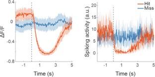
While calcium traces represent an indirect measure of neural activity, deconvolution does not necessarily provide an accurate picture of the spiking underlying those traces and has the potential to introduce additional problems. For instance, deconvolution algorithms tend to perform poorly at inferring the spiking of inhibited neurons (Vanwalleghem et al., 2021). Given that suppression is such a prominent feature of IC activity and is evident both in our calcium data as well as in the electrophysiology data of others (Franceschi and Barkat, 2021), we decided against using deconvolved spikes in our analyses. See also the side-by-side comparison below of the hit and miss trial activity of one example neuron based on either the calcium trace (left) or deconvolved spikes (right) (extracted using the OASIS algorithm (Friedrich et al., 2017) incorporated into suite2p (Pachitariu et al., 2016).
Author response image 1.
(3) Along the same line, the very small proportion of really sensory driven neurons (cluster 3) is not discussed. Is it what on would expect in typical shell or core IC neurons?
As requested by reviewer #2 and mentioned in response to the previous point, we have now quantified the number of neurons in the dataset that produced significant responses to sound (97 / 2649). For a given imaging area, the fraction of neurons that show a statistically significant change in neural activity following presentation of a click of between 53 dB SPL and 65 dB SPL rarely exceeded ten percent. While that number is low, it is not necessarily surprising given the moderate intensity and very short duration of the stimuli. For comparison: Using the same transgenics, labeling approach and imaging setup and presenting 200-ms long pure tones at 60 dB SPL with frequencies between 2 kHz and 64 kHz, we typically find that between a quarter and a third of neurons in a given imaging area exhibit a statistically significant response (data not shown).
Changes to manuscript.
Line 219: “..., while only a small fraction (97 / 2649) exhibited a significant response to the sound.”
Line 658: “Sound-driven neurons were identified by comparing the mean miss trial activity before and after stimulus presentation. Specifically, we performed a Mann-Whitney U test to assess whether there was a statistically significant difference (Benjamini-Hochberg adjusted p-value of 0.05) between the mean activity over the 2 seconds preceding the stimulus and the mean activity over the 1 second period following stimulus presentation. This analysis was performed using miss trials with click intensities from 53 dB SPL to 65 dB SPL (many sessions contained very few or no miss trials at higher sound levels).”
Line 220: “While the number of sound-responsive neurons is low, it is not necessarily surprising given the moderate intensity and very short duration of the stimuli. For comparison: Using the same transgenics, labeling approach and imaging setup and presenting 200-ms long pure tones at 60 dB SPL with frequencies between 2 kHz and 64 kHz, we typically find that between a quarter and a third of neurons in a given imaging area exhibit a statistically significant response (data not shown).”
(4) In the discussion, the interpretation of different transient and permanent cortical inactivation experiment is very interesting and well balanced given the complexity of the issue. There is nevertheless a comment that is difficult to follow. The authors state:
If cortical lesioning results in a greater weight being placed on the activity in spared subcortical circuits for perceptual judgements, we would expect the accuracy with which trial-by-trial outcomes could be read out from IC neurons to be greater in mice without auditory cortex. However, that was not the case.
However, there is no indication that the activity they observe in shell IC is causal to the behavioral decision and likely it is not. There is also no indication that the behavioral signals seen by the authors reflect the weight put on the subcortical pathway for behavior. I find this argument handwavy and would remove it.
While we are happy to amend this section, we would not wish to remove it because a) we believe that the point we are trying to make here is an important and reasonable one and b) because it is consistent with the reviewer’s comment. Hopefully, the following will make this clearer: In order for the mouse to make a perceptual judgment and act upon it - in the context of our task, hearing a sound and then licking a spout - auditory information needs to be read out and converted into a motor command. If the auditory cortex normally plays a key role in such perceptual judgments, cortical lesions would require the animal to base its decisions on the information available from the remaining auditory structures, potentially including the auditory midbrain. This might result in a greater correspondence between the mouse’s behavior and the neural activity in those structures. That we did not observe this outcome for the IC could mean that the auditory cortex did not contribute to the relevant perceptual judgments (sound detection) in the first place. Therefore, no reweighting of signals from the other structures is necessary. Alternatively, greater weight might be placed exclusively on structures other than the auditory midbrain, e.g. the thalamus. The latter would imply that the contribution of the IC remains the same. This includes the possibility that the IC shell does not play a causal role in the behavioral decision – in either control mice or mice with cortical lesions – as suggested by the reviewer.
Changes to manuscript.
Line 471: “This could imply that, following cortical lesions, greater weight is placed on structures other than the IC, with the thalamus being the most likely candidate, ..”
(5) In Fig. 5 the two colors used in B and C are the same although they describe different categories.
The dark green and ‘deep orange’ we used to distinguish between non-lesioned and lesioned in Figure 5C are slightly lighter than the colors used to distinguish between these two categories in other figures and therefore might be more easily confused with the blue and red in Figure 5B. This has been changed.


